. 2003 Sep;77(18):10106-12.
doi: 10.1128/jvi.77.18.10106-10112.2003.

Development of pseudorabies virus strains expressing red fluorescent proteins: new tools for multisynaptic labeling applications

Affiliations

Development of pseudorabies virus strains expressing red fluorescent proteins: new tools for multisynaptic labeling applications

Bruce W Banfield et al. J Virol. 2003 Sep.

Abstract

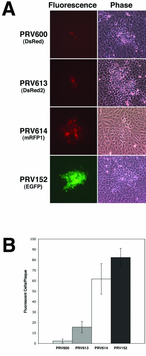
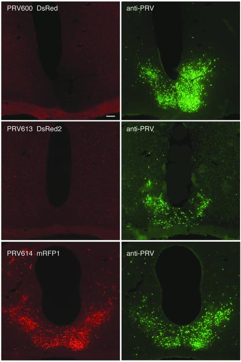
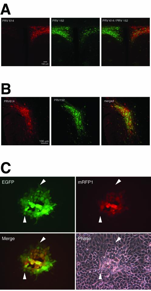
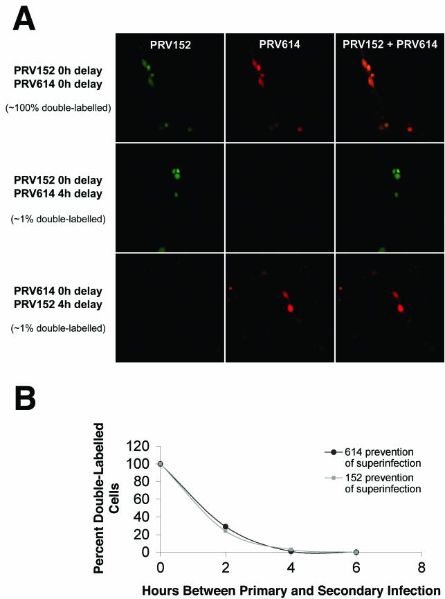
The transsynaptic retrograde transport of the pseudorabies virus Bartha (PRV-Bartha) strain has become an important neuroanatomical tract-tracing technique. Recently, dual viral transneuronal labeling has been introduced by employing recombinant strains of PRV-Bartha engineered to express different reporter proteins. Dual viral transsynaptic tracing has the potential of becoming an extremely powerful method for defining connections of single neurons to multiple neural circuits in the brain. However, the present use of recombinant strains of PRV expressing different reporters that are driven by different promoters, inserted in different regions of the viral genome, and detected by different methods limits the potential of these recombinant virus strains as useful reagents. We previously constructed and characterized PRV152, a PRV-Bartha derivative that expresses the enhanced green fluorescent protein. The development of a strain isogenic to PRV152 and differing only in the fluorescent reporter would have great utility for dual transsynaptic tracing. In this report, we describe the construction, characterization, and application of strain PRV614, a PRV-Bartha derivative expressing a novel monomeric red fluorescent protein, mRFP1. In contrast to viruses expressing DsRed and DsRed2, PRV614 displayed robust fluorescence both in cell culture and in vivo following transsynaptic transport through autonomic circuits afferent to the eye. Transneuronal retrograde dual PRV labeling has the potential to be a powerful addition to the neuroanatomical tools for investigation of neuronal circuits; the use of strain PRV614 in combination with strain PRV152 will eliminate many of the pitfalls associated with the presently used pairs of PRV recombinants.


Figures

FIG. 1.
FIG. 1.
Detection of infected PK15 cells by microscopy. (A) Fluorescence and phase-contrast images of PK15 cells 17 h after inoculation with strain PRV-Bartha derivatives expressing different fluorescent proteins. Cells were infected with strains PRV600, PRV613, PRV614, and PRV152. In the plaque assay, virus is added to monolayers of susceptible cells at a concentration of roughly 100 PFU per dish. The circular plaque that forms in the monolayer is a result of the spread of virus from a single infected cell to neighboring uninfected cells. PRV600 expresses DsRed, PRV613 expresses DsRed2, PRV614 expresses mRFP1, and PRV152 expresses EGFP. (B) Quantitation of the number of fluorescent cells per plaque at 17 h postinfection. Data were obtained through the analysis of 12 plaques per virus strain tested.
FIG. 2.
FIG. 2.
Coronal sections through the SCN, illustrating a red fluorescent signal at 96 h after inoculation of the right eye with strain PRV-Bartha recombinants. The left panels illustrate the expression of red fluorescent reporters in SCN-infected neurons. The right panels illustrate the same sections stained with an anti-PRV antibody and a secondary antibody conjugated to Alexa-488 (green). Note the absence of a red fluorescent signal in strains PRV600 (top left panel) and PRV613 (middle left panel), whereas virtually all PRV614-infected SCN cells produced a red fluorescent signal (bottom left panel).
FIG. 3.
FIG. 3.
Kinetics of mRFP1 and EGFP expression directed by PRV strains. (A) A coronal section of the PVN 96 h after injection of strain PRV614 into the right eye and strain PRV152 into the left eye as viewed using a Texas Red filter set to identify PRV614-infected neurons (left panel) and an EGFP filter set to identify PRV152-infected neurons (middle panel). The right panel image was captured using a dual-band EGFP-Texas Red filter set to identify PRV614- and PRV152-infected neurons simultaneously. (B) A coronal section of the IGL 96 h after a PRV152/PRV614 cocktail was injected into one eye. Images were captured with a Texas Red filter set (left panel) to identify PRV614-infected neurons and an EGFP filter set (right panel) to identify PRV152-infected neurons. A merged image is shown in the right panel. Approximately 75% of infected IGL neurons were labeled with both viruses. (C) Kinetics of fluorescent protein expression in PK15 cells infected with PRV615. PRV615 was engineered to express both mRFP1 and EGFP. Four images of the same PRV615 plaque are shown: top left panel, EGFP signal; top right panel, mRFP1 signal; bottom left panel, merge of EGFP and mRFP1 signals; bottom right panel, phase-contrast image. Arrowheads point to cells that are EGFP positive and mRFP1 negative.
FIG.4.
FIG.4.
Analysis of dual viral infection of cultured DRG cells. (A) DRG cells were infected with strains PRV152 and PRV614 simultaneously (top panels), PRV152 4 h prior to PRV614 infection (middle panels), or PRV614 4 h prior to PRV152 infection (bottom panels). The PRV152 signal (left panels), PRV614 signal (center panels), and merged signals (right panels) are shown. (B) Kinetics of superinfection inhibition. DRG cells were infected with strains PRV152 and PRV614 simultaneously, infected with PRV614 2, 4, or 6 h prior to PRV152 infection (closed circles), or infected with PRV152 2, 4, or 6 h prior to PRV614 infection (closed squares). The percentage of double-labeled cells was determined under each set of conditions and plotted as a function of time. A minimum of 196 cells was scored for each data point. Simultaneous infections were performed in duplicate, 2-h-delay experiments were performed in quadruplicate, 4-h-delay experiments were performed in duplicate (with PRV614 infection performed first) or triplicate (PRV152 first), and 6-h-delay experiments were performed in duplicate.

References

    1. Baird, G. S., D. A. Zacharias, and R. Y. Tsien. 2000. Biochemistry, mutagenesis, and oligomerization of DsRed, a red fluorescent protein from coral. Proc. Natl. Acad. Sci. USA 97:11984-11989. - PMC - PubMed
    1. Bevis, B. J., and B. S. Glick. 2002. Rapidly maturing variants of the Discosoma red fluorescent protein (DsRed). Nat. Biotechnol. 20:83-87. - PubMed
    1. Billig, I., J. M. Foris, L. W. Enquist, J. P. Card, and B. J. Yates. 2000. Definition of neuronal circuitry controlling the activity of phrenic and abdominal motoneurons in the ferret using recombinant strains of pseudorabies virus. J. Neurosci. 20:7446-7454. - PMC - PubMed
    1. Brandimarti, R., T. Huang, B. Roizman, and G. Campadelli-Fiume. 1994. Mapping of herpes simplex virus 1 genes with mutations which overcome host restrictions to infection. Proc. Natl. Acad. Sci. USA 91:5406-5410. - PMC - PubMed
    1. Brideau, A. D., J. P. Card, and L. W. Enquist. 2000. Role of pseudorabies virus Us9, a type II membrane protein, in infection of tissue culture cells and the rat nervous system. J. Virol. 74:834-845. - PMC - PubMed

Publication types

Substances