

doi: 10.1128/jvi.77.18.10131-10138.2003.
Hepatitis C virus-like particle budding: role of the core protein and importance of its Asp111
Affiliations
- PMID: 12941925
- PMCID: PMC224611
- DOI: 10.1128/jvi.77.18.10131-10138.2003
Item in Clipboard
Hepatitis C virus-like particle budding: role of the core protein and importance of its Asp111
J Virol.
2003 Sep.
Abstract
In the absence of a hepatitis C virus (HCV) culture system, the use of a Semliki Forest virus replicon expressing genes encoding HCV structural proteins that assemble into HCV-like particles provides an opportunity to study HCV morphogenesis. Using this system, we showed that the HCV core protein constitutes the budding apparatus of the virus and that its targeting to the endoplasmic reticulum by means of the signal sequence of E1 protein is essential for budding. In addition, the aspartic acid at position 111 in the HCV core protein sequence was found to be crucial for virus assembly, demonstrating the usefulness of this system for mapping amino acids critical to HCV morphogenesis.
Figures
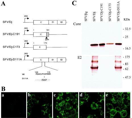
Description of the SFV RNAs encoding HCV structural proteins and expression in BHK-21 cells. (A) RNAs transcribed from the four SFV DNA constructs. The SFV/Dj construct is our original Dj6.4 clone, encoding HCV core-E1-E2, and is described elsewhere (4). The horizontal line in each construct indicates the segment of the SFV genome. The position of the subgenomic SFV promoter is indicated (26S). Boxes show the inserted genes, and the shaded box in the SFV/Dj-C191 construct indicates the signal sequence targeting the E1 envelope glycoprotein to the ER. SPP, signal peptide peptidase processing between residues 173 and 174. The SFV/Dj-D111A construct was produced by introducing a D111-to-A substitution in the original SFV/Dj construct, i.e., introducing a viral L domain, PTAP. (B) Immunofluorescence staining, using a monoclonal anti-core antibody, of BHK-21 cells electroporated with SFV RNA β-Gal (a), Dj (b), Dj-C191 (c), Dj-C173 (d), and Dj-D111A (e). This assay was performed by following a standard procedure (35), using the mouse monoclonal anti-HCV core antibody MAb 1856 (Virostat, Portland, Maine). (C) Western blotting of these cells with monoclonal anti-core and anti-E2 antibodies. For this standard assay (4), the anti-core MAb 1856 gave poor results. We therefore carried out immunoblotting with the human monoclonal anti-core antibody B12.F8 (11) and the mouse monoclonal anti-E2 (10). Size markers (in kilodaltons) were obtained from New England Biolabs (Beverly, Mass.).

Electron micrographs of ultrathin sections of BHK-21 cells electroporated with SFV/Dj (A and B) or SFV/Dj-C173 (C) RNA. The large arrow in panel A indicates ER, revealing a dilated lumen and convoluted membranes, the arrowheads in panel B indicate virus-like particles budding from these convoluted membranes towards the ER lumen, and the arrows in panel C indicate normal, homogeneously distributed ER structures throughout the cytoplasm. Bars, 1 μm (A), 100 nm (B), and 200 nm (C).

Electron micrographs of ultrathin sections of BHK-21 cells electroporated with SFV/Dj-C191 RNA. (A and B) Areas of convoluted ER membranes displaying adhesion via their cytosolic sides (arrows). In panel B, an electron-dense midline similar to those found in intercellular adhesion complexes such as the desmosome can be observed. (C) A typical area of convoluted ER, revealing double-layered membranes and the budding of virus-like particles from these membranes into the ER lumen. (D and E) High-magnification images of some virus-like particles (arrowheads) budding into the ER. In panel D, note the virus-like budding (arrowhead) occurring close to a region in which ER membranes are adhering via their cytosolic sides (arrow). Bars, 100 nm (A, B, D, and E) and 200 nm (C).

Electron micrographs of ultrathin sections of BHK-21 cells electroporated with SFV/Dj (A) or SFV/Dj-D111A (B) RNA. (A) A typical area shows convoluted ER membranes in which the budding of virus-like particles occurs. (B) In the presence of the D111A mutation, the formation of these convoluted ER membranes is retained but no virus particles are detected in these membranes or in the ER lumen. Bars, 1 μm (A) and 500 nm (B).
References
-
- Acosta-Rivero, N., J. C. Aguilar, A. Musacchio, V. Falcon, A. Vina, M. C. de la Rosa, and J. Morales. 2001. Characterization of the HCV core virus-like particles produced in the methylotrophic yeast Pichia pastoris. Biochem. Biophys. Res. Commun. 287:122-125. - PubMed
-
- Beard, M. R., G. Abell, M. Honda, A. Carroll, M. Gartland, B. Clarke, K. Suzuki, R. Lanford, D. V. Sangar, and S. M. Lemon. 1999. An infectious molecular clone of a Japanese genotype 1b hepatitis C virus. Hepatology 30:316-324. - PubMed
-
- Blight, K.-J., A. A. Kolykhalov, and C. M. Rice. 2000. Efficient initiation of HCV RNA replication in cell culture. Science 290:1972-1974. - PubMed
Publication types
MeSH terms
Substances
LinkOut - more resources
Full Text Sources
