. 2003 Sep;77(18):10168-71.
doi: 10.1128/jvi.77.18.10168-10171.2003.

Interleukin-15 and natural killer and NKT cells play a critical role in innate protection against genital herpes simplex virus type 2 infection

Affiliations

Interleukin-15 and natural killer and NKT cells play a critical role in innate protection against genital herpes simplex virus type 2 infection

Ali A Ashkar et al. J Virol. 2003 Sep.

Abstract

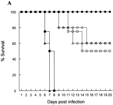
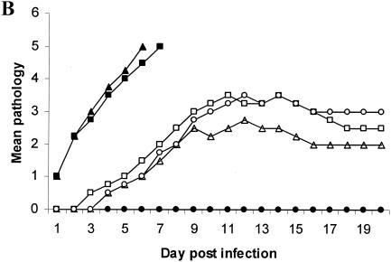
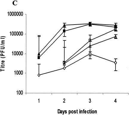
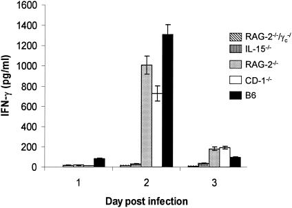
Interleukin-15 (IL-15), natural killer (NK) cells, and NK T (NKT) cells, components of the innate immune system, are known to contribute to defense against pathogens, including viruses. Here we report that IL-15(-/-) (NK(-) and NKT(-/+)) mice and RAG-2(-/-)/gamma(c)(-/-) (NK(-) and NKT(-)) mice that lack all lymphoid cells were very susceptible to vaginal infection with a low dose of herpes simplex virus type 2 (HSV-2). IL-15(-/-) and RAG-2(-/-)/gamma(c)(-/-) mice were 100-fold more susceptible and RAG-2(-/-), CD-1(-/-) (NKT(-)), and gamma interferon (IFN-gamma)(-/-) mice were 10-fold more susceptible to vaginal HSV-2 infection than control C57BL/6 mice. NK and/or NKT cells were the early source of IFN-gamma in vaginal secretions following genital HSV-2 infection. This study demonstrates that IL-15 and NK-NKT cells are critical for innate protection against genital HSV-2.


Figures

FIG. 1.
FIG. 1.
Susceptibility to vaginal HSV-2 infection in mice lacking all or some of the lymphocyte lineages, IL-15, or IFN-γ−/−. Naïve mice were infected IVAG with a low dose (100 PFU) of HSV-2 and monitored daily for vaginal pathology, survival, and vaginal virus titer. Statistical differences in the viral titers were determined by analysis of variance followed by Tukey's test. The statistical significance of the survival rates was determined by log rank statistic (P of <0.05 was considered statistically significant). (A and B) RAG-2−/−c−/− (▪) and IL-15−/− (▴) mice (n = 24) were extremely susceptible to low-dose challenge and showed the most severe pathology compared to B6 control mice (•), which showed 100% survival (n = 20). RAG-2−/− (□), CD-1−/− (▵) and IFN-γ−/− (○) mice (n = 10) were significantly (P < 0.01) more susceptible to vaginal HSV-2 than B6 mice but significantly (P < 0.01) less susceptible than RAG-2−/−c−/− and IL-15−/− mice. (C) Vaginal HSV-2 titers (n = 8 to 10/group) were examined on days 1 to 4 after viral challenge. While no viral shedding was detected in B6 control mice (♦), RAG-2−/−c−/− (▪) and IL-15−/− (▴) mice had the highest viral titers compared to the mice of all other groups. RAG-2−/− (□), CD-1−/− (Δ), and IFN-γ−/− (⋄) mice were significantly more susceptible to vaginal HSV-2 than B6 mice but less susceptible than RAG-2−/−c−/− and IL-15−/− mice.
FIG. 1.
FIG. 1.
Susceptibility to vaginal HSV-2 infection in mice lacking all or some of the lymphocyte lineages, IL-15, or IFN-γ−/−. Naïve mice were infected IVAG with a low dose (100 PFU) of HSV-2 and monitored daily for vaginal pathology, survival, and vaginal virus titer. Statistical differences in the viral titers were determined by analysis of variance followed by Tukey's test. The statistical significance of the survival rates was determined by log rank statistic (P of <0.05 was considered statistically significant). (A and B) RAG-2−/−c−/− (▪) and IL-15−/− (▴) mice (n = 24) were extremely susceptible to low-dose challenge and showed the most severe pathology compared to B6 control mice (•), which showed 100% survival (n = 20). RAG-2−/− (□), CD-1−/− (▵) and IFN-γ−/− (○) mice (n = 10) were significantly (P < 0.01) more susceptible to vaginal HSV-2 than B6 mice but significantly (P < 0.01) less susceptible than RAG-2−/−c−/− and IL-15−/− mice. (C) Vaginal HSV-2 titers (n = 8 to 10/group) were examined on days 1 to 4 after viral challenge. While no viral shedding was detected in B6 control mice (♦), RAG-2−/−c−/− (▪) and IL-15−/− (▴) mice had the highest viral titers compared to the mice of all other groups. RAG-2−/− (□), CD-1−/− (Δ), and IFN-γ−/− (⋄) mice were significantly more susceptible to vaginal HSV-2 than B6 mice but less susceptible than RAG-2−/−c−/− and IL-15−/− mice.
FIG. 2.
FIG. 2.
NK and/or NKT cells are the early source of IFN-γ in vaginal washes following IVAG HSV-2 infection. Significant levels of IFN-γ were detected on day 2 postinfection only in the vaginal washes from mice having NK and/or NKT cells (B6, RAG-2−/−, and CD-1−/− mice), whereas no or very low levels of IFN-γ were detected in the washes from mice lacking NK and NKT cells (RAG-2−/−c−/− and IL-15−/− mice). Vaginal washes from IFN-γ−/− mice were used as a negative control and had values no higher than those of the PBS control, showing the sensitivity of the test for the vaginal fluids.

References

    1. Ahmad, A., E. Sharif-Askari, L. Fawaz, and J. Menezes. 2000. Innate immune response of the human host to exposure with herpes simplex virus type 1: in vitro control of the virus infection by enhanced natural killer activity via interleukin-15 induction. J. Virol. 74:7196-7203. - PMC - PubMed
    1. Biron, C. A., and L. Brossay. 2001. NK cells and NKT cells in innate defense against viral infections. Curr. Opin. Immunol. 13:458-464. - PubMed
    1. Biron, C. A., K. B. Nguyen, G. C. Pien, L. P. Cousens, and T. P. Salazar-Mather. 1999. Natural killer cells in antiviral defense: function and regulation by innate cytokines. Annu. Rev. Immunol. 17:189-220. - PubMed
    1. Blauvelt, A., H. Asada, V. Klaus-Kovtun, D. J. Altman, D. R. Lucey, and S. I. Katz. 1996. Interleukin-15 mRNA is expressed by human keratinocytes Langerhans cells, and blood-derived dendritic cells and is downregulated by ultraviolet B radiation. J. Investig. Dermatol. 106:1047-1052. - PubMed
    1. Fawaz, L. M., E. Sharif-Askari, and J. Menezes. 1999. Up-regulation of NK cytotoxic activity via IL-15 induction by different viruses: a comparative study. J. Immunol. 163:4473-4480. - PubMed

Publication types