. 2003 Jun;4(2):164-75.
doi: 10.1007/s10162-002-2056-4.

Effects of chronic furosemide treatment and age on cell division in the adult gerbil inner ear

Affiliations

Effects of chronic furosemide treatment and age on cell division in the adult gerbil inner ear

H Lang et al. J Assoc Res Otolaryngol. 2003 Jun.

Abstract

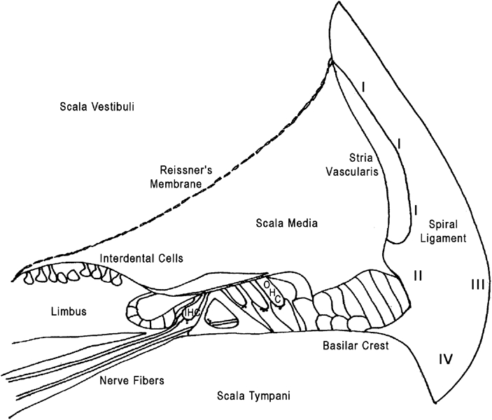
Atrophy of the stria vascularis and spiral ligament and an associated decrease in the endocochlear potential (EP) are significant factors in age-related hearing loss (presbyacusis). To model this EP decrease, furosemide was delivered into the round-window niche of young adult gerbils by osmotic pump for seven days, chronically reducing the EP by 30-40 mV. Compound action potential (CAP) thresholds were correspondingly reduced by 30-40 dB SPL at high frequencies. Two weeks after withdrawal of furosemide, the treated ears showed an EP recovery of up to 20-30 mV along with a similar recovery of CAP thresholds. The influence of cell division on furosemide-induced and age-related decline of the EP was examined using a mitotic tracer, bromodeoxyuridine (BrdU). Cell proliferation was examined in three groups: young control, furosemide-treated, and aged cochleas. Sections immunostained for BrdU were bleached with H2O2 to eliminate ambiguities with melanin pigment in the inner ear. Cell types positively labeled for BrdU in all three groups included Schwann cells in Rosenthal's canal; glial cells in the osseous spiral lamina; fibrocytes in the limbus, sacculus, and spiral ligament (SL); epithelial cells in Reissner's and round-window membranes; intermediate cells in the stria vascularis; and vascular endothelial cells. Quantitative analysis showed that the mean number of BrdU-positive (BrdU+) intermediate cells in the stria did not differ significantly among the three groups. In contrast, there was a significant increase of BrdU + fibrocytes in the SL of furosemide-treated animals as compared to the young control group. Moreover, there was a significant decrease in labeled fibrocytes in the aged versus the young ears, particularly among the type II and type IV subtypes. The results suggest that the increased fibrocyte turnover in the SL after furosemide treatment may be related to the recovery of EP and CAP thresholds, supporting the hypothesis that fibrocyte proliferation may be essential for maintaining the EP and cochlear function in normal and damaged cochleas. Moreover, the decreased turnover of SL fibrocytes with age may be a contributing factor underlying the lateral wall pathology and consequent EP loss that often accompanies presbyacusis.


Figures

Figure 1
Figure 1
Timeline of the experimental protocols. Downward arrows indicate pump implantation or removal; downward arrowheads indicate BrdU injection.
Figure 2
Figure 2
Mean CAP thresholds and EP recorded in control and furosemide-treated ears. Error bars represent standard errors of the mean (SEM). Control data (solid squares) were obtained from the untreated left ears of both furosemide groups. Data from the seven-day treatment are plotted as filled circles, whereas data from animals similarly treated but allowed to recover for two weeks are plottedas open circles. The CAP thresholds and EP of the recovered group are similar to control data, with the largest differences seen at the highest frequencies.
Figure 3
Figure 3
A. Numerous melanosomes were present in strial intermediate cells, which made identifying BrdU+ cells problematic. B. Same section asshown in A after bleaching the melanin pigment with H2O2. C. Nuclei of two intermediate cells (arrows) are stained forBrdU in the bleached stria vascularis. D. Small intestine as a positive control shows a high-BrdU labeling index after bleaching. Bars = 25 µm.
Figure 4
Figure 4
BrdU staining of different cell types in the gerbil labyrinth. A. Low-magnification view of Rosenthal's canal. A′. Arrowsin higher magnification of boxed area in A point to three BrdU+ cells. B. Arrow points to a positively labeled cell in the osseous spiral lamina beneath the limbus. C′. Higher magnification of boxed area in C shows three BrdU+ cells (arrows). D′. Higher magnification of boxed area in D shows a BrdU+ cell (arrow) in Reissner's membrane. E′. Higher magnification of boxed areain E shows four BrdU+ nuclei (arrows) in connective tissue under the utricular macula. F. Arrow points to a BrdU+ cell in the round-window membrane. Bars = 50 µm.
Figure 5
Figure 5
BrdU+ cells in the spiral ligament (arrows). All sections are from the first turn. A. Five BrdU+ nuclei are present in the spiral ligament of a furosemide-treated cochlea. Higher-magnification inset of boxed area shows two pairs of BrdU+ cells. B. A BrdU+ nucleus in the spiral ligament of a young control cochlea is more clearly seen in the higher-magnification inset at bottom right. C. No BrdU+ cells are present in this section of spiral ligament from an aged cochlea. Bar = 50 µm
Figure 6
Figure 6
A. Intermediate cell-labeling indices in the bleached stria vascularis. Means (±SEM) are from BrdU+ cell counts in six furosemide-treated, five aged, and six young control ears. There were no significant differences among the furosemide-treated, aged, and young control groups (ANOVA, p > 0.05). B. Quantification of fibrocyte proliferation in the spiral ligament after bleaching. Means (±SEM) are from same animals shown in A. Note the scale compared with A. The mean number of dividing fibrocytes in the furosemide group was significantly greater than in young controls; conversely, the mean number of proliferating fibrocytes in the aged group was significantly less than in young controls (ANOVA, p < 0.05).
Figure 7
Figure 7
Schematic diagram of a cross section of the cochlear duct showing the location criteria for classification of fibrocytes in the spiral ligament. Type I fibrocytes (I), beneath the stria vascularis; type II fibrocytes (II), superficial between the basilar crest and the stria vascularis; type III fibrocytes (III), adjacent to the otic capsule; type IV fibrocytes (IV), inferior to basilarmembrane near scala tympani.
Figure 8
Figure 8
A. Average numbers of BrdU+ cells within various fibrocyte subtypes in the spiral ligament. Data are from the same ears as Fig. 6B and have been normalized to one ear. Means (±SEM) are from six furosemide-treated, six young control, and five aged ears. Numbers of proliferating cells in all fibrocyte subtypes in the furosemide-treated group were significantly greater than those in young controls; however, only type II and IV fibrocytes were significantly smaller in the aged group as compared with young controls (ANOVA, p < 0.05). B. Average numbers of BrdU+ type II and IV fibrocytes in the spiral ligament as normalized to one ear. Data are from the same animals shown in A.Numbers of proliferating type II and IV fibrocytes in the furosemide-treated group were increased significantly over those from young controls; conversely, the numbers of proliferating type II and IV fibrocytes in the aged group were significantly less than those found in young controls (ANOVA, p < 0.05).
Figure 9
Figure 9
A. Cell densities of type II and IV fibrocytes in the spiral ligaments of furosemide-treated, control, and aged gerbils. Data are from the same animals shown in Fig. 8. Cell density of type II and IV fibrocytes in the aged group was significantly less than that in young controls (ANOVA, p < 0.05); however, there was no significant differencebetween the furosemide-treated and young control groups (ANOVA, p > 0.05). B. The cell-proliferating index of type II and IV fibrocytes as normalized to cell density. Even when cell density is controlled, there was still a significant increase in cell proliferation in type II and IV fibrocytes in the furosemide-treated group compared with young controls (ANOVA, p < 0.05). Similarly, the proliferation of type II and IV fibrocytes was significantly reduced in the aged group as compared to young controls (ANOVA, p < 0.05).

Similar articles

Cited by

References

    1. Conlee JW, Gerity LC, Bennett ML. Ongoing proliferation of melanocytes in the stria vascularis of adult guinea pigs. Hear. Res. 1994;79:115–122. doi: 10.1016/0378-5955(94)90133-3. - DOI - PubMed
    1. Crouch JJ, Sakaguchi N, Lytle C, Schulte BA. Immunohistochemical localization of the Na–K–Cl co-transporter (NKCC1) in the gerbil inner ear. J. Histochem. Cytochem. 1997;45:773–778. - PubMed
    1. Forge A. Observations on the stria vascularis of the guinea pig cochlea and the changes resulting from the administration of the diuretic furosemide. Clin. Otolaryngol. 1976;1:211–219. - PubMed
    1. Franz P, Aharinejad S, Firbas W. Melanocytes in the modiolus of the guinea pig cochlea. Acta Otolaryngol. 1990;109:221–227. - PubMed
    1. Gratacap B, Charachon R, Stoebner P. Results of an ultrastructural study comparing stria vascularis with organ of Corti in guinea pigs treated with kanamycin. Acta Otolaryngol. (Stockh.) 1985;99:339–342. - PubMed

Publication types

MeSH terms