. 2003 Jun;4(2):176-95.
doi: 10.1007/s10162-002-2036-8.

Distribution of gentamicin in the guinea pig inner ear after local or systemic application

Affiliations

Distribution of gentamicin in the guinea pig inner ear after local or systemic application

Shun-Ichi Imamura et al. J Assoc Res Otolaryngol. 2003 Jun.

Abstract

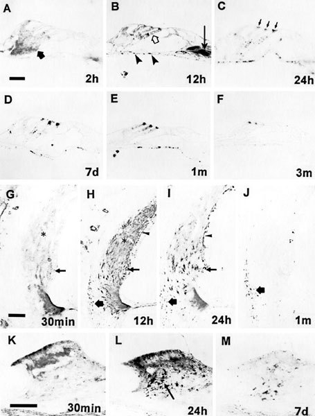
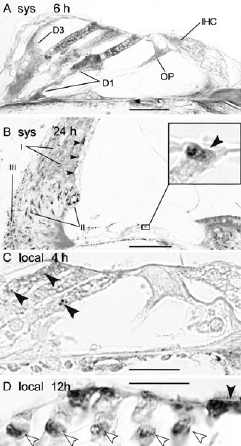
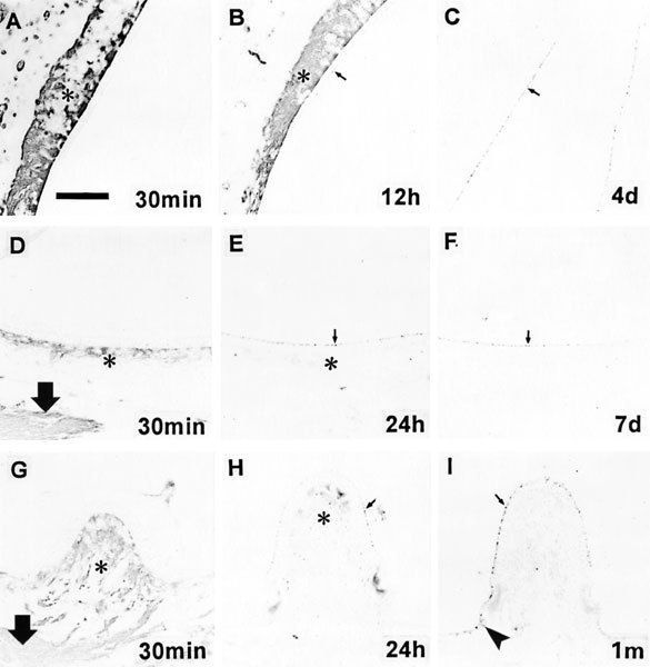
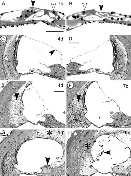
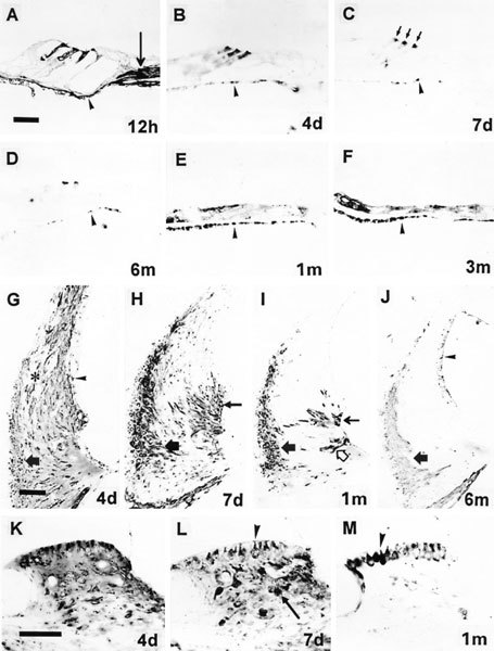
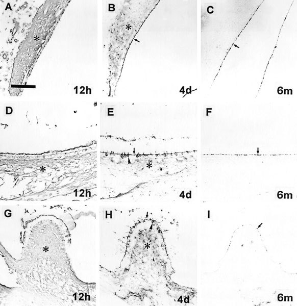
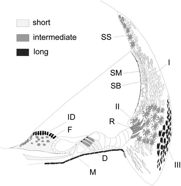
Uptake and retention of gentamicin by cells in the guinea pig inner ear after a single peritoneal injection or local application on the round window were investigated using immunocytochemistry to localize the drug. The cells that accumulated the drug under the two conditions were the same, but staining for the drug was more intense and was often accompanied by widespread cochlear degeneration following local application. Soon after drug administration by either route, there was diffuse staining for the drug throughout all tissue within the labyrinth, including bone. At later times when distinct cell staining became evident, virtually all cell types were found to be positive, with several cell types staining more darkly for the drug than hair cells, indicating that hair cells were not the most avid in accumulating gentamicin. The infracuticular portion of auditory and vestibular hair cells as well as type III fibrocytes of the spiral ligament were positively stained in almost all cases and these sites were found to be positive for as long as six months post administration. In animals with loss of the organ of Corti, there was unusually intense staining for gentamicin in root cells of the spiral ligament, in marginal cells of the stria vascularis, and in cells of the spiral limbus. Dark staining of surviving cells in cases with overt tissue destruction suggests that variability in the extent of damage caused by the drug was determined more by the degree of its local uptake than by differences in animals' capacities to metabolize the drug systemically. The present results show that gentamicin may damage or destroy all cochlear cells following a single round window application. The findings broaden the scope of our knowledge of cochlear gentamicin uptake and damage and have implications for treatment of patients with vestibular disorders by infusion of aminoglycosides into the middle ear, as well as implications for prospects of rehabilitating patients that have been deafened by aminoglycosides.


Figures

Figure 1
Figure 1
Immunostaining for gentamicin of the cochlea following systemic application. Numbers on the photos indicate survival periods. A. Deiters' cells (filled arrow) are immunoreactive for gentamicin. B. Immunostaining for gentamicin in the cytosol of outer hair cells (OHCs) shows a reticular pattern (open arrow). The arrow indicates stained radial fibers. C–F. The subcuticular plate region of OHCs (arrows in C) shows spotty reaction product for gentamicin. G–J. Immunostaining for gentamicin in the lateral wall of the cochlea shows different patterns depending upon survival periods. The four images are the same view of the cochlea represented at different survival times. In G, there is faint, indistinct immunostaining for gentamicin in the spiral ligament and stria vascularis with no clearly delineated immunoreactive cells. (To compare this staining within background levels, compare with J.) In H, type I (asterisk), type II (small arrow), and type III fibrocytes (large arrow) of the spiral ligament are positive for gentamicin. Basal cells (arrowhead) of the stria vascularis are positive. In I, type II fibrocytes and root cells (small arrow) and type III fibrocytes (large arrow) of the spiral ligament are immunoreactive for gentamicin. Basal cells (arrowhead) of the stria vascularis are also positive. In J, only type III fibrocytes (large arrow) of the spiral ligament are positive for gentamicin. The reaction product in the stria vascularis is an artifact produced by the enzymatic activity of erythrocytes. K–M. Immunostaining for gentamicin of the spiral limbus shows diverse patterns depending upon survival periods. In K, there is diffuse immunostaining for gentamicin in the spiral limbus. In L, fibrocytes (arrow) of the spiral limbus are positive for gentamicin and there is diffuse staining in the upper surface of the limbus. In M, only fibrocytes of the spiral limbus are positive for gentamicin. E and F are from basal turn, others are from second turn of the cochlea. The scale bar in A = 20 µm and applies to the B–F. The scale bar in G = 50 µm and applies to the G–J. The calibration bar in K = 50 µm and applies to the K–M.
Figure 2
Figure 2
Patterns of hair cell immunostaining in organ of Corti. A. Systemic injection case showing a reticular pattern of staining in OHCs and diffuse staining of IHC and first and third row Deiters' cells (D1, D3). Outer pillar cells (OP) and mesenchymal cells of the basilar membrane are also stained. Scale bar = 25 µm. B. Systemic injection case with 24 h survival. Type II and type III fibrocytes are darkly stained in comparison with the organ of Corti. Type I fibrocytes are weakly stained. Arrowheads indicate darkly stained strial basal cells. The inset is a high magnification view of the OHC apex indicated by the small box. In this view the reaction product is confined to the region below the cuticular plate, which is indicated by the arrowhead. Scale bar = 100 µm. C. Local application case with 4 h survival showing granular reaction product in the OHC cytoplasm (arrowheads). Scale bar = 25 µm. D. Local injection case with 12 h survival showing dense staining in the bases (open arrowheads) and apices of OHCs. The plane of section was not parallel to the long axis of the cells and the section was somewhat tangential so that there appear to be 5 rows of OHCs. The paired bases (open arrowheads) and apices (above) are from different cells. The closed arrowhead indicates a clear cuticular plate above the dense apical reaction product. Scale bar = 25 µm.
Figure 3
Figure 3
Immunostaining for gentamicin in the vestibular organs following systemic application. (Numbers on the photos indicate survival periods.) Images in each row are of the same organs showing progressively less staining with increasing survival times. A, D, G. Fibrocytes of the connective tissue (asterisks) of the saccule (A), the utricle (D), and the lateral semicircular canal (G) are immunoreactive for gentamicin, but hair cells are not positive. B, E, H. The apical regions (arrows) of hair cells of the saccule (B), the utricle (E), and the crista of the lateral canal (H) are positive for gentamicin. Fibrocytes (asterisk in B and H) are also positive. C, F, I. Only the apical portions (arrows) of hair cells of the saccule (C), the utricle (F), and the lateral canal (I) are immunopositive for gentamicin. In addition, apical staining of transitional cells (arrowhead in I) was sometimes present. Large arrows in D and G indicate the vestibular nerve. Scale bar in A =100 µm.
Figure 4
Figure 4
H&E staining of cochleas treated with local application of gentamicin. The pair of images at the top are from the upper basal turn and show a gentamicin-treated ear (B) and the corresponding image of the contralateral control ear (A) in a case with minimal degeneration. The open arrowheads indicate IHCs, closed arrowheads indicate OHCs. On the treated side both OHCs and Deiters' cells are missing and the height of the organ is considerably reduced. Scale bar = 50 µm. C, D. Treated (C) and untreated control (D) cochleas from a case with minimal overt tissue damage but endolymphatic hydrops, as indicated by the bulging Reissner's membrane (closed arrowhead in C). The white arrowhead indicates a site of apparent cell loss within the spiral ligament. The artificial perilymph formulation of gentamicin was used in this case. In all other cases shown in this figure except for E, the gentamicin for injection formulation was applied to the round window. Calibration bar in D = 100 µm and applies to C and D. E. A 4 day survival case with complete destruction of the organ of Corti and disruption of other epithelial cells except for the stria vascularis, which is shrunken. In E and F the organ of Corti has been replaced by a squamous epithelium. The nuclei of the squamous cells are present on the superior surface of the basilar membrane. The arrowheads in the spiral ligament of E and F indicate sites of cell loss, which shows obvious cell loss (compare with E and F with G). This was the most extreme case of degeneration seen in animals treated with the gentamicin formulated in artificial perilymph. Scale bar =100 µm and applies to E–H. F. A case in which the membranous labyrinth was reduced to a squamous epithelium, except for strial marginal cells. There may be mild cell loss in the spiral ligament (region indicated by the arrowhead). G. A case with less destruction of epithelial cells than shown in E and F as evidenced by pillar cells in the organ of Corti and interdental cells, indicated by the arrowhead. There was little apparent cell loss in the spiral ligament but there was endolymphatic hydrops and the scala vestibuli was completely filled with fibrotic tissue (asterisk). H. The most advanced degeneration seen following gentamicin administration. The membranous labyrinth has been reduced to a squamous epithelium, including the stria vascularis. The debris indicated by the arrowhead appears to be the remains of the stria vascularis. There was endolymphatic hydrops, fibrosis of the scala vestibuli, and new bone formation. The white asterisk indicates new bone formed on the middle ear side of the otic capsule. The black asterisk indicates new bone that has replaced part of the spiral ligament. Numbers in the photos indicate survival periods.
Figure 5
Figure 5
Immunostaining for gentamicin in the cochlea following local application. Numbers on the photos indicate survival periods. A. The cytosol of OHCs shows intense immunoreaction for gentamicin. The arrow indicates heavily stained radial nerve fibers. B. OHC cuticular plates and supranuclear cytosol are immunoreactive for gentamicin. C, D. Only the subcuticular plate regions are positive for gentamicin. E, F. The partially collapsed organ of Corti shows inmunostaining for gentamicin. In A–F, the mesenchymal cells beneath the basilar membrane (arrowheads) show intense immunostaining. In G–J, immunostaining for gentamicin in the lateral wall of the cochlea shows different patterns depending upon survival periods. G. All fibrocytes in the spiral ligament show intense immunostaining. The asterisk indicates the type I fibrocyte region. Basal cells and intermediate cells (arrowhead) of the stria vascularis are immunoreactive. H. Type II fibrocytes and root cells (small arrow) and type III fibrocytes (large arrow) of the spiral ligament are immunoreactive. I. Root cells (open arrow and small arrow) and type III fibrocytes (large closed arrow) of the spiral ligament are immunoreactive. J. Type III fibrocytes (large arrow) are immunoreactive. The marginal cells of the stria vascularis (arrowhead) are positive in this case, which had organ of Corti destruction. K, L, M. The spiral limbus shows different patterns of immunostaining for gentamicin depending upon survival periods. K. There is diffuse staining within the entire spiral limbus. L. The fibrocytes (arrow) and interdental cells (arrowhead) are immunoreactive. M. Interdental cells (arrowhead) are positive. A, K, L, and M are from second turn, others are from basal turn of the cochlea. The scale bar in A = 20 µm and applies to A–F. The calibration bar in G = 50 µm and applies to the G–J. Scale bar in K = 50 µm and applies to the K–M.
Figure 6
Figure 6
Immunostaining for gentamicin in the vestibular system following local application. Numbers on the photos indicate survival periods. Each row shows the progression of staining with increasing survival time for a given tissue. A, D, G. Hair cells and fibrocytes (asterisk) in the subepithelial connective tissue of the saccule (A), the utricle (D), and crista of the lateral canal (G) are intensely positive for gentamicin. B, E, H. The apical portions of hair cells (arrows) of the saccule (B), the utricle (E), and crista of the lateral canal (H) show intense immunoreaction. Fibrocytes (asterisks) are weakly immunoreactive. In the utricle (E) and lateral canal (H), the cytosol of hair cells (arrowheads) is immunostained. C, F, I. The apical portions of hair cells (arrowheads) of the saccule (C), the utricle (F), and crista of the lateral canal (I) are immunoreactive. In C the vestibular membrane is also positive. Scale bar = 100 µm.
Figure 7
Figure 7
A schematic illustration of gentamicin uptake and retention in cells within the basal turn following local application. The affinity of cells for gentamicin was strongly correlated with duration of retention. The shading code indicates the duration after administration for which gentamicin immunostaining was present. The cytosol of inner hair cells and most supporting cells showed weak retention of gentamicin. The infracuticular plate region of OHCs, the mesenchymal cells (M) beneath the basilar membrane, interdental cells (ID) of the spiral ligament, and type III fibrocytes (III) showed high affinity and long-term retention. Outer sulcus cells, adjacent root cells (R), type II fibrocytes (II), fibrocytes (F) of the spiral limbus, and strial marginal cells (SM) showed intermediate affinity and retention of gentamicin. Type I fibrocytes (I), strial basal cells (SB), strial intermediate cells (SI), and Deiters' cells (D) showed low affinity for and brief retention of gentamicin.

Similar articles

Cited by

References

    1. Adams JC. Biotin amplification of biotin and horseradish peroxidase signals in histochemical stains. J. Histochem. Cytochem. 1992;40:1457–1463. - PubMed
    1. Aran JM, Erre JP, Lima da Costa D, Debbarh I, Dulon D. Acute and chronic effects of aminoglycosides on cochlear hair cells. Ann. N. Y. Acad. Sci. 1999;884:60–68. - PubMed
    1. Balogh K, Hiraide F, Ishi D. Distribution of radioactive dihydrostreptomycin in the cochlea—an autoradiographic study. Ann. Otol. (St. Louis) 1970;79:641–652. - PubMed
    1. Blakley BW. Update on intratympanic gentamicin for Meniere's disease. Laryngoscope. 2000;110:236–240. - PubMed
    1. Brummett RE, Traynor J, Brown R, Himes D. Cochlear damage resulting from kanamycin and furosemide. Acta Otolaryngol. 1975;80:86–92. - PubMed

Publication types

MeSH terms