. 2003 Jun;4(2):196-218.
doi: 10.1007/s10162-002-2037-7.

Changes in cytochemistry of sensory and nonsensory cells in gentamicin-treated cochleas

Affiliations

Changes in cytochemistry of sensory and nonsensory cells in gentamicin-treated cochleas

Shun-Ichi Imamura et al. J Assoc Res Otolaryngol. 2003 Jun.

Abstract

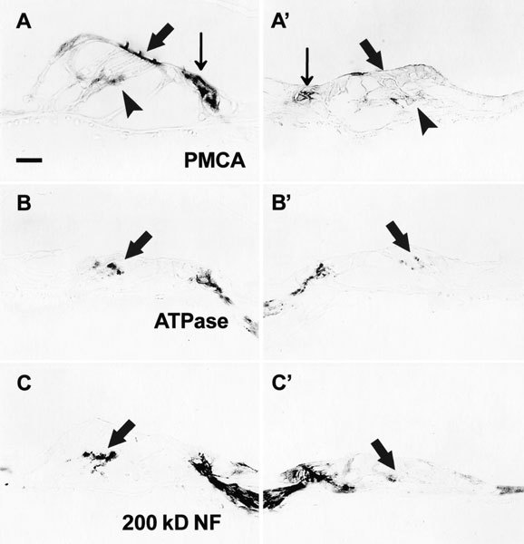
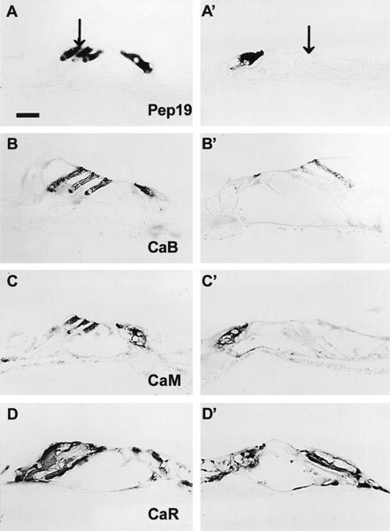
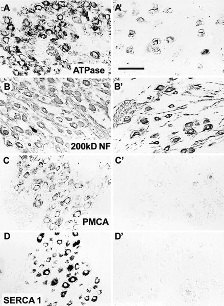
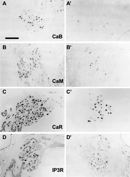
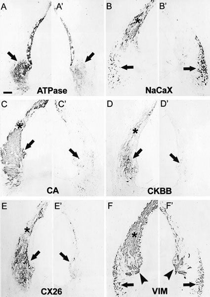
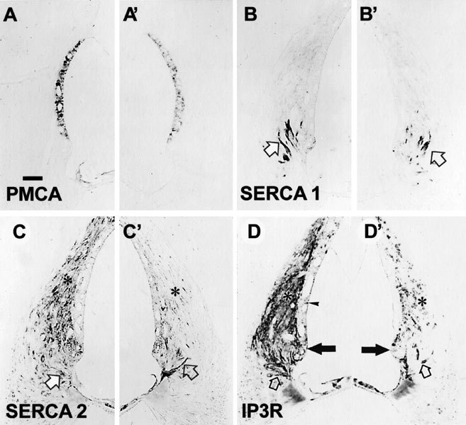
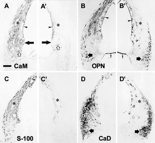
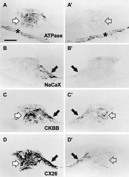
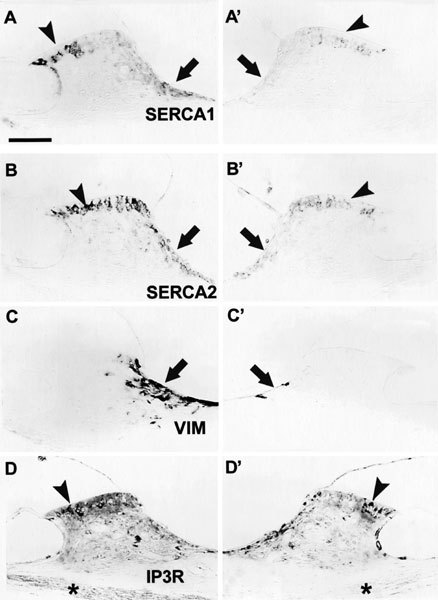
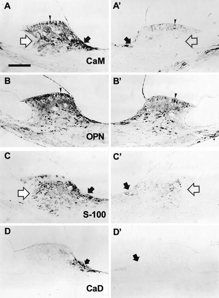
Effects of a single local dose of gentamicin upon sensory and nonsensory cells throughout the cochlea were assessed by changes in immunostaining patterns for a broad array of functionally important proteins. Cytochemical changes in hair cells, spiral ganglion cells, and cells of the stria vascularis, spiral ligament, and spiral limbus were found beginning 4 days post administration. The extent of changes in immunostaining varied with survival time and with cell type and was not always commensurate with the degree to which individual cell types accumulated gentamicin. Outer hair cells, types I and II fibrocytes of the spiral ligament, and fibrocytes in the spiral limbus showed marked decreases in immunostaining for a number of constituents. In contrast, inner hair cells, type III fibrocytes and root cells of the spiral ligament, cells of the stria vascularis, and interdental cells in the spiral limbus showed less dramatic decreases, and in some cases they showed increases in immunostaining. Results indicate that, in addition to damaging sensory cells, local application of gentamicin results in widespread and disparate disruptions of a variety of cochlear cell types. Only in the case of ganglion cells was it apparent that the changes in nonsensory cells were secondary to loss or damage of hair cells. These results indicate that malfunction of the ear following gentamicin treatment is widespread and far more complex than simple loss of sensory elements. The results have implications for efforts directed toward detecting, preventing, and treating toxic effects of aminoglycosides upon the inner ear.


Figures

Figure 1
Figure 1
Changes of immunostaining in the organ of Corti. In this and all other figures, pairs of images are presented from a given specimen. In each case the image on the left is the untreated control cochlea and that on the right is the gentamicin-treated cochlea. Corresponding portions of the cochleas are shown so that the images appear as mirror images of each other. A,A′. The cytosol of inner hair cells (IHCs) (small vertical arrow) and the stereocilia and cuticular plate of the outer hair cells (OHCs) (large diagonal arrow) on control side (A) are positive for PMCA-ATPase. On the treated side (A′), immunostaining of OHCs is missing and IHCs show weaker staining. Arrowheads indicate staining of Deiters′ cell cups. B,B′. Nerve terminals beneath OHCs and IHCs in the control side (B) are positive for Na+, K+-ATPase. On the treated side (B′), the staining of terminal of OHCs is decreased (arrow), but that of IHCs is not clearly decreased. C,C′. Nerve fibers beneath OHCs and IHCs in the control side (C) are positive for 200kD neurofilaments. On the treated side (C′), only nerve terminals beneath IHCs are immunopositive. Those beneath OHCs are not stained. Survival periods: A = 4 days, B,C = 1 month. Scale bar in A = 20 µm and applies to all images.
Figure 2
Figure 2
Changes of immunostaining in hair cells with 7 days survival following local application of gentamicin. A, A′. Arrows indicate third row of OHCs. OHCs (arrows) and (IHCs) on the control side (A) are positive for peptide 19. On the treated side (A′), only IHCs are stained. Arrow in A′ indicates the location of unstained OHCs. B,B′. IHCs and OHCs of the control side (B) are immunoreactive for calbindin. There is decreased staining on the treated side (B′). C,C′. IHCs and OHCs on the control side (C) are positive for calmodulin. On the treated side (C′), only IHCs are positive. D,D′. IHCs, Hensen's cells, and Deiters' cells of the control side (D) are positive for calretinin. On the treated side (D′), staining of Deiters' cells and Hensen's cells is decreased. Scale bar in A = 20 µm.
Figure 3
Figure 3
Changes of immunostaining in the spiral ganglion cells. A,A′. The periphery of spiral ganglion cells in the control ear (A) are positive for Na+,K+-ATPase. The number of cells and intensity of the immunostaining is decreased on the treated side (A′). B,B′. The cytosol of the spiral ganglion cells and nerve fibers in the control ear (B) are immunostained for 200kD neurofilaments. On the treated side (B′), the numbers of stained cells and nerve fibers are less. C,C′. The periphery of spiral ganglion cells in the control side (C) are positive for PMCA. On the treated side (C′), the number of cells and intensity of staining for PMCA are obviously decreased. D,D′. The cytosol of spiral ganglion cells in the control side (D) is positive for SERCA1. There is much reduced staining for SERCA1 in the treated (D′) side. Survival periods: A = 6 months, B = 1 month, C,D = 3 months. Scale bar in A' = 50 µm.
Figure 4
Figure 4
Changes of immunostaining in spiral ganglion cells following a local application of gentamicin. A,A′. Some spiral ganglion cells on the control side (A) are positive for calbindin. The number of cells and the intensity of immunostaining are decreased on the treated side (A′). B,B′. Spiral ganglion cells on the control side (B) are positive for calmodulin. The number of cells and the intensity of immunostaining are decreased on the treated side (B′). C,C′. Ganglion cells and nerve fibers of the control side (C) are positive for calretinin. The number of cells and fibers and the intensity of immunostaining are decreased on the treated side (C′). D,D′. Spiral gangilion cells are positive for IP3R. The number of cells and the intensity of immunostaining are decreased on the treated side (D′). Survival periods: B = 1 month, C,D = 3 months, A = 6 months. Scale bar in A = 100 µm.
Figure 5
Figure 5
Changes of immunostaining in the lateral wall of the cochlea (basal turn) with 1 month survival (1). A,A′. The stria vascularis and type II fibrocytes (arrow) in the spiral ligament of the control side (A) are immunoreactive for Na+,K+-ATPase. The staining is decreased on the treated side (A′). B,B′. Type I (asterisks) and III fibrocytes (arrows) in the spiral ligament on the control side (B) are immunoreactive for the Na+, Ca++ exchanger. On the treated side (B′), staining of type I fibrocytes is decreased. In contrast, staining of type III fibrocytes is increased. C,C′. Types I (asterisk) and II (arrows) fibrocytes of the spiral ligament in the control side (C) are positive for carbonic anhydrase. There was decreased immunostaining of the types I and II fibrocytes for carbonic anhydrase on the treated side (C′). D,D′. Types I and II fibrocytes of the spiral ligament in the control side are positive for creatinine kinase BB. In the treated ear (D′), this staining is clearly decreased. E,E′. Types I and II fibrocytes of spiral ligament in the control ear (E) are positive for connexin 26. There is decreased immunostaining in the treated side (E′). F,F′. Types I (asterisk) and III (arrows) fibrocytes and root cells of the spiral ligament (arrowheads) and basal cells in the stria vascularis in the control side (F) are positive for vimentin. In the treated ear (F′), immunostaining of type I fibrocytes and basal cells are obviously decreased, but immunostaining of root cells and type III fibrocytes still remains. Scale bar in A = 50 µm.
Figure 6
Figure 6
Changes of immunostaining in the lateral wall of the cochlea (basal turn) with 1 month survival. A,A′. Marginal cells of stria vascularis in the control side (A) are positive for PMCA. There was decreased immunostaining in treated side (A′). B,B′. Root cells (arrow) in the control ear (B) are positive for SERCA1. In the treated ear (B′), immunostaining of root cells is slightly decreased. C,C′. Type I fibrocytes of the spiral ligament in the control ear (C) are positive for SERCA2, and root cells (arrow) are slightly positive. In the treated ear (C′), there is decreased immunostaining of type I fibrocytes but there is no apparent decease in immunostaining of root cells. The survival period: A = 7 days, B = 6 months, C = 3 months. Scale bar in A = 50 µm.
Figure 7
Figure 7
Changes of immunostaining in the lateral wall of the cochlea (basal turn) with 1 month survival following a local application of gentamicin. A,A′. The lumenal surface of the stria vascularis (arrowhead) and types I (asterisk) and II fibrocytes (arrow) and root cells (open arrow) in the spiral ligament of the control side (A) are positive for calmodulin. Staining of type I and type II fibrocytes is obviously decreased on the treated side (A′). B,B′. The stria vascularis (arrowhead), types I (asterisk) and III fibrocytes (filled arrow) in the spiral ligament, and mesenchymal cells beneath the basilar membrane (small vertical arrow) of the control side (B) are immunoreactive for osteopontin. On the treated side (B′), there is decreased staining of type I fibrocytes. In contrast, there is increased immunostaining of type III fibrocytes. C,C′. Type I fibrocytes (asterisk) in the spiral ligament are positive for S-100 protein. Staining is decreased on the treated side (C′). D,D′. Types I (asterisk) and III flbrocytes (arrow) in the spiral ligament are immunoreactive for caldesmon. Staining of type I fibrocytes is decreased on the treated side (D′). Scale bar in A = 50 µm.
Figure 8
Figure 8
Changes in immunostaining of the spiral limbus with 1 month survival. A,A′. fibrocytes (arrow) in the spiral limbus and nerve fibers (asterisk) of the control side (A) are positive for Na+,K+-ATPase. Staining of fibrocytes is decreased in the treated side (A′). Cells in the supralimbal area (arrow) of the control side (B) are positive for the Na+,Ca++ exchanger. Immunostaining on the treated side (B′) is decreased. Fibrocytes (open arrow) and supralimbal cells (closed arrow) in the spiral limbus of the control side (C) are positive for creatine kinase isozyme BB. Staining for both cell types on the treated side (C′) is decreased. Fibrocytes (open arrow) and supralimbal cells (closed arrow) in the spiral limbus of the control side (D) are positive for connexin 26. There is decreased immunostaining of both cell types on the treated side (D′). Scale bar in A = 50 µm.
Figure 9
Figure 9
Changes in immunostaining of the spiral limbus (2). Interdental cells (arrowhead) located on the organ of Corti side in the spiral limbus and fibrocytes (arrow) in the supralimbal area of the control side (A) are positive for SERCA1. There is decreased unmunostaining of both cell types on the treated side (A′). Interdental cells (arrowhead) in the spiral limbus and fibrocytes (arrow) in the supralimbal area of the control side (B) are immunoreactive for SERCA2. This staining is decreased on the treated side (B′). Cells in the supralimbal area (arrow) of the control side (C) are positive for the vimentin. Staining on the treated side (C′) is decreased. Interdental cells (arrowhead) are immunostained for IP3R (D). On the treated side (D′), a decrease in staining is apparent on the modiolar side of the limbus, but less change, if any, is present on the organ of Corti side (arrowhead). Survival times: A = 3 months, B = 1 month, C = 6 months. Scale bar in A = 50 µm.
Figure 10
Figure 10
Changes in immunostaining of the spiral limbus (basal turn) following local application of gentamicin. Fibrocytes (open arrow) in the spiral limbus, supralimbal cells (closed arrow), and interdental cells (arrowhead) of the control side (A) are immunoreactive for calmodulin. There is decreased staining of all these cells on the treated side (A′). Fibrocytes in the spiral limbus and interdental cells (arrowhead) of the control side (B) are immunoreactive for osteopontin. There is decreased staining on the treated side (B′). Fibrocytes (open arrow) in the spiral limbus and supralimbal cells (closed arrow) of the control side (C) are positive for S-100. There is decreased immunostaining of both cell types on the treated side (C′). Supralimbal cells (arrow) of the control side (D) are positive for caldesmon. There is markedly decreased immunostaining on the treated side (D′). The survival periods: A,B = 7 days, C = 1 month, D = 6 months. Scale bar in A = 50 µm.

Similar articles

Cited by

References

    1. Adams JC. Biotin amplification of biotin and horseradish peroxidase signals in histochemical stains. J. Histochem. Cytochem. 1992;40:1457–1463. - PubMed
    1. Anniko M, Thornell L-E, Virtanen I. Cytoskeletal organization of the human inner ear. Acta Otolaryngol. Suppl. (Stockh.) 1987;437:29–54. - PubMed
    1. Apicella S, Chen S, Bing R, Penniston JT, Llinas R, Hillman DE. Plasmalemmal ATPase calcium pump localizes to inner and outer hair bundles. Neuroscience. 1997;79:1145–1151. doi: 10.1016/S0306-4522(97)00035-3. - DOI - PubMed
    1. Bareggi R, Grill V, Narducci P, Zweyer M, Tesei L, Russolo M. Gentamicin ototoxicity: histological and ultrastructural alterations after transtympanic administration. Pharmacol. Res. 1990;22:635–644. - PubMed
    1. Berggren D, Anniko M, Ramaekers F, Virtanen I. Intermediate filament proteins in the embryonic inner ear of mice under normal conditions and after exposure to ototoxic drugs. Acta Otolaryngol. 1990;109:57–65. - PubMed

Publication types

MeSH terms

LinkOut - more resources