. 2003 Jun;4(2):219-34.
doi: 10.1007/s10162-002-3022-x.

Neomycin-induced hair cell death and rapid regeneration in the lateral line of zebrafish (Danio rerio)

Affiliations

Neomycin-induced hair cell death and rapid regeneration in the lateral line of zebrafish (Danio rerio)

Julie A Harris et al. J Assoc Res Otolaryngol. 2003 Jun.

Abstract

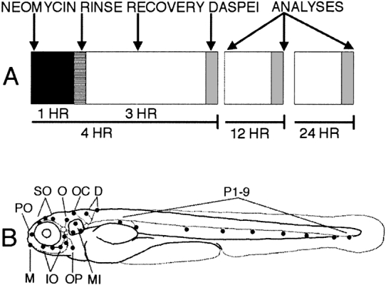
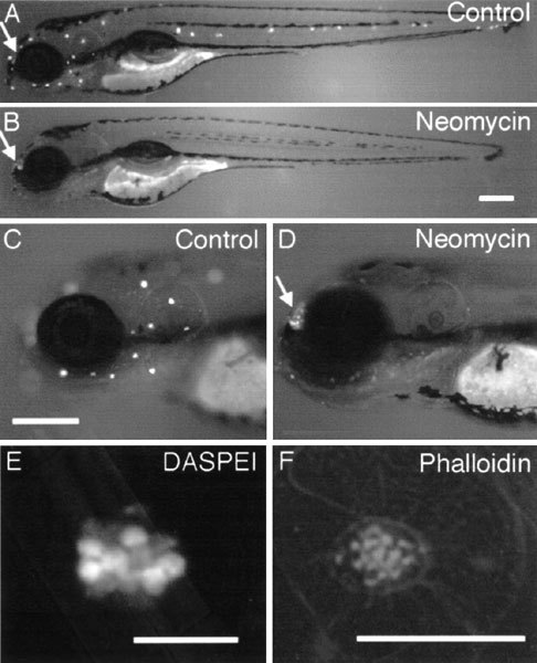
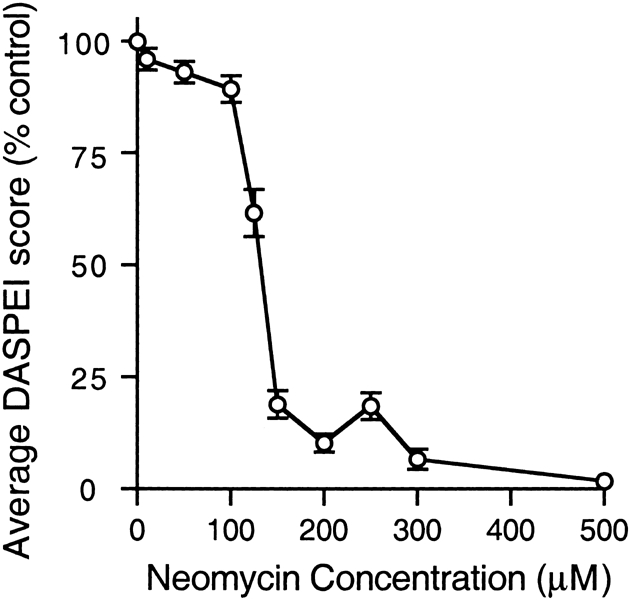
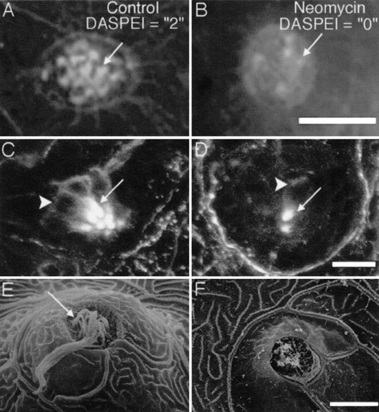
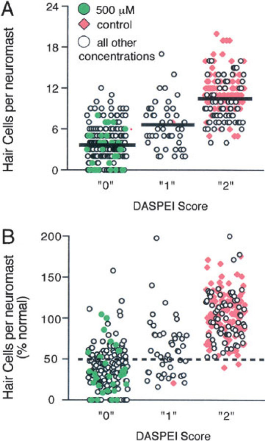
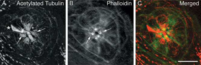
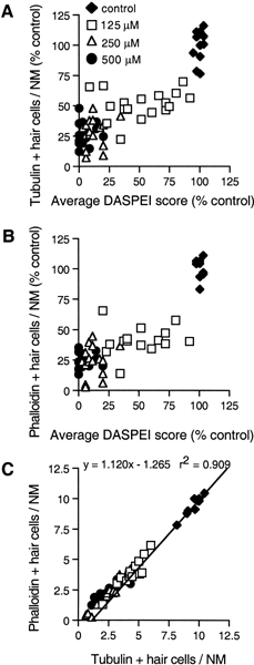
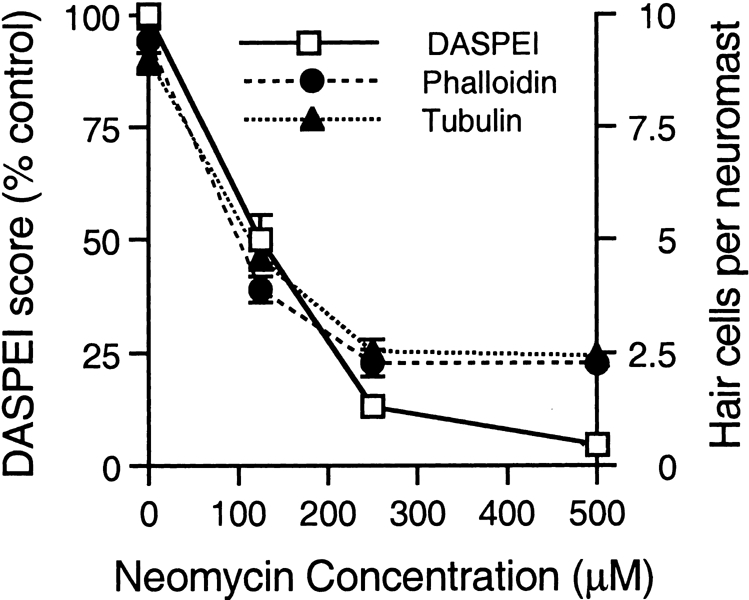
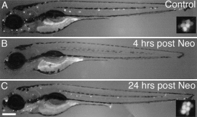
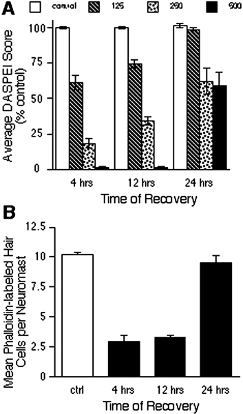
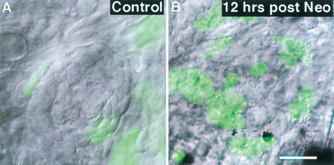
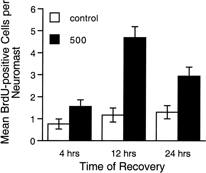
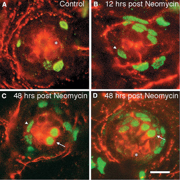
Mechanoreceptive hair cells are extremely sensitive to aminoglycoside antibiotics, including neomycin. Hair cell survival was assessed in larval wild-type zebrafish lateral line neuromasts 4 h after initial exposure to a range of neomycin concentrations for 1 h. Each of the lateral line neuromasts was scored in live fish for the presence or absence of hair cells using the fluorescent vital dye DASPEI to selectively label hair cells. All neuromasts were devoid of DASPEI-labeled hair cells 4 h after 500 microM neomycin exposure. Vital DASPEI staining was proportional to the number of hair cells per neuromast identified in fixed larvae using immunocytochemistry for acetylated tubulin and phalloidin labeling. The time course of hair cell regeneration in the lateral line neuromasts was also analyzed following neomycin-induced damage. Regenerated hair cells were first observed using live DASPEI staining 12 and 24 h following neomycin treatment. The potential role of proliferation in regenerating hair cells was analyzed. A 1 h pulse-fix protocol using bromodeoxyuridine (BrdU) incorporation was used to identify S-phase cells in neuromasts. BrdU incorporation in neomycin-damaged neuromasts did not differ from control neuromasts 4 h after drug exposure but was dramatically upregulated after 12 h. The proliferative cells identified during a 1 h period at 12 h after neomycin treatment were able to give rise to new hair cells by 24-48 h after drug treatment. The results presented here provide a standardized preparation for studying and identifying genes that influence vertebrate hair cell death, survival, and regeneration following ototoxic insults.


Figures

Figure 1
Figure 1
Experimental protocol. A. Larvae were exposed to neomycin for 1 h. Neuromast hair cell survival was analyzed 4, 12, and 24 h following initial neomycin exposure. B. The stereotyped positions of lateral line neuromasts in p4 and p5 zebrafish larvae (adapted from Metcalfe et al. 1985; Raible and Kruse 2000). The position names are based on the head and trunk lateral lines. Abbreviations: D = dorsal trunk, IO = infraorbital, M = mandibular, MI = middle, O = otic, OC = occipital, OP = opercular, P = posterior, PO = preoptic, SO =supraorbital.
Figure 2
Figure 2
Lateral line neuromasts stained with DASPEI. A, C. Live untreated p5 zebrafish larvae. B, D. Representative live p5 larvae 4 h after initial treatment with 500 µM neomycin for 1 h. A–D are lateral views with anterior to the left and dorsal up. The nasal epithelium is also stained with DASPEI in both groups (arrows). E. A higher-magnification view of a DASPEI-stained neuromast from a live control animal shows the rosette pattern of hair cells. F. A neuromast from a fixed larva containing hair cells labeled for f-actin with phalloidin. The neuromasts shown in E and F are not identical neuromasts and thus may have a different average number of hair cells (see Table 1). Scale bars = 0.5 mm (A and B in B; C and D in C) and 25 µm (E and F).
Figure 3
Figure 3
The dose–response relationship between neuromast hair cell survival as indicated by DASPEI staining in live fish and neomycin concentration. Zebrafish larvae (n = 232; 18–41/treatment group) treated for 1 h with various doses of neomycin were examined for the viability of hair cells in neuromasts stained with DASPEI. The doses used were 0, 10, 50, 100, 125, 150, 200, 250, 300, and 500 µM neomycin in embryo medium. DASPEI staining first significantly decreased after 50 µM neomycin. Average DASPEI scores were approximately 60% of control levels in larvae exposed to 125 µM neomycin. An exposure level of 500 µM neomycin eliminated DASPEI-labeled hair cells in neuromasts. The overall decrease in average DASPEI score with increasing neomycin concentrations was highly significant (p < 0.001).
Figure 4
Figure 4
DASPEI scores are proportional to the number of hair cells present within a neuromast analyzed three ways. In panels A and B, hair cell stereocilia bundles were labeled with fluorescent phalloidin. A. Representative phalloidin-labeled hair cells from a neuromast given the DASPEI score of 2. B. There are fewer phalloidin-labeled hair cells in a representative neuromast given the DASPEI score of 0. In panels C and D, neuromast hair cell bodies and stereocilia were labeled with antiacetylated tubulin. C. A representative control neuromast containing acetylated tubulin-positive hair cells (DASPEI score = 2). D. A representative neuromast from an animal fixed 4 h after initial treatment with 500 µM neomycin for 1 h contains fewer acetylated tubulin-labeled hair cells (DASPEI score = 0). Lateral line neuromasts were examined qualitatively using scanning electron microscopy. E. SEM of a representative neuromast from an untreated larva (DASPEI score = 2) showing intact kinocilia and stereocilia bundles of its hair cells. F. A damaged neuromast (DASPEI score = 0) from a larva 4 h after being treated with 500 µM neomycin. Kinocilia and stereocilia bundles were eliminated. Arrows indicate stereocilia or cuticular plate in A–D. Arrow in E is pointing to kinocilia. Arrowheads in C and D indicate hair cell bodies. Scale bars = 10 µm (A and B in B, C and D in D) and 5 µm (E and F in F).
Figure 5
Figure 5
Correspondence between DASPEI and phalloidin staining. Expanded scatterplots of live DASPEI scores from neuromasts (n = 388) that were also stained with phalloidin after fixation. A. Neuromasts are grouped by DASPEI score and the number of phalloidin-labeled hair cells counted in each neuromast is shown on the Y axis. The mean number of hair cells per neuromast for each scoring category is shown by the black lines. These means (± SD) are 3.9 ± 2.70, 6.5 ± 3.37, and 10.8 ± 3.07 hair cells, respectively. The means are all significantly different from each other by ANOVA (p < 0.001). B. Neuromasts are grouped by DASPEI score. The percent of the normal number of hair cells per neuromast is shown on the Y axis. This percentage was determined by dividing the number of phalloidin-labeled hair cells observed in each neuromast (data in A) by the normal mean number of hair cells in each specific neuromast based on its lateral line location (from Table 1).
Figure 6
Figure 6
A representative neuromast from a control zebrafish double-labeled with antiacetylated tubulin and phalloidin. A. Acetylated tubulin labels the kinocilia (indicated with an asterisk) and extends toward the nuclei of the cells. Two labeled hair cells are indicated with arrows. B. Phalloidin labels the stereocilia bundles. The same two hair cells indicated in A are shown here labeled with phalloidin indicated by the arrows. C. A composite image of acetylated tubulin label (red) and phalloidin (green). Scale bar = 10 µM.
Figure 7
Figure 7
Scatterplot comparisons of three methods used to assess neuromast hair cell loss. A. The average DASPEI score is shown plotted against the average number of acetylated tubulin-positive (+) hair cells per neuromast for each larva at all concentrations (0, 125, 250, and 500 µM). Both measures are expressed as a percentage of the mean control value for that analysis. B. The average DASPEI score is shown plotted against the average number of phalloidin-positive (+) hair cells per neuromast for each larva at all concentrations. Both measures are expressed as a percentage of the mean control value for that analysis. C. The average number of acetylated tubulin-positive (+) hair cells per neuromast is plotted against the number of phalloidin-positive (+) hair cells per neuromast for larvae at all concentrations. This plot was fitted with a linear regression. There is a high correlation between the number of hair cells counted using phalloidin and acetylated tubulin labeling (r2 = 0.909).
Figure 8
Figure 8
Dose–response relationships of neuromast hair cell survival and neomycin concentration determined by three different methods (from the same data as shown in Fig. 7). The average DASPEI score expressed as a percentage of the control value is plotted on the left Y axis. The average number of hair cells per neuromast counted using phalloidin and acetylated tubulin labeling is plotted using the right Y axis. The overall decrease in DASPEI scores and hair cell counts was highly significant (p < 0.001) and all doses were significantly different from each other (p's < 0.001), except scores for 250 vs. 500 µM using phalloidin and acetylated tubulin staining. There was no significant difference between phalloidin and acetylated tubulin cell counts at any concentration. DASPEI scores differed significantly from both other methods at the highest doses, 250 and 500 µM (p's < 0.001), but they did not differ at the intermediate dose, 125 µM. Hence the change in DASPEI scores is proportional to hair cell counts across the dose–response range but slightly overestimates hair cell loss at the high doses.
Figure 9
Figure 9
Hair cell regeneration in neomycin-treated neuromasts. A. An untreated larva showing typical DASPEI staining of neuromasts. B. A DASPEI-stained larva 4 h after exposure to 500 µM neomycin. DASPEI staining is eliminated in neuromasts. C. A DASPEI-stained larva 24 h following exposure to 500 µM neomycin. Note the return of a normal DASPEI staining pattern. A,C insets: DASPEI-labeled control (A) and regenerated (C) neuromasts appear similar at higher magnification. Scale bar = 0.5 mm for all panels excluding insets.
Figure 10
Figure 10
Time course of neuromast hair cell regeneration following neomycin exposure. A. Neuromast hair cell survival quantified with DASPEI scores relative to control are plotted at 4, 12, and 24 h postexposure for larvae treated with various doses of neomycin (0, 125, 250, and 500 µM, n =11–29 fish/group/time point). DASPEI scores significantly increased as a function of recovery time following neomycin exposure (p < 0.001). B. Mean phalloidin-labeled hair cell counts per neuromast are plotted at 4, 12, and 24 h postexposure to 500 µM neomycin (n > 26 neuromasts/group). Similar to the DASPEI scores, there is significant recovery of hair cells between 4 and 24 h and between 12 and 24 h (p < 0.001). The mean number of hair cells per neuromast is also plotted from a control, untreated group for comparison (white bar). Bars are mean ± SEM.
Figure 11
Figure 11
Neomycin-induced upregulation of proliferation in neuromasts. A. A representative neuromast from an untreated control larva that was exposed to BrdU 12 h after the initiation of the experiment contains few BrdU-labeled cells. B. A neuromast from an animal treated with 500 µM neomycin contains many more S-phase cells than the control neuromast when exposed to BrdU for 1 h 12 h after treatment. The BrdU nuclei (green) are overlaid onto a gray DIC image of the neuromast. Each fluorescent image is from a flattened z-series through the neuromast. Scale bar = 10 µm.
Figure 12
Figure 12
Time course of neuromast cells entering S-phase following 500 µM neomycin exposure. Mean BrdU-positive cells per neuromast are plotted for neuromasts exposed to BrdU in the embryo medium for 1 h 4, 12, and 24 h after neomycin exposure (n = 173 neuromasts, 19–41 neuromasts/group and 3–7 neuromasts/fish). The number of S-phase cells in a neuromast is upregulated at 12 and 24 h after neomycin exposure. The time of BrdU exposure and neomycin treatment had significant effects on the number of proliferating cells (two-way ANOVA, p< 0.005). Bars are mean ± SEM.
Figure 13
Figure 13
New hair cells produced by neuromast cells in S-phase at 12 h after 500 µM neomycin exposure. Hair cells were identified using immunocytochemistry (ICC) for acetylated tubulin (shown in red). BrdU-positive cells were also detected with ICC (shown in green). A. A representative neuromast from a control animal pulsed with BrdU for 1 h 12 h after the experiment was initiated and fixed immediately. There are few S-phase cells within the neuromast. B. A representative neuromast exposed to 500 µM neomycin, pulsed with BrdU 12 h later, and then immediately fixed. This neuromast contains many more S-phase cells but few hair cells. C, D. Two examples of neuromasts treated with 500 µM neomycin, exposed to BrdU for 1 h 12 h after neomycin exposure, and then fixed 36 h later (48 h after neomycin treatment). These neuromasts contained new hair cells double-labeled for acetylated tubulin and BrdU as well as hair cells without BrdU labeling and BrdU-labeled cells without acetylated tubulin labeling. Asterisks indicate hair cells, arrowheads indicate BrdU labeling, and the arrows indicate “new” hair cells. Scale bar = 10 µm.

Similar articles

Cited by

References

    1. Adler HJ, Raphael Y. New hair cells arise from supporting cell conversion in the acoustically damaged chick inner ear. Neurosci. Lett. 1996;205:17–20. doi: 10.1016/0304-3940(96)12367-3. - DOI - PubMed
    1. Alexandre D, Ghysen A. Somatotopy of the lateral line projection in larval zebrafish. Proc. Natl. Acad. Sci. U.S.A. 1999;96:7558–7562. doi: 10.1073/pnas.96.13.7558. - DOI - PMC - PubMed
    1. Baird RA, Steyger PS, Schuff NR. Mitotic and nonmitotic hair cell regeneration in the bullfrog vestibular otolith organs. Ann. N.Y. Acad. Sci. 1996;781:59–70. - PubMed
    1. Bang PI, Sewell WF, Malicki JJ. Morphology and cell type heterogeneities of the inner ear epithelia in adult and juvenile zebrafish (Danio rerio). J. Comp. Neurol. 2001;438:173–190. doi: 10.1002/cne.1308. - DOI - PubMed
    1. Carey JP, Fuchs AF, Rubel EW. Hair cell regeneration and recovery of the vestibuloocular reflex in the avian vestibular system. J. Neurophysiol. 1996;76:3301–3312. - PubMed

Publication types

MeSH terms

LinkOut - more resources