. 2003 Jun;4(2):264-75.
doi: 10.1007/s10162-002-3033-7.

Expression of aquaporin 1 and 5 in the developing mouse inner ear and audiovestibular assessment of an Aqp5 null mutant

Affiliations

Expression of aquaporin 1 and 5 in the developing mouse inner ear and audiovestibular assessment of an Aqp5 null mutant

Michele Merves et al. J Assoc Res Otolaryngol. 2003 Jun.

Abstract

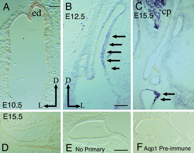
To examine the potential roles of aquaporins 1 and 5 (AQP1 and AQP5, respectively) in inner ear development and function, we defined their spatial and temporal expression patterns in the developing mouse inner ear and examined the morphologic and physiologic effects of loss of Aqp5 function. Standard in situ hybridization (ISH) and immunohistochemical (IHC) assays were used for expression studies with routine morphologic, behavioral, and physiologic assessments of hearing and balance in Aqp5 null mutant mice. AQP1 was first detected at embryonic day 10.5 (E10.5) in the otocyst but eventually localized to specific nonsensory portions of the inner ear and connective tissue cells surrounding the membranous labyrinth. AQP5 displayed specific cochlear expression, first detectable at E15.5 in the nonsensory epithelium and later restricted to the lateral wall of the cochlear duct near the spiral prominence. AQP5 expression continued through postnatal periods with a change of expression domain to the stria vascularis between postnatal day 7 (P7) and P14. By in situ hybridization and immunohistochemical techniques, subtle differences between transcript and protein expression patterns were noted for both AQP1 and 5. Although AQP5 is dynamically expressed in the developing mouse inner ear, adult Aqp5 knockout mice show normal hearing when tested and normal inner ear structural development. These results suggest redundant or alternative mechanisms that likely regulate water homeostasis in the developing and mature inner ear.


Figures

Figure 1
Figure 1
IHC controls and AQP1 expression at E10.5–E15.5. A. IHC. AQP1 protein is expressed heterogeneously throughout the epithelium of the otocyst (E10.5) and particularly intensely in the forming endolymphatic duct. AQP1 transcripts are not seen until E12.5. B. ISH. At E12.5, AQP1 transcripts are expressed only in the lateral wallof the common crus (arrows) and vestibule. C. ISH. By E15.5, AQP1 mRNA is expressed in the lateral wall of the common crus, in the nonsensory portion of the saccule (arrows), and in the connective tissue surrounding the membranous labyrinth of the inner ear. It is also expressed in the choroid plexus. D. AQP1 protein expression is similar to AQP1 mRNA at E15.5 and immunostaining surrounding the lateral ampulla is shown. (E, F). Representative cochlear sections from an E18.5 embryo. These late embryonic specimens were chosen because AQP1 is robustly expressed at this developmental stage in these regions of the inner ear. The absence of any signal on these sections supports the specificity of the antisera used in this study. No primary antibody negative control (E) and AQP1 preimmune control (F). The axes in A apply to D, E, and F, and axes in B also apply to C. Scale bar in A=50 µm. Scale bar in B=100 µm and applies to C--F. ed = endolymphatic duct; cp = choroid plexus.
Figure 2
Figure 2
Aquaporin 1 protein expression at postnatal day 2. A. AQP1 is expressed in the connective tissue surrounding the cochlea including the fibrocytes of the spiral ligament (arrows) and mesenchymal cells below the basilar membrane (arrowheads). It is not detected in the epithelial tissue. B. AQP1 protein is not detected in the epithelium of the endolymphatic sac but in a discrete layer immediately surrounding the epithelium. C. AQP1 is also expressed in the connective tissue surrounding the vestibular portion of the inner ear and robustly in nonsensory portion of the saccule (large arrows in C) and in the epithelium of the lateral wall of the common crus (not shown). The small arrow labels the macula of the saccule. D. Lateral ampulla. Arrow labels the crista. Scale bars in A–D=100 µm. els= endolymphatic sac; sac mac = saccule macula.
Figure 3
Figure 3
Aquaporin 5 controls and early expression patterns. A. No signal was detected in control experiments with an AQP5 sense probe performed on E15.5 cochlear sections. B. ISH. AQP5 E18.5 mouse salivary gland. AQP5 has been reported to be expressed in mouse salivary glands and accordingly was used as a positive control (Krane et al. 1999). From E10 (15 somites, data not shown) through E15.0, AQP5 mRNA or protein is not detected. Sample negative in situ data from an E12.5 embryo is shown in C. Starting at E15.5, signals for AQP5 transcripts (arrows in D) and protein (arrows in E) were detected in the lateral wall of all turns of the cochlear duct (in a region corresponding to the presumptive stria vascularis) and were particularly intense in the basal turn. Scale bars in A–E=100 µm.
Figure 4
Figure 4
AQP5 expression between E15.5 and E18.5. Comparison of the AQP5 signal in the lateral cochlear wall at E15.5 under higher magnification (arrows in A and adjacentsection A′) shows the transcript and protein signals to overlap without a demonstrable difference. AQP5 transcripts (B) and protein (B′) then restricted to a region inferior to the presumptive stria vascularis by E18.5 (arrows in B and B′) and remains in this domain through P2. Scale bars=100 µm. Scale bar in A also applies to A′. Scale bar in B applies to B′.
Figure 5
Figure 5
Postnatal AQP5 expression and histology of aquaporin 5 deficient mice. A. At P7, AQP5 protein was detected in the type II fibrocytes surrounding the spiral prominence. By P14, the expression pattern of AQP5 protein again shifted from the spiral prominence region to the stria vascularis. The more apical turns continued to show signal in the cells near the spiral prominence (not shown), while the basal turns demonstrated signal only in the stria vascularis. At 4 weeks (not shown) and B. 5–8 months, AQP5 protein is expressed only in the stria vascularis. C. IHC. No anti-AQP5 staining was demonstrable in AQP5 −/− inner ears at 5–8 months of age. D. AQP5 −/− hematoxylin and eosin-stained tissue sections. Note the normal appearance of the cochlear duct with a well-preserved organ of Corti and normal-appearing Reissner's membrane. Areas of normal AQP5 expression (along the lateral cochlear wall) similarly appear unaffected compared with controls. Scale bar in A=100 µm and appliesto B, C, and D.
Figure 6
Figure 6
Auditory brainstem responses (ABR). A. Representative ABR recordings from Aqp5+/+ (left) and Aqp5−/− (right) mice exposed to a broadband click in the 40–70 dB SPL range. Both Aqp5+/+ and Aqp5−/− mice showed observable responses to click sounds of 40 dB. B. Summary of ABR thresholds for Aqp5+/+ (open bars, n = 6) and Aqp5−/− (hatched bars, n = 6) mice in response to the clicks and the three pure tones at 8, 16, and 32 kHz. The difference in mean ABR thresholds between Aqp5+/+ and Aqp5−/− mice was not statistically significant. Values shown are mean ± standard deviation.
Figure 7
Figure 7
Postnatal functional differentiation of the lateral cochlear wall. A. H&E cochlea. Building upon the work of Everett et al. (1999) and data reproduced in ourstudies, (B) P2 AQP5 expression localizes to a subset of cells expressing (C) Pendrin (Pds) immediately inferior to the stria vascularis. D. Tyrosinase-related protein 2 (TRP-2) marks the melanocytes in the stria vascularis while (E) bone morphogenetic protein 4 (BMP4) was used to mark the presumptive Hensen and Claudius cells of the external sulcus. F. Schematic drawing of molecular markers. Hatchings along the epithelium of the cochlear duct indicatethe expression domain of each gene. Black cross-hatchings along the SV, Trp2; red cross-hatchings along the SV, AQP5 at P14; orange cross-hatchings, AQP5 at P7; yellow cross-hatchings, PDS; blue cross-hatchings, AQP5 at P2; red cross-hatchings at the external sulcus, BMP4; black cross-hatchings along the inferior edge of the basilar membrane area and along the spiral ligament, AQP1. Although data from adjacent sections stained with AQP1 are not included in this diagram, the hatchings along the inferior margin of the basilar membrane and along the fibrocytes of the spiral ligament are labeled to show the relative domains of AQP1 expression. PDS, pendrin; Trp2, tyrosinase-related protein 2; BMP4, bone morphogenetic protein 4. (Probe for PDS kindly provided by L. Everett; Trp2 probe provided by K. Steel; BMP4 probe courtesy of D. Wu). OC = organ of Corti; RM = Reissner's membrane; SP = spiral prominence; SV = stria vascularis.

Similar articles

Cited by

References

    1. Arenberg I. Results of the first 300 consecutive endolymphatic system-mastoid shunts with valve implants for hydrops. Otolaryngol. Clin. North Am. 1982;16:153–174. - PubMed
    1. Beitz E, Kumagami H, Krippeit–Drews P, Ruppersberg J, Schultz J. Expression pattern of aquaporin water channels in the inner ear of the rat. The molecular basis for a water regulation system in the endoylmphatic sac. Hear. Res. 1999;132:76–84. doi: 10.1016/S0378-5955(99)00036-2. - DOI - PubMed
    1. Belyantseva IA, Frolenkov GI, Wade JB, Mammano F, Kachar B. Water permeability of cochlear outer hair cells: characterization and relationship to electromotility. J. Neurosci. 2000;20:8996–9003. - PMC - PubMed
    1. Borgnia MJ, Kozono D, Calamita G, Maloney PC, Agre P. Functional reconstitution and characterization of AqpZ, the E. coli water channel protein. J. Mol. Biol. 1999;291:1169–1179. doi: 10.1006/jmbi.1999.3032. - DOI - PubMed
    1. Choo D, Sanne J, Wu D. The differential sensitivities of inner ear structures to retinoic acid during development. Dev. Biol. 1998;204:136–150. doi: 10.1006/dbio.1998.9095. - DOI - PubMed

Publication types

MeSH terms