

Organization of skin stratum corneum extracellular lamellae: diffraction evidence for asymmetric distribution of cholesterol
- PMID: 12944282
- PMCID: PMC1303341
- DOI: 10.1016/S0006-3495(03)74597-4
Organization of skin stratum corneum extracellular lamellae: diffraction evidence for asymmetric distribution of cholesterol
Abstract
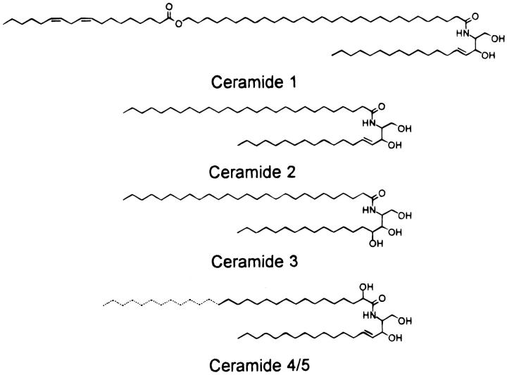
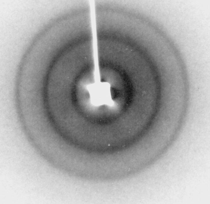
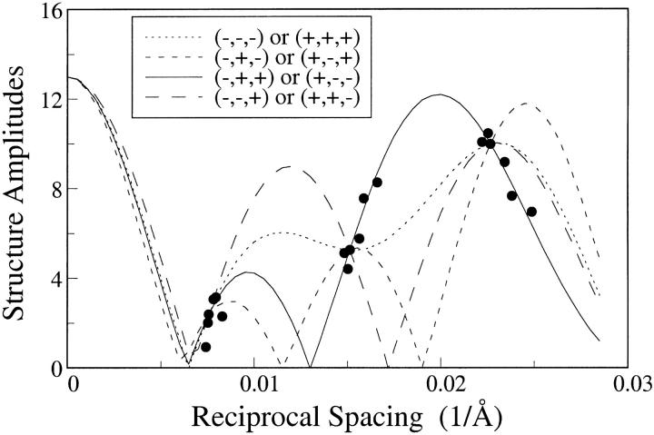
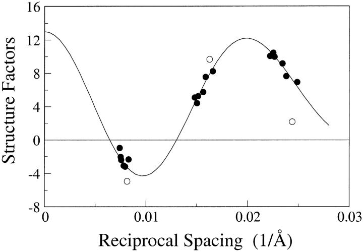
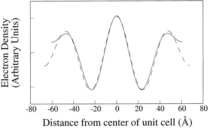
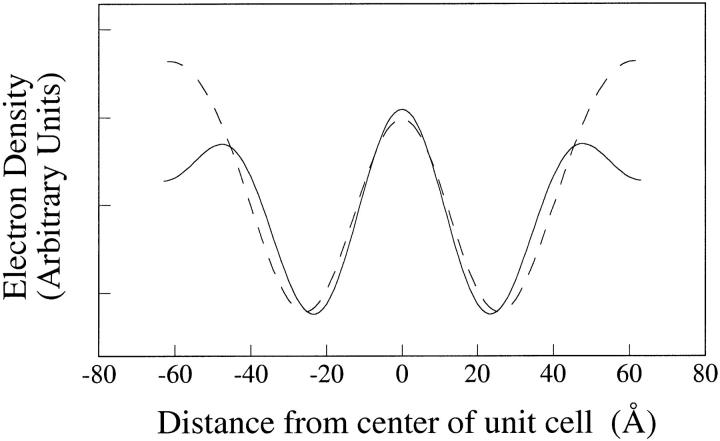
Lipid suspensions containing 2:1:1 skin ceramides:palmitic acid:cholesterol, similar to the lipid composition found in the extracellular matrix of skin stratum corneum, were analyzed by X-ray diffraction methods. These suspensions gave a sharp wide-angle reflection at 4.1 A, indicating tight hydrocarbon chain packing that would function as a water barrier, and low-angle lamellar diffraction with a repeat period near 130 A, similar to that previously recorded from intact stratum corneum. The lamellar repeat increased from 121 A at pH 6 to 133 A at pH 8.5, allowing phase angles of the lamellar data to be obtained by a sampling theorem "swelling" analysis. Electron density profiles showed that each repeating unit contained two asymmetric bilayers, with a fluid space on one side of the bilayer that increased with increasing pH, due to electrostatic repulsion between bilayers because of ionization of the palmitic acid. Profiles obtained from lamellae with cholesterol sulfate partially substituted for cholesterol showed large density increases on that same side of the bilayer, indicating that cholesterol is asymmetrically distributed in each bilayer. A molecular model was developed postulating that this asymmetry is due to the exclusion of cholesterol from lipid monolayers containing the ester-linked unsaturated (linoleic) hydrocarbon chain of skin ceramide 1. This model can explain the altered organization of extracellular lamellae in epidermal cysts (P. W. Wertz, D. C. Swartzendruber, K. C. Madison, D. T. Downing. 1987. J. Invest. Dermatol. 89:419-425) where the ester-linked chains have a higher percentage of saturated fatty acids than found in normal epidermis.
Figures
Similar articles
-
X-ray diffraction analysis of isolated skin lipids: reconstitution of intercellular lipid domains.Biochemistry. 1996 Mar 26;35(12):3649-53. doi: 10.1021/bi952762q. Biochemistry. 1996. PMID: 8619983
-
Mesophase formation by ceramides and cholesterol: a model for stratum corneum lipid packing?Biochim Biophys Acta. 1993 Apr 22;1147(2):273-6. doi: 10.1016/0005-2736(93)90013-p. Biochim Biophys Acta. 1993. PMID: 8476922
-
Phase behavior of stratum corneum lipids in mixed Langmuir-Blodgett monolayers.Biophys J. 1996 Sep;71(3):1389-99. doi: 10.1016/S0006-3495(96)79341-4. Biophys J. 1996. PMID: 8874014 Free PMC article.
-
Stratum corneum lipid organization as observed by atomic force, confocal and two-photon excitation fluorescence microscopy.Int J Cosmet Sci. 2008 Dec;30(6):391-411. doi: 10.1111/j.1468-2494.2008.00458.x. Int J Cosmet Sci. 2008. PMID: 19099542 Review.
-
The skin barrier in healthy and diseased state.Biochim Biophys Acta. 2006 Dec;1758(12):2080-95. doi: 10.1016/j.bbamem.2006.06.021. Epub 2006 Jul 11. Biochim Biophys Acta. 2006. PMID: 16945325 Review.
Cited by
-
Is Deuterium Sequestering by Reactive Carbon Atoms an Important Mechanism to Reduce Deuterium Content in Biological Water?FASEB Bioadv. 2025 May 14;7(6):e70019. doi: 10.1096/fba.2025-00032. eCollection 2025 Jun. FASEB Bioadv. 2025. PMID: 40496345 Free PMC article. Review.
-
Evaluation of Constrained and Restrained Molecular Dynamics Simulation Methods for Predicting Skin Lipid Permeability.ACS Omega. 2021 Dec 15;6(51):35363-35374. doi: 10.1021/acsomega.1c04684. eCollection 2021 Dec 28. ACS Omega. 2021. PMID: 34984268 Free PMC article.
-
Arrangement of ceramide [EOS] in a stratum corneum lipid model matrix: new aspects revealed by neutron diffraction studies.Eur Biophys J. 2008 Jul;37(6):989-99. doi: 10.1007/s00249-008-0328-6. Epub 2008 Apr 22. Eur Biophys J. 2008. PMID: 18427800
-
The New Challenge of Green Cosmetics: Natural Food Ingredients for Cosmetic Formulations.Molecules. 2021 Jun 26;26(13):3921. doi: 10.3390/molecules26133921. Molecules. 2021. PMID: 34206931 Free PMC article. Review.
-
Extracellular matrix stiffness-The central cue for skin fibrosis.Front Mol Biosci. 2023 Mar 8;10:1132353. doi: 10.3389/fmolb.2023.1132353. eCollection 2023. Front Mol Biosci. 2023. PMID: 36968277 Free PMC article. Review.
References
-
- Abraham, W., and D. T. Downing. 1992. Lamellar structures formed by stratum corneum lipids in vitro: a deuterium nuclear magnetic resonance (NMR) study. Pharm. Res. 9:1415–1421. - PubMed
-
- Blaurock, A. E. 1971. Structure of the nerve myelin membrane: proof of the low-resolution profile. J. Mol. Biol. 56:35–52. - PubMed
-
- Bouwstra, J. A., F. E. Dubbelaar, G. S. Gooris, A. M. Weerheim, and M. Ponec. 1999. The role of ceramide composition in the lipid organization of the skin barrier. Biochim. Biophys. Acta. 1419:127–136. - PubMed
-
- Bouwstra, J. A., G. S. Gooris, W. Bras, and D. T. Downing. 1995. Lipid organization in pig stratum corneum. J. Lipid Res. 36:685–695. - PubMed
-
- Bouwstra, J. A., G. S. Gooris, K. Cheng, A. Weerheim, W. Bras, and M. Ponec. 1996. Phase behavior of isolated skin lipids. J. Lipid Res. 37:999–1011. - PubMed
Publication types
MeSH terms
Substances
Grants and funding
LinkOut - more resources
Full Text Sources
Other Literature Sources
Medical
