. 2003 Sep;85(3):1996-2005.
doi: 10.1016/S0006-3495(03)74627-X.

Organization and adhesive properties of the hyaluronan pericellular coat of chondrocytes and epithelial cells

Affiliations

Organization and adhesive properties of the hyaluronan pericellular coat of chondrocytes and epithelial cells

Miriam Cohen et al. Biophys J. 2003 Sep.

Abstract

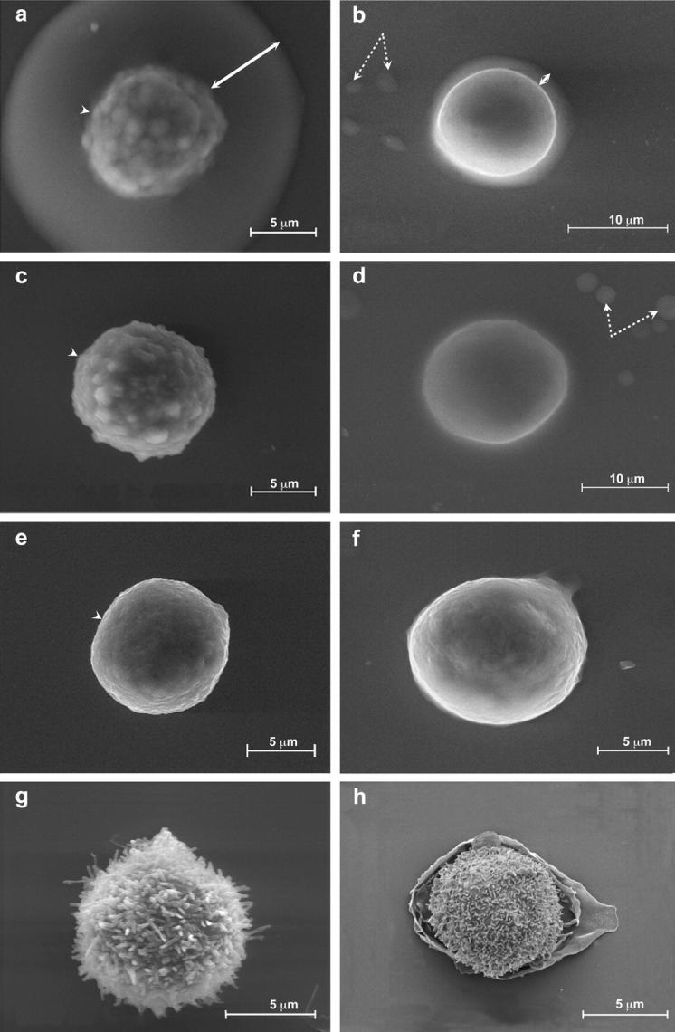
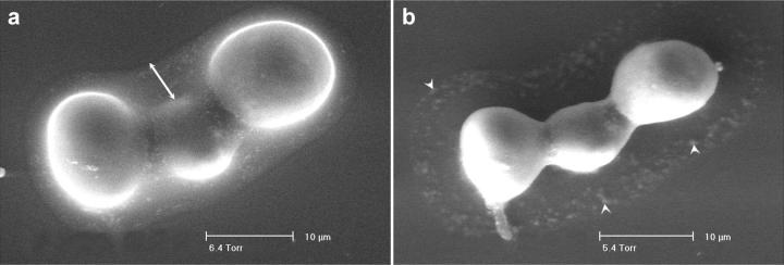
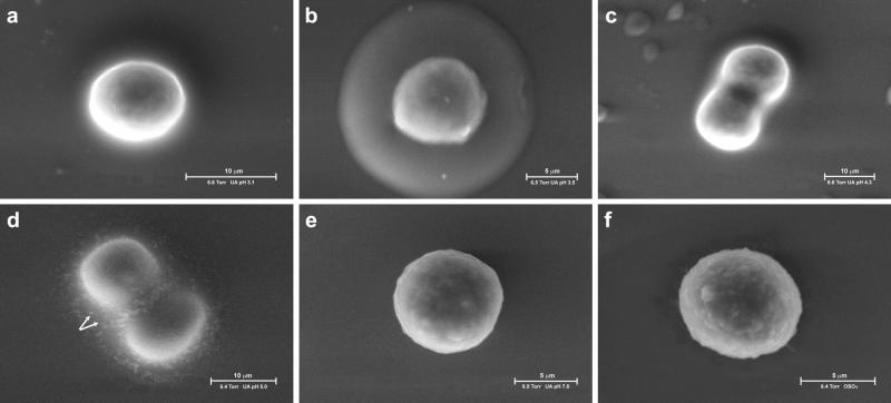
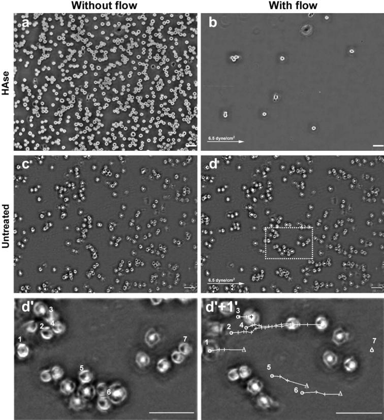
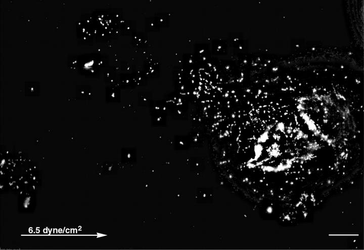
Hyaluronan is a megadalton glycosaminoglycan composed of repeating units of D-N-acetylglucosamine-beta-D-Glucuronic acid. It is known to form a highly hydrated pericellular coat around chondrocytes, fibrosarcoma, and smooth muscle cells. Using environmental scanning electron microscopy we detected fully hydrated hyaluronan pericellular coats around rat chondrocytes (RCJ-P) and epithelial cells (A6). Hyaluronan mediates early adhesion of both chondrocytes and A6 cells to glass surfaces. We show that chondrocytes in suspension establish early "soft contacts" with the substrate through a thick, hyaluronidase-sensitive coat (4.4 +/- 0.7 microm). Freshly-attached cells drift under shear stress, leaving hyaluronan "footprints" on the surface. This suggests that chondrocytes are surrounded by a multilayer of entangled hyaluronan molecules. In contrast, A6 cells have a 2.2 +/- 0.4- microm-thick hyaluronidase-sensitive coat, do not drift under shear stress, and remain firmly anchored to the surface. We consider the possibility that in A6 cells single hyaluronan molecules, spanning the whole thickness of the pericellular coat, mediate these tight contacts.


Figures

FIGURE 1
FIGURE 1
Visualization of hydrated pericellular coats using the environmental scanning electron microscope. Chondrocytes (RCJ-P: a, c, e, g) and epithelial cells (A6: b, d, f, h) examined in the ESEM. (a and b) Untreated cells, labeled with uranyl ions; (c and d) hyaluronidase-treated cells; (e and f) untreated cells, not labeled with uranyl ions; (g and h) critical point dried cells. The cells were labeled with uranyl acetate at pH 3.5 after fixation; the uranyl ions bind to hyaluronan, resulting in visualization of a 4.4 ± 0.7-μm-thick halo around RCJ-P cells (a) and a 2.2 ± 0.4-μm-thick halo around A6 cells (b). Arrows indicate the gel, dashed arrows indicate water droplets in equilibrium with the wet environment. The cell membrane looks blurred through the gel (a, arrowhead). Hyaluronidase-treated cells are not surrounded by halos, and their borders are well defined (c, d, arrowhead). In cells that were not incubated with uranyl acetate (e and f) and in critical point dried cells (e and f), no gel is detected.
FIGURE 2
FIGURE 2
The hyaluronan coat disappears upon dehydration. Chondrocytes were labeled with uranyl acetate, pH 5.0, after fixation and examined in the ESEM. (a) At 6.4 Torr (853 Pascal), the dew point, the pericellular coat is visible around the cells (arrow). (b) Gradual reduction of the pressure to 5.4 Torr (720 Pascal) resulted in dehydration and disappearance of the gel. Arrowheads indicate residual traces of uranyl acetate.
FIGURE 3
FIGURE 3
The mode of binding uranyl ions to the hyaluronan coat of chondrocytes is pH dependent. Chondrocytes were labeled with uranyl acetate at pH 3.1 (a), 3.5 (b), 4.3 (c), 5.0 (d), with uranyl acetate oxalate at pH 7.0 (e), or with osmium tetraoxide (f). Cells labeled with uranyl acetate at pH 3.1 or 4.3 (a and c) were weakly labeled, whereas at pH 3.5 the labeling was strong (b). At pH 5.0 uranyl acetate stained the gel but precipitated inside the fixed gel (d, arrows). No staining of the gel was detected when cells were stained with uranyl acetate oxalate at pH 7.0 (e), or with osmium tetraoxide (f).
FIGURE 4
FIGURE 4
3D reconstruction of the pericellular hyaluronan coat by particle exclusion assay. (a and b) Fluorescence micrographs of rhodamine-labeled chondrocytes immersed in medium containing FITC-labeled silica beads. Cells were allowed to adhere to glass coverslips for 25 min before fixation, and labeled with tetramethyl rhodamine isothiocyanate (red). They were then incubated with FITC-labeled 0.4-μm silica beads (green). Micrographs were taken with a digital microscope (DeltaVision) able to generate 3D images by image reconstruction from a series of z-sections at 0.5-μm resolution. The excluded volume is dark. Untreated cells have 5- to 6-μm wide excluded zone around them (a), whereas beads reach up to the surface of hyaluronidase treated cells (b). (c and d) Deconvoluted and reconstituted images along the z axes. Untreated cells (c) have a 1.2-μm excluded zone also on the apical region, whereas hyaluronidase-treated cells (d) have no excluded volume. We note that imaging from oil into water reduces the height of the sample by a factor equal to the ratio of refractive index between oil and water. In addition, imaging with an oil objective deep into a water sample introduces depth dependent aberration. This, for 10μm depth, may reach up to at least half the resolution of the objective (Kam et al., 1997). Everything considered, the excluded volume in the apical region may reach up to ∼2-μm thickness. The beads appear as segments because of Brownian motion. Scale bar, 5 μm.
FIGURE 5
FIGURE 5
Hyaluronan-mediated adhesion: the role of hyaluronan in early adhesion and its resistance to shear stress. Chondrocytes (RCJ-P) were allowed to adhere to serum-coated glass for 25 min, then washed with a continuous flow of medium which exerted shear force of 6.5 dyne/cm2. Cell movement was recorded by a time-lapse phase microscope. (a and c) Cells before applying flow; (b and d) cells 2 s within the flow. (a and b) Hyaluronidase treated cells. (c) and (d) Untreated cells. The arrows indicate the flow direction. Hyaluronidase-treated cells washed away immediately after applying the flow (compare a to b). In contrast, untreated cells remained attached to the surface (compare c to d) and moved 43.10 ± 10.79 μm before detaching from the surface. (d′) Enlargement of the area marked with a dashed line in d. (d′+1′) The same frame as in d′, 60 s later. The circles mark the original position of seven selected cells; the crosses mark the cell position at 4-s intervals; the triangles mark the detachment position; the lines mark the cell paths. Cells 1, 5, and 6 moved 37.57–41.71 μm at the speed of 3.13–3.48 μm/s before detaching from the surface. Cells 2 and 4 traveled longer (66.12–82.10 μm) but slower (1.10–1.47 μm/s) and did not detach within 60 s of flow. Cell 3 started to move only 36 s after applying the flow, covering 13.46 μm, and remained attached to the surface. Cell 7 did not move but detached 2 s after applying flow. Scale bar, 50 μm.
FIGURE 6
FIGURE 6
Hyaluronan “footprints” of chondrocytes are left after application of shear stress. Chondrocytes were treated with flow as described in Fig. 5, then fixed and incubated with biotinylated hyaluronan binding proteins followed by incubation with strepavidin-CY3. Tracks of hyaluronan are visible upstream to the cells. Cell with hyaluronan “footprints” of 75 μm. The arrow marks the flow direction. Scale bar, 10 μm.

References

    1. Adams, J. C. 2001. Cell-matrix contact structures. Cell. Mol. Life Sci. 58:371–392. - PMC - PubMed
    1. Albersdorfer, A., and E. Sackmann. 1999. Swelling behavior and viscoelasticity of ultrathin grafted hyaluronic acid films. Eur. Phys. J. B. 10:663–672.
    1. Bajorath, J. 2000. Molecular organization, structural features, and ligand binding characteristics of CD44, a highly variable cell surface glycoprotein with multiple functions. Proteins. 39:103–111. - PubMed
    1. Bard, J. B., W. H. McBride, and A. R. Ross. 1983. Morphology of hyaluronidase-sensitive cell coats as seen in the SEM after freeze-drying. J. Cell Sci. 62:371–383. - PubMed
    1. Borland, G., J. A. Ross, and K. Guy. 1998. Forms and functions of CD44. Immunology. 93:139–148. - PMC - PubMed

Publication types

MeSH terms

LinkOut - more resources