Reconstituted fusion pore
- PMID: 12944316
- PMCID: PMC1303375
- DOI: 10.1016/S0006-3495(03)74631-1
Reconstituted fusion pore
Abstract
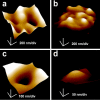
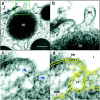
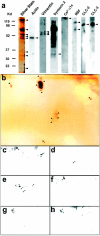
Fusion pores or porosomes are basket-like structures at the cell plasma membrane, at the base of which, membrane-bound secretory vesicles dock and fuse to release vesicular contents. Earlier studies using atomic force microscopy (AFM) demonstrated the presence of fusion pores at the cell plasma membrane in a number of live secretory cells, revealing their morphology and dynamics at nm resolution and in real time. ImmunoAFM studies demonstrated the release of vesicular contents through the pores. Transmission electron microscopy (TEM) further confirmed the presence of fusion pores, and immunoAFM, and immunochemical studies demonstrated t-SNAREs to localize at the base of the fusion pore. In the present study, the morphology, function, and composition of the immunoisolated fusion pore was investigated. TEM studies reveal in further detail the structure of the fusion pore. Immunoblot analysis of the immunoisolated fusion pore reveals the presence of several isoforms of the proteins, identified earlier in addition to the association of chloride channels. TEM and AFM micrographs of the immunoisolated fusion pore complex were superimposable, revealing its detail structure. Fusion pore reconstituted into liposomes and examined by TEM, revealed a cup-shaped basket-like morphology, and were functional, as demonstrated by their ability to fuse with isolated secretory vesicles.
Figures


References
-
- Bennett, V. 1990. Spectrin-based membrane skeleton: a multipotential adaptor between plasma membrane and cytoplasm. Physiol. Rev. 70:1029–1065. - PubMed
-
- Bradford, M. M. 1976. A rapid and sensitive method for the quantitation of microgram quantities of protein utilizing the principle of protein-dye binding. Anal. Biochem. 72:248–254. - PubMed
-
- Cho, S.-J., K. Jeftinija, A. Glavaski, S. Jeftinija, B. P. Jena, and L. L. Anderson. 2002a. Structure and dynamics of the fusion pores in live GH-secreting cells revealed using atomic force microscopy. Endocrinology. 143:1144–1148. - PubMed
-
- Cho, S.-J., A. Wakade, G. D. Pappas, and B. P. Jena. 2002b. New structure involved in transient membrane fusion and exocytosis. Ann. New York Acad. Sci. 971:254–256. - PubMed
-
- Cho, S.-J., A. S. Quinn, M. H. Stromer, S. Dash, J. Cho, D. J. Taatjes, and B. P. Jena. 2002c. Structure and dynamics of the fusion pore in live cells. Cell Biol. Int. 26:35–42. - PubMed
Publication types
MeSH terms
Substances
Grants and funding
LinkOut - more resources
Full Text Sources
Other Literature Sources
Miscellaneous

