. 2003 Sep;23(18):6385-95.
doi: 10.1128/MCB.23.18.6385-6395.2003.

Werner syndrome protein phosphorylation by abl tyrosine kinase regulates its activity and distribution

Affiliations

Werner syndrome protein phosphorylation by abl tyrosine kinase regulates its activity and distribution

Wen-Hsing Cheng et al. Mol Cell Biol. 2003 Sep.

Abstract

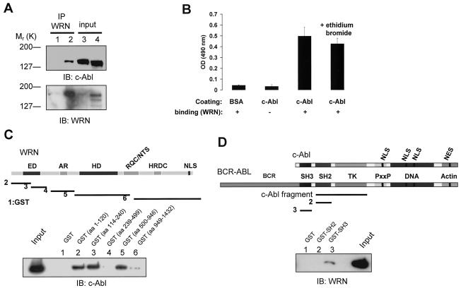
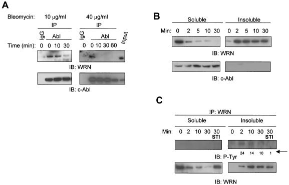
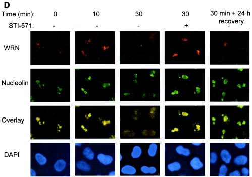
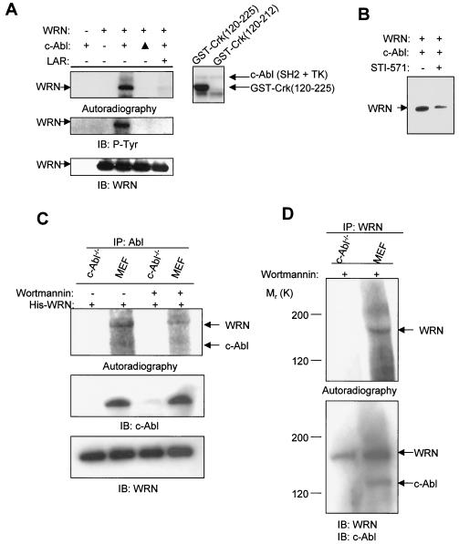
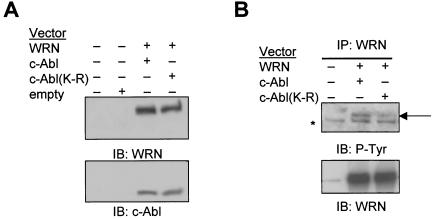
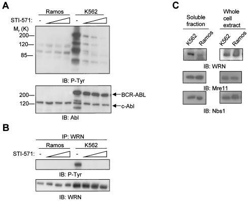
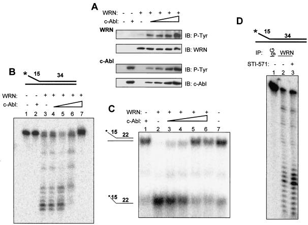
The Werner syndrome protein (WRN) is a caretaker of the human genome, and the Abl kinase is a regulator of the DNA damage response. Aberrant DNA repair has been linked to the development of cancer. Here, we have identified a direct binding between WRN and c-Abl in vitro via the N-terminal and central regions of WRN and the Src homology domain 3 of c-Abl. After bleomycin treatment in culture, WRN and c-Abl are dissociated and followed by an Abl kinase-dependent WRN relocalization to the nucleoplasm. WRN is a substrate of c-Abl in vitro and in vivo. WRN is tyrosine phosphorylated either transiently by treatment of HeLa cells with bleomycin or constitutively in cells from chronic myeloid leukemia (CML) patients, and these phosphorylations are prevented by treatment with the Abl kinase inhibitor STI-571. Tyrosine phosphorylation of WRN results in inhibition of both WRN exonuclease and helicase activities. Furthermore, anti-WRN immunoprecipitates from CML cells treated with STI-571 show increased 3'-->5' exonuclease activity. These findings suggest a novel signaling pathway by which c-Abl mediates WRN nuclear localization and catalytic activities in response to DNA damage.


Figures

FIG. 1.
FIG. 1.
WRN interacts with c-Abl. (A) Lysates from WS (lanes 1 and 3) and control (lanes 2 and 4) fibroblasts were immunoprecipitated (IP) with polyclonal anti-WRN antibodies (lanes 1 and 2). The immunoprecipitates were analyzed by immunoblotting (IB) with anti-Abl and anti-WRN antibodies. Lysates containing 10% of the amount for immunoprecipitation were loaded as the input controls (lanes 3 and 4). (B) Direct interaction between WRN and c-Abl by ELISA. The purified full-length c-Abl was coated onto ELISA plates and followed by incubation with purified WRN. Rabbit antibodies against WRN (Ab200; Novus) were used to detect the bound WRN. Values are means ± standard errors (n = 3). OD, optical density; +, present; −, absent. (C) Known structural motifs and domains of WRN. ED, exonuclease domain; AR, acidic regions; HD, helicase domain; RQC/NTS, RecQ C-terminal/nucleolar targeting sequence; HRDC, helicase-related domain; NLS, nuclear localization signal. Bacterially expressed GST and GST-WRN fragments were bound to glutathione beads and incubated with 100 ng of purified c-Abl in the presence of ethidium bromide. After extensive washing, proteins were eluted, separated, and analyzed by IB with anti-Abl antibody. The input lane was loaded with 10 ng of c-Abl protein. (D) Schematic diagram of c-Abl domains in c-Abl and BCR-ABL proteins. TK, tyrosine kinase; PxxP, PXXP motif; DNA and actin, DNA- and actin-binding domains; NLS, nuclear localization signal; NES, nuclear export signal. The c-Abl fragment used for the in vitro kinase and WRN catalytic activity assays contains SH2 and TK domains. The binding assays were conducted as described except that 100 ng of WRN protein was used and followed by IB with anti-WRN antibody.
FIG.2.
FIG.2.
Bleomycin-induced WRN tyrosine phosphorylation and dissociation from c-Abl and c-Abl tyrosine kinase-dependent WRN relocalization. (A) HeLa cells were incubated with bleomycin (10 or 40 μg/ml for 0 to 60 min). Soluble cell lysates were immunoprecipitated with anti-immunoglobulin G (IgG) or anti-Abl antibodies. The anti-Abl immunoprecipitates (IP) or input (10% of the amount for IP) were analyzed by immunoblotting (IB) with anti-WRN antibodies and anti-Abl antibodies. (B and C) HeLa cells were incubated with bleomycin (40 μg/ml) with or without STI-571 pretreatment (1 μM, 24 h), and the soluble and insoluble lysates were IB with anti-WRN or anti-Abl antibodies. Alternatively, the anti-WRN IPs were analyzed by IB by using anti-phosphotyrosine (P-Tyr) antibodies followed by anti-WRN antibodies. The numbers indicated by an arrow are relative intensities of the bands. (D) HeLa cells were pretreated in the absence or presence of STI-571 (1 μM, 24 h) and then incubated with bleomycin (40 μg/ml) for 0 to 30 min. Some cells were replaced with normal medium 30 min after the bleomycin treatment and were stained 24 h thereafter. The cells were costained with rabbit anti-WRN (red) (Novus) and mouse anti-nucleolin (green) (MS-3; Santa Cruz) antibody and were visualized by a fluorescent microscope. The nuclei were stained with DAPI. +, present; −, absent.
FIG.2.
FIG.2.
Bleomycin-induced WRN tyrosine phosphorylation and dissociation from c-Abl and c-Abl tyrosine kinase-dependent WRN relocalization. (A) HeLa cells were incubated with bleomycin (10 or 40 μg/ml for 0 to 60 min). Soluble cell lysates were immunoprecipitated with anti-immunoglobulin G (IgG) or anti-Abl antibodies. The anti-Abl immunoprecipitates (IP) or input (10% of the amount for IP) were analyzed by immunoblotting (IB) with anti-WRN antibodies and anti-Abl antibodies. (B and C) HeLa cells were incubated with bleomycin (40 μg/ml) with or without STI-571 pretreatment (1 μM, 24 h), and the soluble and insoluble lysates were IB with anti-WRN or anti-Abl antibodies. Alternatively, the anti-WRN IPs were analyzed by IB by using anti-phosphotyrosine (P-Tyr) antibodies followed by anti-WRN antibodies. The numbers indicated by an arrow are relative intensities of the bands. (D) HeLa cells were pretreated in the absence or presence of STI-571 (1 μM, 24 h) and then incubated with bleomycin (40 μg/ml) for 0 to 30 min. Some cells were replaced with normal medium 30 min after the bleomycin treatment and were stained 24 h thereafter. The cells were costained with rabbit anti-WRN (red) (Novus) and mouse anti-nucleolin (green) (MS-3; Santa Cruz) antibody and were visualized by a fluorescent microscope. The nuclei were stained with DAPI. +, present; −, absent.
FIG. 3.
FIG. 3.
c-Abl phosphorylates WRN in vitro. (A) Kinase assays were conducted by incubating purified WRN protein (left panels) or GST-Crk (aa 120 to 225 or 120 to 212) (right panel) with the c-Abl fragment (Fig. 1D) as described in Materials and Methods. The phosphorylated proteins were separated, transferred to a polyvinylidene difluoride membrane, and followed by autoradiography. After satisfactory exposures, the membrane was used for immunoblotting (IB) to detect phosphotyrosine and WRN. ▴, heat-inactivated c-Abl; LAR, a tyrosine phosphatase; +, present; −, absent. (B) Kinase assays were conducted with purified WRN and the c-Abl kinase in the presence of STI-571 (50 nM). (C) Kinase assays were conducted with anti-Abl immunoprecipitates (IP) from c-Abl−/− and wild-type MEFs, with the addition of the purified WRN protein, in the absence or presence of wortmannin (100 nM). Autoradiography and immunoblotting were performed by using the respective antibodies against individual proteins. (D) Kinase assays were conducted by using anti-WRN immunoprecipitates from c-Abl−/− and wild-type MEFs in the presence of wortmannin (100 nM) followed by autoradiography and immunoblotting with the respective antibodies.
FIG. 4.
FIG. 4.
c-Abl phosphorylates WRN in vivo. (A) 293T cells were cotransfected with WRN and c-Abl or c-Abl(K-R) expression vectors. Cell lysates were immunoblotted (IB) with anti-WRN and anti-Abl antibodies. (B) The anti-WRN immunoprecipitates (IP) were subjected to immunoblotting with antibodies against phosphotyrosine and WRN. The arrow indicates the tyrosine-phosphorylated WRN, and the star indicates a cross-reacting nonspecific protein. +, present; −, absent.
FIG. 5.
FIG. 5.
Levels of Abl and WRN proteins and the effects of STI-571 on total protein and WRN tyrosine phosphorylation in K562 and Ramos lymphoblasts. (A) Ramos and K562 lymphoblasts were treated with STI-571 (0, 0.6, 1.3, or 2.5 μM) (53) for 24 h. The lysates (50 μg of protein) were subjected to immunoblotting (IB) with antibodies against phosphotyrosine and Abl. (B) The lysates were immunoprecipitated (IP) with anti-WRN antibodies followed by immunoblotting with the respective antibodies. (C) Lysates from soluble fractions (left panels) and whole-cell extracts (right panels) of the untreated K562 and Ramos lymphoblasts were subject to immunoblotting with antibodies against WRN, Mre11, or Nbs1. −, absent.
FIG. 6.
FIG. 6.
Tyrosine phosphorylation of WRN by c-Abl inhibits its exonuclease and helicase activities. (A) WRN was incubated with the c-Abl fragment by using the same protein concentrations and reaction conditions as those for the exonuclease assay. The proteins were analyzed by immunoblotting (IB) with anti-phosphotyrosine (P-Tyr) antibodies followed by anti-WRN or anti-Abl antibodies. (B) The structure of the exonuclease substrate is shown at the top. WRN (13 nM, lanes 3 to 7) was incubated in a 10-μl reaction volume with a 0, 0.02, 0.04, 0.09 (lanes 3 to 6), or 0.18 (lanes 2 and 7) nM concentration of the c-Abl fragment for 5 min at 28°C prior to the addition of the DNA substrate (0.5 nM, final concentration) under the conditions described in Materials and Methods, and products were analyzed on 14% denaturing polyacrylamide gels. (C) The structure of the helicase substrate is shown on the left. Purified WRN (1.0 nM, lanes 2 to 6) was incubated with the c-Abl fragment at a 0, 0.02, 0.04, 0.09 (lanes 2 to 5), or 0.18 (lanes 1 and 6) nM concentration in a 20-μl reaction volume for 5 min at 28°C prior to the addition of the DNA substrate (0.5 nM, final concentration) under the conditions described in Materials and Methods. Products were analyzed on 12% native polyacrylamide gels. Lane 7, no enzyme control. (D) Lysates from K562 cells with (0.6 μM, lane 3) or without (lanes 1 and 2) STI-571 treatment were immunoprecipitated with antibodies against immunoglobulin G (IgG) (lane 1) or WRN (lanes 2 and 3). The washed immunocomplexes were incubated with the exonuclease substrate (top, 0.5 nM, final concentration) as described. A representative was shown (n = 2). +, present; −, absent.

Similar articles

Cited by

References

    1. Agami, R., G. Blandino, M. Oren, and Y. Shaul. 1999. Interaction of c-Abl and p73α and their collaboration to induce apoptosis. Nature 399:809-813. - PubMed
    1. Andersen, J. S., C. E. Lyon, A. H. Fox, A. K. Leung, Y. W. Lam, H. Steen, M. Mann, and A. I. Lamond. 2002. Directed proteomic analysis of the human nucleolus. Curr. Biol. 12:1-11. - PubMed
    1. Baskaran, R., L. D. Wood, L. L. Whitaker, C. E. Canman, S. E. Morgan, Y. Xu, C. Barlow, D. Baltimore, A. Wynshaw-Boris, M. B. Kastan, and J. Y. Wang. 1997. Ataxia telangiectasia mutant protein activates c-Abl tyrosine kinase in response to ionizing radiation. Nature 387:516-519. - PubMed
    1. Blander, G., N. Zalle, Y. Daniely, J. Taplick, M. D. Gray, and M. Oren. 2002. DNA damage-induced translocation of the Werner helicase is regulated by acetylation. J. Biol. Chem. 277:50934-50940. - PubMed
    1. Brosh, R. M., Jr., and V. A. Bohr. 2002. Roles of the Werner syndrome protein in pathways required for maintenance of genome stability. Exp. Gerontol. 37:491-506. - PubMed

Publication types

MeSH terms