. 2003 Sep;23(18):6396-405.
doi: 10.1128/MCB.23.18.6396-6405.2003.

Nucleocytoplasmic shuttling of p53 is essential for MDM2-mediated cytoplasmic degradation but not ubiquitination

Affiliations

Nucleocytoplasmic shuttling of p53 is essential for MDM2-mediated cytoplasmic degradation but not ubiquitination

Kevin O'Keefe et al. Mol Cell Biol. 2003 Sep.

Abstract

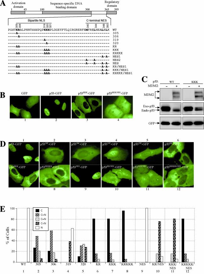
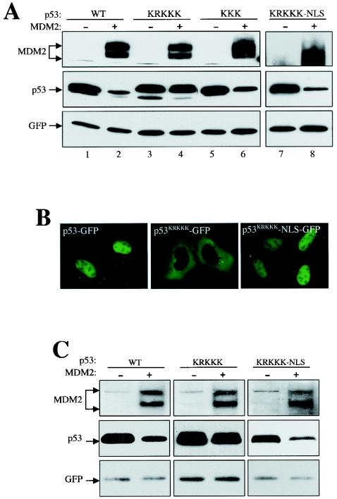
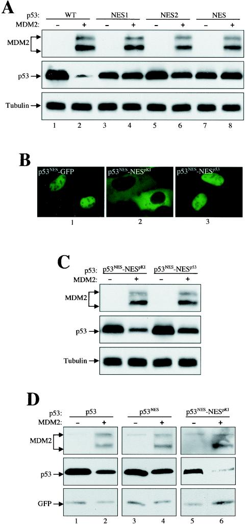
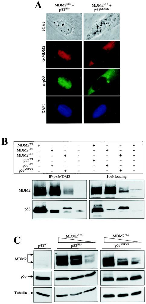
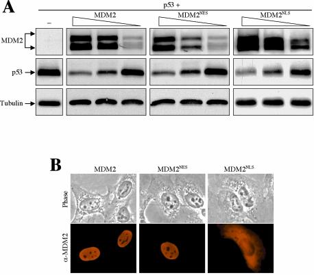
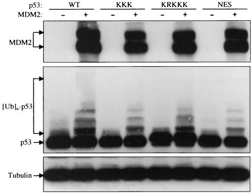
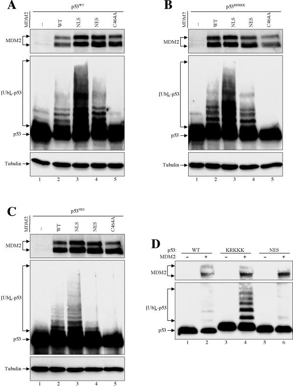
As a shuttling protein, p53 is constantly transported through the nuclear pore complex. p53 nucleocytoplasmic transport is carried out by a bipartite nuclear localization signal (NLS) located at its C-terminal domain and two nuclear export signals (NES) located in its N- and C-terminal regions, respectively. The role of nucleocytoplasmic shuttling in p53 ubiquitination and degradation has been a subject of debate. Here we show that the two basic amino acid groups in the p53 bipartite NLS function collaboratively to import p53. Mutations disrupting individual amino acids in the NLS, although causing accumulation of p53 in the cytoplasm to various degrees, reduce but do not eliminate the NLS activity, and these mutants remain sensitive to MDM2 degradation. However, disrupting both parts of the bipartite NLS completely blocks p53 from entering the nucleus and causes p53 to become resistant to MDM2-mediated degradation. Similarly, mutations disrupting four conserved hydrophobic amino acids in the p53 C-terminal NES block p53 export and prohibit it from MDM2 degradation. We also show that colocalization of a nonshuttling p53 with MDM2 either in the nucleus or in the cytoplasm is sufficient for MDM2-induced p53 polyubiquitination but not degradation. Our data provide new insight into the mechanism and regulation of p53 nucleocytoplasmic shuttling and degradation.


Figures

FIG.1.
FIG.1.
Disruption of both basic amino acid parts but not individual amino acids in the bipartite NLS abolishes p53 nuclear import. (A) Diagram of p53 with positions and amino acid sequences of the bipartite NLS and C-terminal NES indicated. Amino acid substitutions used in the study are indicated in bold face. (B) Combining an NES mutation revealed a nuclear import activity in the p53KKK mutant. U2-OS cells were transfected with the indicated plasmids, and pictures were taken 24 h after transfection with living cells. (C) Degradation of p53KKK mutant by MDM2. U2-OS cells were transiently transfected with indicated p53 and MDM2 plasmid DNAs along with a GFP plasmid. Twenty-four hours after transfection, total cell lysate was prepared from each transfected cell population, electrophoretically separated by SDS-PAGE, and immunoblotted with antibodies specific to MDM2 (SMP14), p53 (DO1), and GFP (Ab2). (D) Various contributions of the basic amino acids in the bipartite NLS to the nuclear import of p53. The conserved basic amino acids in the bipartite NLS of p53 were replaced with alanine residues individually or in combination as indicated in panel A. The plasmid DNA was transfected into U2-OS cells, and the pictures were taken of living cells 24 h after transfection. (E) Quantification of GFP-positive cells. The GFP fluorescence pattern of each transfected cell population expressing the indicated p53 constructs was scored for at least 200 cells. The graph shows the percentage of cells with the indicated GFP patterns.
FIG. 2.
FIG. 2.
Nuclear import of p53 is required for MDM2-mediated degradation. (A) U2-OS cells were transiently transfected with plasmid DNA expressing various p53 and MDM2 mutants, as indicated. Cell lysates were collected 24 h after transfection, separated by SDS-PAGE, and transferred onto a nitrocellulose membrane. Different portions of the membrane were blotted with the indicated antibodies. (B) Attachment of the simian virus 40 NLS to the C terminus of the nuclear import-deficient p53KRKKK restored its nuclear localization and its sensitivity to MDM2 degradation (A). (C) Mouse embryo fibroblast cells lacking both MDM2 and p53 (2KO) were transiently transfected, and the cell lysate was blotted as for panel A.
FIG. 3.
FIG. 3.
Nuclear export of p53 is also required for MDM2-mediated degradation. (A) The NES mutant p53 was resistant to MDM2 degradation. Plasmid DNAs encoding wild-type and various NES mutant p53s were either singly transfected or cotransfected with MDM2 into U2-OS cells. Cell lysates were prepared 24 h after transfection, separated by SDS-PAGE, and transferred onto a nitrocellulose membrane. Different portions of the membrane were blotted with antibodies, as indicated. (B) Localization of p53-GFP fusion proteins. The pKI NES or p53 NES was attached to the C terminus of the p53NES mutant to create p53NES-NESpKI and p53NES-NESp53, respectively. The constructs were then fused C-terminally with GFP and expressed in U2-OS cells. Pictures were taken of living cells 24 h after transfection. (C) Attachment of the pKI NES or p53 NES to the to the C terminus of the p53NES mutant restored its sensitivity to MDM2 degradation. (D) 2KO cells were transfected and the cell lysate was blotted as for panel A.
FIG. 4.
FIG. 4.
Colocalization with MDM2 either in the nucleus or in the cytoplasm is not sufficient to mediate nonshuttling p53 degradation. (A) Colocalization of MDM2 mutants with p53 mutants. U2-OS cells were transiently cotransfected with the indicated MDM2 and p53 DNAs. Twenty-four hours after transfection, cells were fixed and coimmunostained with a mouse anti-MDM2 antibody (SMP14) and a rabbit anti-p53 antibody (FL393) followed by staining with Texas red-conjugated donkey anti-mouse and fluorescein isothiocyanate-conjugated donkey anti-rabbit immunoglobulin antibodies. Phase contrast and 4′,6′-diamidino-2-phenylindole (DAPI) pictures are also shown. (B) Interaction of mutant p53 and MDM2 in vivo. U2-OS cells were transiently transfected with the indicated plasmid, and cells were lysed and immunoprecipitated with antibodies recognizing MDM2. Different portions of the membrane were blotted with antibodies, as indicated. (C) Resistance of nonshuttling p53 to MDM2 degradation. Plasmid DNA encoding nuclear export-deficient p53NES or nuclear import-deficient p53KRKKK were coexpressed with decreasing amountsof the nuclear export-deficient MDM2NES or nuclear import-deficient MDM2NLS mutant in U2-OS cells, respectively. Cell lysates were prepared 24 h after transfection, separated by SDS-PAGE, and transferred onto a nitrocellulose membrane. Different portions of the membrane were blotted with antibodies, as indicated.
FIG. 5.
FIG. 5.
Nucleocytoplasmic shuttling of MDM2 is not essential for promoting p53 degradation. (A) Plasmid DNA (1 μg) encoding wild-type p53 was coexpressed with decreasing amounts (5, 3, and 1 μg) of a wild-type, an NES mutant, or an NLS mutant MDM2 in U2-OS cells. Cell lysates were prepared 24 h after transfection, separated by SDS-PAGE, and transferred onto a nitrocellulose membrane. Different portions of the membrane were blotted with antibodies, as indicated. (B) Localization of MDM2. U2-OS cells were transiently transfected with the indicated MDM2 DNAs. Twenty-four hours after transfection, cells were fixed and immunostained with an anti-MDM2 antibody (SMP14) and a Texas red-conjugated donkey anti-mouse immunoglobulin antibody. Phase contrast pictures are also shown.
FIG. 6.
FIG. 6.
Nonshuttling, nondegradable p53 mutants can be ubiquitinated by MDM2. U2-OS cells were transiently transfected with plasmid DNA encoding wild-type MDM2, wild-type p53, or various p53 mutants, as indicated. Twenty-four hours after transfection, cells were treated with 10 μM MG132 for 6 h and then harvested in SDS lysis buffer. The cell lysate was separated by SDS-PAGE and transferred onto a nitrocellulose membrane. Different portions of the membrane were blotted with antibodies to MDM2 (SMP14), p53 (DO1), and β-tubulin, as indicated.
FIG. 7.
FIG. 7.
MDM2-mediated p53 ubiquitination occurs preferentially in the cytoplasm. (A to C) Wild-type and mutant MDM2s were cotransfected with plasmid DNA encoding wild-type p53 (A), nuclear import mutant p53KRKKK (B), and nuclear export mutant p53NES (C) into U2-OS cells. Twenty-four hours after transfection, cells were treated with 10 μM MG132 for 6 h and then harvested in SDS lysis buffer. The cell lysate was separated by SDS-PAGE and transferred onto a nitrocellulose membrane. Different portions of the membrane were blotted with antibodies to MDM2 (SMP14), p53 (DO1), and β-actin, as indicated. (D) H1299 cells were transiently transfected with plasmid DNA encoding wild-type MDM2, wild-type p53, and various p53 mutants, as indicated. Twenty-four hours after transfection, cells were lysed with 0.5% NP-40 lysis buffer. The cell lysate was separated by SDS-PAGE and transferred onto a nitrocellulose membrane. Different portions of the membrane were blotted with antibodies to MDM2 (SMP14) and p53 (DO1), as indicated.

Similar articles

Cited by

References

    1. Ashcroft, M., and K. H. Vousden. 1999. Regulation of p53 stability. Oncogene 18:7637-7643. - PubMed
    1. Barak, Y., T. Juven, R. Haffner, and M. Oren. 1993. Mdm-2 expression is induced by wild-type p53 activity. EMBO J. 12:461-468. - PMC - PubMed
    1. Benditt, J. O., C. Meyer, H. Fasold, F. C. Barnard, and N. Riedel. 1989. Interaction of a nuclear location signal with isolated nuclear envelopes and identification of signal-binding proteins by photoaffinity labeling. Proc. Natl. Acad. Sci. USA 86:9327-9331. - PMC - PubMed
    1. Blattner, C., T. Hay, D. W. Meek, and D. P. Lane. 2002. Hypophosphorylation of Mdm2 augments p53 stability. Mol. Cell. Biol. 22:6170-6182. - PMC - PubMed
    1. Boyd, S. D., K. Y. Tsai, and T. Jacks. 2000. An intact HDM2 RING-finger domain is required for nuclear exclusion of p53. Nat. Cell Biol. 2:563-568. - PubMed

Publication types

MeSH terms