. 2003 Sep;23(18):6442-54.
doi: 10.1128/MCB.23.18.6442-6454.2003.

Nuclear factor of activated T cells c is a target of p38 mitogen-activated protein kinase in T cells

Affiliations

Nuclear factor of activated T cells c is a target of p38 mitogen-activated protein kinase in T cells

Chia-Cheng Wu et al. Mol Cell Biol. 2003 Sep.

Abstract

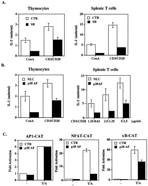
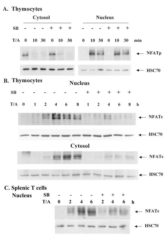
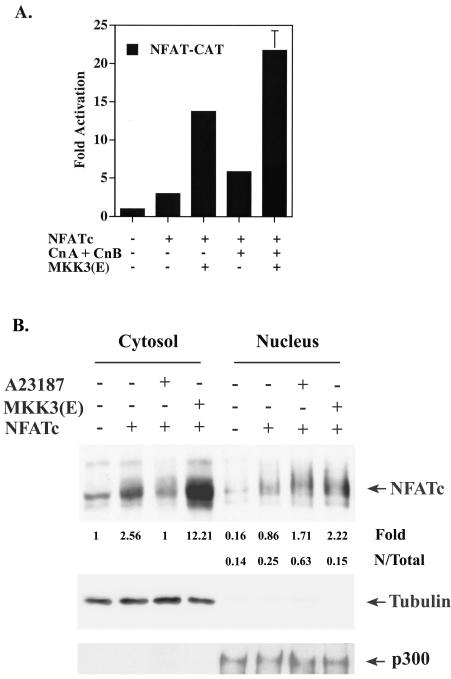
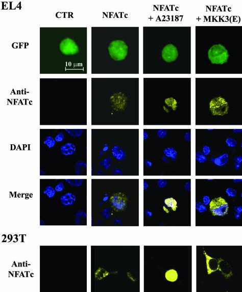
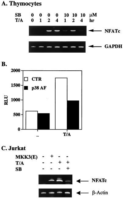
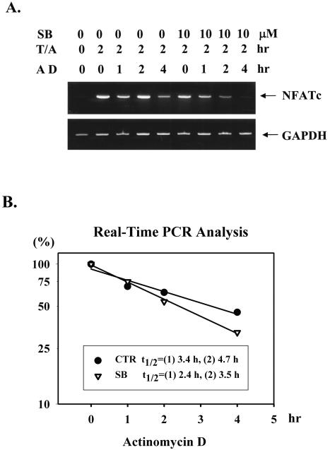
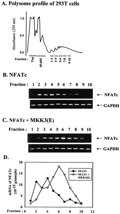
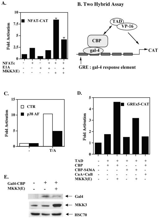
p38 mitogen activated protein kinase (MAPK) is essential for T-cell activation. Here we demonstrated that nuclear factor of activated T cells (NFAT) is a direct target of p38 MAPK. Inhibition of p38 MAPK led to selective inactivation of NFAT in T cells. We further linked a strict requirement of p38 MAPK to activation of NFATc. A stimulatory effect of p38 MAPK on at least four other stages of NFATc activation was found. First, the p38 MAPK cascade activated the NFATc promoter and induced the transcription of NFATc mRNA. Second, p38 MAPK mildly increased the mRNA stability of NFATc. Third, p38 MAPK enhanced the translation of NFATc mRNA. Fourth, p38 MAPK promoted the interaction of NFATc with the coactivator CREB-binding protein. In contrast, p38 MAPK moderately enhanced the expulsion of NFATc from the nucleus in T cells. Therefore, p38 MAPK has opposite effects on different stages of NFATc activation. All together, the overall effect of p38 MAPK on NFATc in T cells is clear activation.


Figures

FIG. 1.
FIG. 1.
NFAT is a target of p38 MAPK on the IL-2 gene promoter. (A) SB 203580 inhibited IL-2 production. Thymocytes and splenic T cells (5 × 10⁵/well) from C57BL/6 mice were stimulated with ConA (2.5 μg/ml) plus anti-CD3 (10 μg/ml) or anti-CD28 (2.5 μg/ml) in the presence (SB) or absence (CTR [control]) of SB 203580 (10 μM). IL-2 produced was quantitated 24 h later with indicator cell line HT-2. (B) Transgenic p38α(AF) suppressed IL-2 production in T cells. Thymocytes and splenic T cells (5 × 10⁵/well) from p38α(AF)-transgenic mice and NLC mice were stimulated with ConA (2.5 μg/ml) and CD3 plus CD28 at the concentrations indicated. IL-2 levels were determined 24 h after activation. (C) p38α(AF) inhibited the activation of NFAT-CAT in T cells. EL4 T cells were transfected with CAT reporters containing AP-1, NFAT, or NF-κB elements (18) and with or without 1 μg of p38α(AF) by the DEAE-dextran method. T cells were stimulated with TPA (10 ng/ml) plus A23187 (80 ng/ml) (T/A) 24 h later (or not stimulated [−]), and CAT activities were determined after another 8 h. Data are reported as means and standard errors of the means.
FIG. 2.
FIG. 2.
p38 MAPK is required for the activation of NFATc but not NFATp. Thymocytes (A and B) and splenic T cells (C) were stimulated with TPA (10 ng/ml) and A23187 (80 ng/ml) (T/A) in the absence or presence of SB 203580 (10 μM) (SB), and cytosol extracts and nuclear extracts were prepared. (A) Nuclear translocation of NFATp was not affected by p38 MAPK suppression. Cytoplasmic and nuclear NFATp contents in thymocytes 10 and 30 min after activation were determined by immunoblotting with anti-NFATp (4G6-G5; BD-PharMingen). (B) Inhibition of p38 MAPK reduced nuclear and cytoplasmic presence of NFATc in thymocytes. Nuclear and cytosolic NFATc contents of thymocytes isolated 1, 2, 4, 6, and 8 h after TPA and A23187 activation were determined with anti-NFATc (7A6; BD-PharMingen). (C) SB 203580 inhibited the nuclear appearance of NFATc in splenic T cells. The amounts of NFATc in nuclear extracts of splenic T cells activated for 2, 4, and 6 h were determined as described for panel B.
FIG. 3.
FIG. 3.
p38 MAPK promotes NFATc activation but increases nuclear export of NFATc in T cells. (A) NFAT transcription activation mediated by NFATc was enhanced by MKK3b(E). EL4 T cells were transfected with NFAT-CAT in the presence of CMV-NFATc, calcineurin catalytic A subunit plus regulatory B subunit (CnA + CnB), and MKK3b(E) as indicated. CAT activities were determined 24 later. Data are reported as means and standard errors of the means. (B) EL4 T cells were similarly transfected with CMV-NFATc and/or MKK3b(E) or treated with A23187. Cytosolic and nuclear extracts were prepared 24 h after transfection, and the amounts of NFATc in the nucleus and cytoplasm were assessed by immunoblotting. α-Tubulin was used as a marker for the cytosol, and p300 was used as a marker for the nucleus. The quantity of NFATc was determined by densitometry and normalized by densitometric reading of the respective internal control (α-tubulin or p300). For ease of comparison, the quantity of cytoplasmic NFATc in untreated T cells was set at 1. N/Total, ratio of nuclear NFATc content to total (nucleus plus cytosol) NFATc content.
FIG. 4.
FIG. 4.
Confocal image of intracellular distribution of NFATc stimulated with MKK3(E) or A23187. EL4 T cells and 293T cells were transfected with EGFP only (CTR [control]); EGFP and CMV-NFATc (NFATc); EGFP, CMV-NFATc, and A23187 (NFATc + A23187); and EGFP, CMV-NFATc, and MKK3b(E) [NFATc + MKK3(E)]. At 24 h after transfection, cells were fixed with 3.7% paraformaldehyde, followed by methanol permeabilization. The cells were stained with DAPI (DAPI) and with anti-NFATc followed by phycoerythrin-conjugated anti-mouse immunoglobulin (anti-NFATc). The NFATc expression of cells was analyzed by use of Zeiss confocal laser scanning microscope LSM 510 with a ×63 objective lens. Green cells (GFP) indicate transfected cells, while DAPI indicates the nucleus. The nuclear localization of NFATc was examined by overlapping the anti-NFATc-stained image with the DAPI-stained image (merge).
FIG. 5.
FIG. 5.
NFATc mRNA induction requires p38 MAPK. (A) NFATc mRNA induction in thymocytes was inhibited by SB 203580. Thymocytes were activated with TPA and A23187 (T/A) in the absence or presence of SB 203580 (10 μM) (SB). Thymocytes were harvested for RNA isolation 1, 2, and 4 h after activation. The amount of NFATc mRNA was determined by RT-PCR. GAPDH, glyceraldehyde-3-phosphate dehydrogenase. (B) Activation of the NFATc promoter was p38 MAPK dependent. A pGL2 reporter containing the NFATc promoter (−752 to −21 bp) was transfected into EL4 T cells without (CTR [control]) or with p38α(AF). T cells were activated with TPA and A23187 24 h later, and luciferase activity was determined 24 h after activation. −, no activation. RLU, relative light units. (C) MKK3b(E) alone induced NFATc expression in Jurkat cells. Jurkat T cells were transfected with active MKK3b(E) or pcDNA3 as a control. Cells transfected with pcDNA3 were then left untreated or were treated with TPA amd A23187 24 h later. The amount of NFATc mRNA was determined by RT-PCR.
FIG. 6.
FIG. 6.
NFATc mRNA stability was moderately enhanced by p38 MAPK. (A) RNA stability was determined by adding actinomycin D (10 μg/ml) (AD) to thymocyte cultures preactivated with TPA and A23187 (T/A) for 2 h and with or without SB 203580 (10 μM) (SB). The RNA was isolated 1, 2, and 4 h after actinomycin D addition. (B) The kinetics of NFATc mRNA degradation were plotted, and the half-life (t1/2) was calculated with a second-order polynomial curve fit on CA-Cricket Graph III (Computer Associates, Islandia, N.Y.). CTR, control. Half-lives of NFATc mRNA from two independent experiments are indicated. The amounts of NFATc mRNA were determined by RT-PCR (A) and by real-time PCR (B).
FIG. 7.
FIG. 7.
Increased NFATc mRNA translation by p38 MAPK. 293T cells were collected 24 h after transfection with CMV-NFATc or CMV-NFATc plus MKK3b(E). Cell lysates were analyzed by sucrose gradient centrifugation. (A) Typical profile of the sucrose gradient monitored by measuring the absorbance at 254 nm. The top of the gradient, the 40S and 60S fractions, and polysome-containing fractions (1 to 10) are indicated. (B and C) Amounts of NFATc mRNA and glyceraldehyde-3-phosphate dehydrogenase (GAPDH) mRNA in a polysome-containing fraction from 293T cells transfected with NFATc (B) or MKK3(E) plus NFATc (C) were determined by RT-PCR. (D) The distribution of the NFATc mRNA polysome-containing fraction was enhanced by p38 MAPK. NFATc mRNA in each polysome fraction was quantitated by real-time PCR. The quantity of NFATc mRNA was then plotted against the number of the polysome fraction.
FIG. 8.
FIG. 8.
p38 MAPK promotes the interaction between NFATc and CBP. (A) E1A inhibited p38 MAPK-stimulated NFAT activation. EL4 cells were transfected with NFAT-CAT in the presence of NFATc, E1A, or MKK3(E) as indicated. Cells were harvested 24 h after transfection, and CAT activities were determined. Data are reported as means and standard errors of the means. (B) Interaction of CBP and NFATc, as determined by the illustrated binding of pGAL-CBP to pVP16-NFAT(TAD) and the activation of pG5B-CAT (containing five tandem repeats of the GRE). (C) The NFAT-CBP interaction was p38 MAPK dependent. 293T cells were transfected with pGAL-CBP, pVP16-NFAT(TAD), or pG5B-CAT, with or without (CTR [control]) p38α(AF). After 24 h, 293T cells were stimulated with TPA and A23187 (T/A), and CAT activities were determined after another 24 h. −, no stimulation. (D) MKK3(E) stimulated NFATc-CBP binding. Jurkat T cells were transfected with pGAL-CBP or pGAL-CBP(S436A), pVP16-NFAT(TAD), pG5B-CAT, 1 μg of EGFP with or without calcineurin (both A and B subunits) (CnA+CnB), or MKK3(E) by the DEAE-dextran method. CAT activation was quantitated 24 h later. (E) Enhanced NFATc-CBP interaction was not due to MKK3(E)-stimulated Gal4-CBP expression. Levels of Gal4-CBP and MKK3(E) in panel D were quantitated with anti-Gal4 and anti-MKK3.

Similar articles

Cited by

References

    1. Avots, A., M. Buttmann, S. Chuvpilo, C. Escher, U. Smola, A. J. Bannister, U. R. Rapp, T. Kouzarides, and E. Serfling. 1999. CBP/p300 integrates Raf/Rac-signaling pathways in the transcriptional induction of NF-ATc during T cell activation. Immunity 10:515-524. - PubMed
    1. Ballinger, D. G., and M. L. Pardue. 1983. The control of protein synthesis during heat shock in Drosophila cells involved altered polypeptide elongation rate. Cell 33:103-144. - PubMed
    1. Beals, C. R., C. M. Sheridan, C. W. Turck, P. Gardner, and G. R. Crabtree. 1997. Nuclear export of NF-ATc enhanced by glycogen synthase kinase-3. Science 275:1930-1934. - PubMed
    1. Chen, C. Y., and A. B. Shyu. 1995. AU-rich elements: characterization and importance in mRNA degradation. Trends Biochem. Sci. 20:465-470. - PubMed
    1. Chow, C.-W., M. Rincón, and R. J. Davis. 1999. Requirement for transcription factor NFAT in interleukin-2 expression. Mol. Cell. Biol. 19:2300-2307. - PMC - PubMed

Publication types

MeSH terms

LinkOut - more resources