. 2003 Sep;23(18):6455-68.
doi: 10.1128/MCB.23.18.6455-6468.2003.

Insulation of the chicken beta-globin chromosomal domain from a chromatin-condensing protein, MENT

Affiliations

Insulation of the chicken beta-globin chromosomal domain from a chromatin-condensing protein, MENT

Natalia E Istomina et al. Mol Cell Biol. 2003 Sep.

Abstract

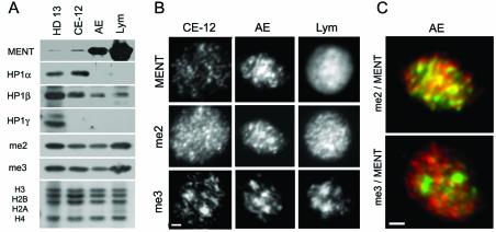
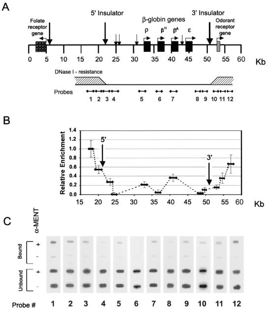
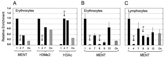
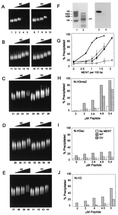
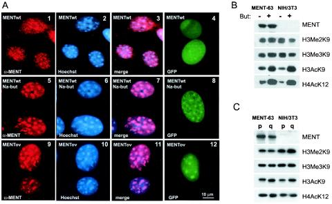
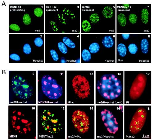
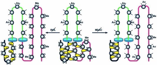
Active genes are insulated from developmentally regulated chromatin condensation in terminally differentiated cells. We mapped the topography of a terminal stage-specific chromatin-condensing protein, MENT, across the active chicken beta-globin domain. We observed two sharp transitions of MENT concentration coinciding with the beta-globin boundary elements. The MENT distribution profile was opposite to that of acetylated core histones but correlated with that of histone H3 dimethylated at lysine 9 (H3me2K9). Ectopic MENT expression in NIH 3T3 cells caused a large-scale and specific remodeling of chromatin marked by H3me2K9. MENT colocalized with H3me2K9 both in chicken erythrocytes and NIH 3T3 cells. Mutational analysis of MENT and experiments with deacetylase inhibitors revealed the essential role of the reaction center loop domain and an inhibitory affect of histone hyperacetylation on the MENT-induced chromatin remodeling in vivo. In vitro, the elimination of the histone H3 N-terminal peptide containing lysine 9 by trypsin blocked chromatin self-association by MENT, while reconstitution with dimethylated but not acetylated N-terminal domain of histone H3 specifically restored chromatin self-association by MENT. We suggest that histone H3 modification at lysine 9 directly regulates chromatin condensation by recruiting MENT to chromatin in a fashion that is spatially constrained from active genes by gene boundary elements and histone hyperacetylation.


Figures

FIG. 1.
FIG. 1.
Heterochromatin proteins in chicken blood cell nuclei. (A) Proteins from the nuclei of chicken HD13 cells, 12-day embryo chicken erythrocytes (CE-12), adult erythrocytes (AE), and chicken lymphocytes (Lym) were analyzed by SDS-PAGE and Western blotting with antibodies against MENT, HP1α, HP1β, HP1γ, H3me2K9 (me2), and H3me3K9 (me3) as indicated. The bottom panel shows electrophoresis of histones from the same material stained with Coomassie blue R-250. (B) Immunofluorescence microscopy of nuclei of 12-day embryo chicken erythrocytes, adult erythrocytes, and chicken lymphocytes stained with antibodies against MENT, H3me2K9, and H3me3K9 as indicated. Bar, 1 μm. (C) Overlays of immunofluorescence micrographs of adult chicken erythrocyte nuclei. Top panel, double staining with antibodies against MENT (red) and H3me2K9 (green). Bottom panel, double staining with antibodies against MENT (red) and H3me3K9 (green). Bar, 1 μm.
FIG. 2.
FIG. 2.
Mapping of MENT on the β-globin domain in chicken erythrocytes. (A) A scheme of the β-globin cluster shows the positions of β-globin genes and the map numbering system from reference . Vertical arrows indicate the positions of DNase I-hypersensitive sites, including those corresponding to the 5′ and 3′ insulators (58). The lower horizontal bar shows a cumulative scheme of DNase I resistance (shaded areas indicate resistance) derived from several experiments (3, 28, 58, 64). The scheme also shows the positions of hybridization probes 1 to 12 used throughout this work. (B) MENT levels on the chicken β-globin cluster. The graph shows the relative enrichment of MENT at the regions corresponding to the β-globin probes 1 to 12. Error bars indicate standard deviations. (C) Slot hybridization analysis of immunoprecipitated MENT-DNA complexes from the chicken β-globin cluster. After formaldehyde cross-linking of isolated nuclei, DNA-MENT complexes were immunoprecipitated either with α-MENT (+) or with preimmune serum (−). DNA was purified and hybridized with probes 1 to 12 derived from the chicken β-globin cluster.
FIG. 3.
FIG. 3.
MENT levels at selected DNA sequences from chicken blood cells. (A) The histograms show relative enrichment of MENT, H3me2K9 (H3me2), and H3acK9,13 (H3ac) detected by ChIP in chicken erythrocytes. (B and C) The histograms show the relative enrichment of MENT (as related to the probe 1) detected by ChIP with selected genome probes in chicken erythrocytes (A) and lymphocytes (B). DNA probes 1, 4, 5, 7, 8, and 12 (black bars) from the β-globin domain (see Fig. 2A) and Ov (gray bars) from the chicken ovalbumin-coding region, positions 2375 to 3285 (17), were used. Arrows show the probes corresponding to the MENT minimums at the 5′ boundary and the 3′ boundary of the chicken β-globin gene (Fig. 2B).
FIG. 4.
FIG. 4.
Cooperative interaction of MENTwt with the dimethylated N-terminal domain of histone H3. (A) Agarose gel electrophoresis of native chicken chromatin reconstituted with MENTwt (lanes 1 to 5) and MENTov (lanes 6 to 10); (B) agarose gel electrophoresis of trypsin-digested chromatin reconstituted with MENTwt (lanes 11 to 15) and MENTov (lanes 16 to 20); (C) agarose gel electrophoresis of trypsin-digested chromatin reconstituted with 5 μM dimethylated N-H3me2 and increasing concentrations of MENTwt (lanes 21 to 24) and MENTov (lanes 25 to 28); (D) agarose gel electrophoresis of trypsin-digested chromatin reconstituted with 5 μM acetylated N-H3ac and increasing concentrations of MENTwt (lanes 29 to 32) and MENTov (lanes 33 to 36); (E) agarose gel electrophoresis of trypsin-digested chromatin reconstituted with 5 μM unmodified N-H3 and increasing concentration MENTwt (lanes 37 to 40) and MENTov (lanes 41 to 44). MENT/DNA ratios (moles per 100 bp) were 0 (lanes 1, 6, 11, 16, 21, 25, 29, 33, 37, and 41), 0.5 (lanes 2, 7, 12, 17, 22, 26, 34, 38, and 42), 1 (lanes 3, 8, 13, 18, 23, 27, 35, 39, and 43), 2 (lanes 4, 9, 14, 19, 24, 28, 36, 40, and 44), and 4 (lanes 5, 10, 15, and 20). (F) SDS-PAGE (lanes 1 and 2) and Western blotting with α-H3me2K9 (lanes 3 and 4) of native chicken erythrocyte chromatin before (lanes 1 and 3) and after (lanes 2 and 4) digestion with trypsin. Note the absence of intact histone H3 and the disappearance of H3me2K9 in the trypsindigested sample. P1 shows the position of the main histone H3 trypsin digestion product (23). (G) Graph showing the percentage of precipitated material in native chromatin reconstituted with MENTwt (•) and MENTov (▪) and in trypsin-digested chromatin reconstituted with MENTwt (○) and MENTov (□). Ratios of MENT/DNA were as indicated. Error bars indicate standard deviations. (H to J) Histograms showing the percentage of precipitated material in trypsin-digested chromatin reconstituted with no MENT (open bars), 1 mol of MENTwt/60 bp (diagonally hatched bars), and 1 mol of MENTov/60 bp (horizontally hatched bars) plus increasing concentrations of N-H3me2 (H), N-H3ac (I), and N-H3 (J). Histone peptide concentrations were as indicated.
FIG. 5.
FIG. 5.
A histone deacetylase inhibitor and a mutation in the RCL domain change the MENT location in nuclear chromatin. (A) Immunofluorescence microscopy of proliferating 3T3/MENT-63 cells (panels 1 to 3), the same cells incubated in the presence of 10 mM sodium butyrate for 48 h (panels 5 to 7), and 3T3/MENTov-16 cells stably expressing a MENT-ovalbumin swap mutant (panels 9 to 11) fixed in acetone-methanol and stained with α-MENT (panels 1, 5, and 9) and Hoechst 33258 (panels 2, 6, and 10). Panels 3, 7, and 11, overlays of images stained with α-MENT and Hoechst. Panels 4, 8, and 12, immunofluorescence microscopy of unfixed, unstained proliferating NIH 3T3 cells transiently expressing GFP fusions with MENTwt (panels 4 and 8) and MENT-ovalbumin swap (panel 12). The cells in panel 8 were incubated for 48 h with 10 mM sodium butyrate. Bar, 10 μm. (B) Protein samples from the nuclei of 3T3/MENT-63 cells stably expressing MENTwt (MENT-63) and control NIH 3T3-lacI cell clones stably transfected with nonexpressing vector (NIH 3T3) incubated in the absence (−) or presence (+) of 10 mM sodium butyrate (But) for 48 h were analyzed by SDS-PAGE and Western blotting with α-MENT, α-H3me2K9, α-H3me3K9, α-H3acK9, and α-H4acK12 as indicated. (C) Protein samples from the nuclei of proliferating (p) and quiescent (q) 3T3/MENT-63 cells and control NIH 3T3-lacI cells were analyzed by SDS-PAGE and Western blotting with α-MENT, α-H3me2K9, α-H3me3K9, α-H3acK9, and α-H4acK12 as indicated.
FIG. 6.
FIG. 6.
Rearrangement of chromatin marked by histone H3 methylation in quiescent MENT-expressing cells. (A) Immunofluorescence microscopy of 3T3/MENT-63 cells (panels 1 to 4), control NIH 3T3-lacI cells (panels 5 and 6), and 3T3/MENTov-16 cells (panels 7 and 8). Cells were fixed in the proliferating state (panels 1 and 2) and the quiescent state (panels 3 to 8) and then stained with α-H3me2K9 (me2) (panels 1, 3, 5, and 7) and Hoechst 33258 (panels 2, 4, 6, and 8). Bar, 10 μm. (B) Immunofluorescence microscopy of 3T3/MENT-63 cells (panels 9 to 14 and 16 to 18) and control NIH 3T3-lacI cells (panel 15). Cells were fixed in the quiescent state and then stained with α-H3me2K9 (green) (panels 9, 12, 14, and 18), α-MENT (red) (panels 10 to 12), α-H4acK12 (red) (panels 13 and 14), α-H3me3K9 (red) (panels 15 and 16), Hoechst 33258 (blue) (panels 9, 11, 15, and 16), and propidium iodine (red) (panels 17 and 18). Bar, 10 μm.
FIG. 7.
FIG. 7.
A model for chromatin condensation regulated by MENT, histone modifications, and gene boundary elements. (Central structure) MENTwt (yellow) binds simultaneously to constitutive heterochromatin (black) and silenced euchromatin (red) marked by H3 dimethylated at lysine 9 (Me). MENT binds to chromatin cooperatively and forms bridges between chromatin fibers that bring euchromatin containing dimethylated histone H3 close to constitutive heterochromatin, thus causing chromatin condensation. A chromosomal domain containing active hyperacetylated chromatin (green) is insulated from MENT spreading by boundary elements (blue disks) that create zones of especially strong hyperacetylation blocking MENT spreading from the domain flanks. (Right structure) Histone deacetylase (HDAC) inhibitors increase the acetylation level of noncentromeric chromatin, interfere with MENT-histone interactions, and promote MENT binding to constitutive heterochromatin. (Left structure) RCL mutations disrupt MENT interaction with chromatin marked by dimethylated histone H3 and also promote MENT binding to constitutive heterochromatin.

Similar articles

Cited by

References

    1. Bannister, A. J., R. Schneider, and T. Kouzarides. 2002. Histone methylation. Dynamic or static? Cell 109:801-806. - PubMed
    1. Bell, A. C., A. G. West, and G. Felsenfeld. 2001. Insulators and boundaries: versatile regulatory elements in the eukaryotic genome. Science 291:447-450. - PubMed
    1. Bellard, M., M. T. Kuo, G. Dretzen, and P. Chambon. 1980. Differential nuclease sensitivity of the ovalbumin and beta-globin chromatin regions in erythrocytes and oviduct cells of laying hen. Nucleic Acids Res. 8:2737-2750. - PMC - PubMed
    1. Bird, C. H., E. J. Blink, C. E. Hirst, M. S. Buzza, P. M. Steele, J. Sun, D. A. Jans, and P. I. Bird. 2001. Nucleocytoplasmic distribution of the ovalbumin serpin pi-9 requires a nonconventional nuclear import pathway and the export factor crm1. Mol. Cell. Biol. 21:5396-5407. - PMC - PubMed
    1. Blanton, J., M. Gaszner, and P. Schedl. 2003. Protein:protein interactions and the pairing of boundary elements in vivo. Genes Dev. 17:664-675. - PMC - PubMed

Publication types

MeSH terms

LinkOut - more resources