. 2003 Sep;23(18):6494-506.
doi: 10.1128/MCB.23.18.6494-6506.2003.

Phagosomes fuse with late endosomes and/or lysosomes by extension of membrane protrusions along microtubules: role of Rab7 and RILP

Affiliations

Phagosomes fuse with late endosomes and/or lysosomes by extension of membrane protrusions along microtubules: role of Rab7 and RILP

Rene E Harrison et al. Mol Cell Biol. 2003 Sep.

Abstract

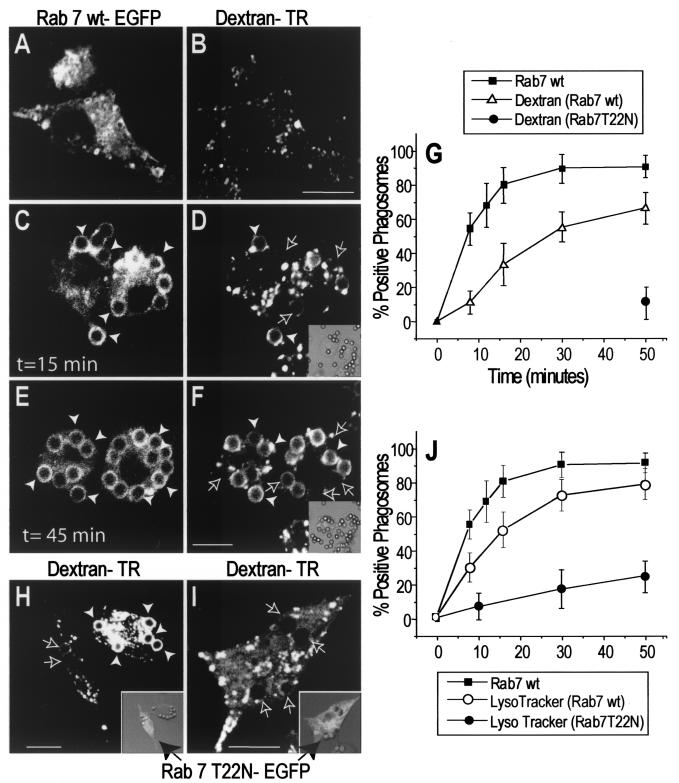
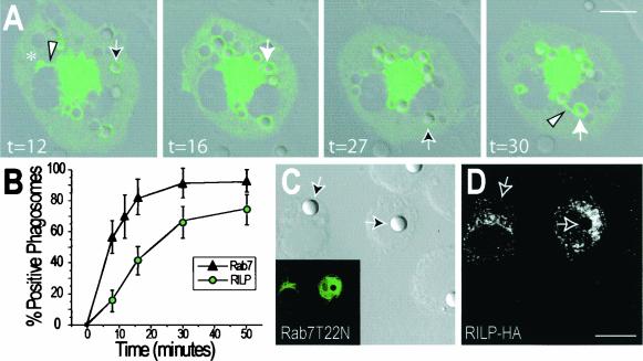
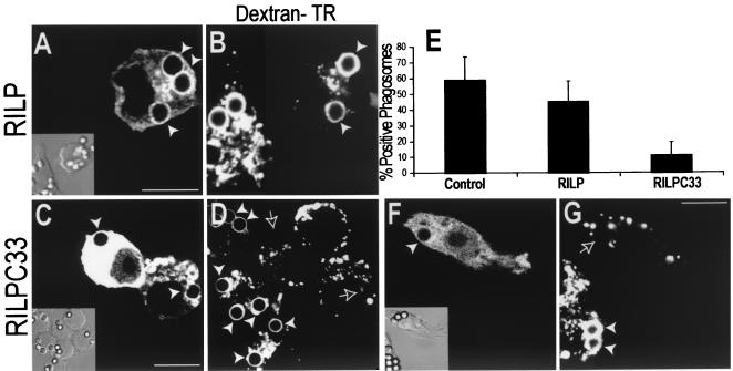
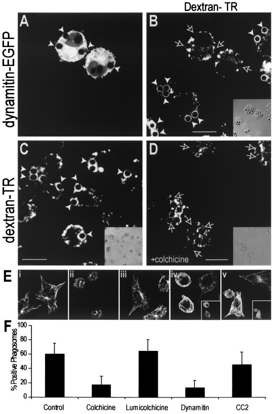
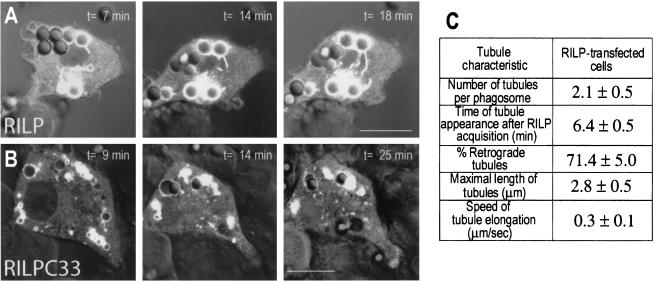
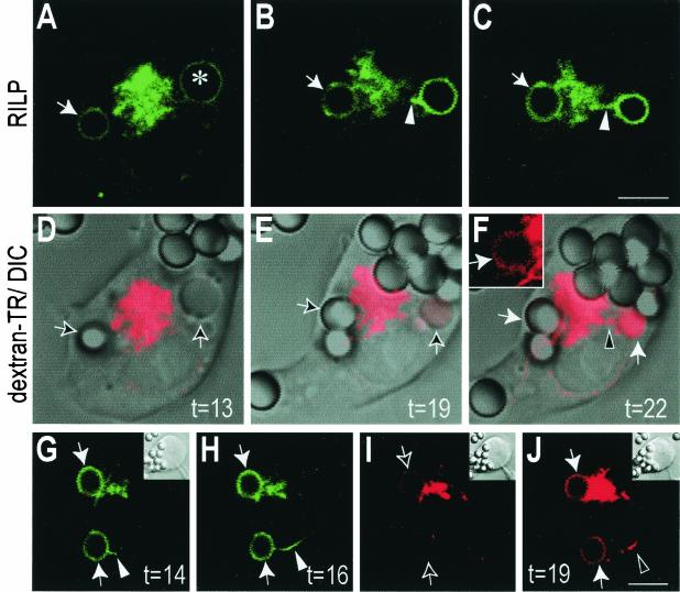
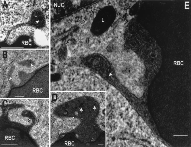
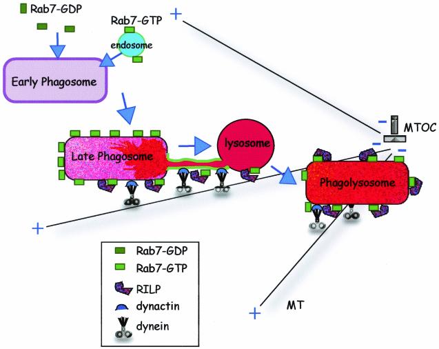
Nascent phagosomes must undergo a series of fusion and fission reactions to acquire the microbicidal properties required for the innate immune response. Here we demonstrate that this maturation process involves the GTPase Rab7. Rab7 recruitment to phagosomes was found to precede and to be essential for their fusion with late endosomes and/or lysosomes. Active Rab7 on the phagosomal membrane associates with the effector protein RILP (Rab7-interacting lysosomal protein), which in turn bridges phagosomes with dynein-dynactin, a microtubule-associated motor complex. The motors not only displace phagosomes in the centripetal direction but, strikingly, promote the extension of phagosomal tubules toward late endocytic compartments. Fusion of tubules with these organelles was documented by fluorescence and electron microscopy. Tubule extension and fusion with late endosomes and/or lysosomes were prevented by expression of a truncated form of RILP lacking the dynein-dynactin-recruiting domain. We conclude that full maturation of phagosomes requires the retrograde emission of tubular extensions, which are generated by activation of Rab7, recruitment of RILP, and consequent association of phagosomes with microtubule-associated motors.


Figures

FIG. 1.
FIG. 1.
Role of Rab7 in phagolysosome fusion and acidification. RAW cells were preloaded with Texas Red-dextran as a marker of lysosomes and late endosomes and were transfected with Rab7-EGFP. Phagocytosis was initiated by addition of IgG-opsonized latex beads (time zero), and the reaction was monitored in live cells by confocal fluorescence (main panels) and DIC microscopy (insets). (A through F) Cells were transfected with wild-type Rab7-EGFP, and images were acquired at the times indicated. Green (Rab7) fluorescence (A, C, and E) and the corresponding red (dextran) fluorescence (B, D, and F) are shown. Panels A and B show representative Rab7-EGFP and dextran labeling in a resting cell prior to phagocytosis. White arrowheads point to Rab7- or dextran-labeled phagosomes, while outlined arrows point to unlabeled phagosomes. Adherent, noninternalized beads were identified by staining with a Cy5-conjugated anti-human antibody. (G) The fraction of Rab7wt- or dextran-positive phagosomes was scored, and data for 50 cells from three experiments are summarized. (H and I) Cells transfected with dominant-negative (T22N) Rab7-EGFP were allowed to ingest particles, and images were acquired at 50 min. Insets show an overlay of green fluorescence and DIC images. A longer exposure was used in panel I to confirm the absence of phagosomal labeling. The fraction of dextran-positive phagosomes in Rab7(T22N)-transfected cells was scored and is summarized in panel G. (J) Staining of phagosomes with LysoTracker Red was scored over a 50-min time course. The percentage of Rab7(T22N) phagosomes containing LysoTracker Red is also shown. Data for Rab7(T22N) phagosomes in panels G and J are means ± standard deviations for 30 cells from three experiments. Bars, 10 μm.
FIG. 2.
FIG. 2.
Phagosomes acquire RILP in a Rab7-GTP-dependent manner. (A) RAW cells were transfected with RILP-EGFP, and after 24 h, phagocytosis was initiated by addition of IgG-opsonized latex beads (time zero). Confocal fluorescence and DIC images were acquired at the indicated times (in minutes) and superimposed. Outlined arrows indicate early phagosomes with little RILP-EGFP, and solid white arrows identify the same phagosomes which had accumulated RILP at a later time. Asterisk denotes a macropinosome. Retrograde tubular extensions often extend from the RILP-positive phagosomes and macropinosomes (arrowheads). (B) Comparison of the time course of acquisition of RILP by phagosomes (from experiments like that for which images are shown in panel A) with that of Rab7 (from Fig. 1). Data are means ± standard deviations for 50 cells from three experiments. (C and D) Cells were cotransfected with Rab7(T22N)-EGFP and epitope-tagged RILP (RILP-HA) and were allowed to internalize particles as in panel A. After 50 min, the cells were fixed and immunostained with anti-HA and Cy3-conjugated secondary antibodies. Arrows point to phagosomes. (C) DIC image (main panel) and corresponding green [Rab7(T22N)-EGFP] fluorescence (inset). (D) Red (RILP-HA) fluorescence. Images in panels A, C, and D are representative of at least 20 cells from three separate experiments. Bars, 10 μm.
FIG. 3.
FIG. 3.
RILP is necessary for phagolysosome formation. (A to D) RAW cells were preloaded with Texas Red-dextran as a marker of late endosomes and/or lysosomes and were transfected either with wild-type RILP-EGFP (A and B) or with truncated RILP-C33-EGFP (C and D). Phagocytosis of IgG-opsonized latex beads was allowed to proceed for 50 min, and green (A and C) and red (B and D) confocal fluorescence and DIC images (insets) were acquired. (E) The fractions of dextran-positive phagosomes in untransfected cells and in cells transfected with either wild-type or truncated RILP were scored. Data are means ± standard deviations for 30 cells from five experiments. (F and G) RAW cells were preloaded with Cy5-dextran as a marker of late endosomes and/or lysosomes and were cotransfected with cyan-labeled truncated RILP (RILP-C33-CFP) and YFP-labeled, constitutively active Rab7 [Rab7(Q67L)-YFP]. (F) Location of RILP-C33-CFP. (Inset) Corresponding DIC image. (G) Distribution of Cy5-dextran. White arrowheads point to labeled phagosomes, and outlined arrows indicate phagosomes devoid of dextran. Images are representative of at least three experiments of each type. Bars, 10 μm.
FIG.4.
FIG.4.
RILP promotes dynamitin recruitment and displacement of phagosomes towards the MTOC. RAW cells were transfected with RILP-EGFP (A, C, and F) or RILP-C33-EGFP (B, D, and G) and allowed to internalize 3.1-μm-diameter (A, B, F, and G) or 0.8-μm-diameter (C and D) IgG-coated beads. After 50 min, the cells were fixed, permeabilized, and immunostained for either α-tubulin (A and B), γ-tubulin (C and D), or p50-dynamitin (F and G). Main panels in A and B show α-tubulin staining overlaid on DIC images. Insets show distribution of RILP-EGFP (A) or RILP-C33-EGFP (B). Bars, 10 μm. Panels C and D show smaller beads (blue) overlaid with γ-tubulin (red) and RILP (C) (green) at 30 min and RILP-C33 (D) (green) at 50 min. Asterisks indicate the location of the MTOC. Bars, 5 μm. Quantitation of bead distance from MTOC is shown in panel E. Data are means from four separate experiments. Main panels in F and G show p50-dynamitin staining. Insets show distribution of RILP-EGFP (F) or RILP-C33-EGFP (G) overlaid on DIC images. Arrowheads point to phagosomes. Bars, 10 μm.
FIG.5.
FIG.5.
Microtubule-mediated dynein activity is necessary for phagolysosome formation. (A and B) RAW cells were preloaded with Texas Red-dextran as a marker of late endosomes and/or lysosomes, and dynamitin-EGFP was overexpressed by transfection to disrupt dynein function. Phagocytosis of IgG-opsonized latex beads was allowed to proceed for 50 min, and green (A) and red (B) confocal fluorescence and DIC images (inset) were acquired. (C and D) RAW cells were preloaded with Texas Red-dextran and allowed to internalize beads for 10 min at 37°C. The cells were then either left untreated (C) or cooled and treated with 10 μM colchicine for 20 min before maturation was allowed to proceed for an additional 30 min at 37°C. The dextran distribution is shown in the main panels, and the corresponding DIC is shown in the insets. White arrowheads indicate dextran-positive phagosomes, whereas outlined arrows mark phagosomes without dextran accumulation. Bars, 10 μm. (E) Tubulin immunostaining in control (i), colchicine-treated (ii), lumicolchicine-treated (iii), dynamitin-overexpressing (iv), and CC2 domain-expressing (v) cells. The transfected cells in panels iv and v are shown in the insets. (F) Phagosomal maturation was measured by the acquisition of Texas Red-dextran in either control cells, colchicine- or lumicolchicine-treated cells, or cells expressing dynamitin or the CC2 domain. Data are means ± standard deviations for 30 cells from three experiments.
FIG. 6.
FIG. 6.
Characterization of phagosomal tubules. The fluorescence of RAW cells transfected with RILP-EGFP (A) or RILP-C33-EGFP (B) was monitored by confocal analysis during phagosome maturation. Confocal fluorescence and DIC images were acquired at the indicated times (in minutes) after the appearance of RILP on the phagosomal membrane, and these images were superimposed. The characteristics of the tubules observed in cells transfected with full-length RILP are summarized (C). Means ± standard errors for 20 individual time lapse experiments are presented. The percentage of retrograde tubules was calculated by assuming that the perinuclear late-endosomal-lysosomal cluster observed in RILP-transfected cells reflects the location of the MTOC (see reference 19).
FIG. 7.
FIG. 7.
Fusion of phagosomes with late endosomes and/or lysosomes proceeds via RILP-containing retrograde extensions. RAW cells were preloaded with Texas Red-dextran as a marker of late endosomes and lysosomes and were transfected with RILP-EGFP. At time zero, phagocytosis was initiated by addition of IgG-opsonized beads, and the reaction was monitored by simultaneous fluorescence confocal and DIC microscopy. (A through C) Green (RILP) fluorescence. (D through F) Corresponding red (dextran) fluorescence, overlaid on DIC images. Inset in panel F shows the fluorescence of phagosome indicated by the horizontal white arrow in the main panel. A macropinosome is identified by an asterisk in panel A. The time after addition of beads (in minutes) is indicated. (G through J) An independent experiment. Green (RILP) fluorescence at the times indicated (G and H) and red (dextran) fluorescence corresponding to 16 and 19 min (I and J) are shown. Note the appearance of lysosomal label in the phagosomal lumen between 16 and 19 min. Phagosomes are indicated by arrows, and arrowheads point to tubular extensions. Bars, 5 μm.
FIG. 8.
FIG. 8.
Transmission electron microscopy of phagosomal tubules. The late-endosomal-lysosomal compartment of RAW cells was labeled by pulsing with colloidal gold-conjugated albumin, followed by an overnight chase. The cells were then allowed to internalize IgG-coated RBC, and after 50 min, they were fixed and processed for transmission electron microscopy. Micrographs show the presence of tubules extending from phagosomes. The tubules are often oriented towards the nucleus (NUC) and in the vicinity of electron-dense lysosomes (L). Some of the tubules contain gold-labeled particles (arrows). In panel E the tubule has seemingly fused with a multivesicular body. Bars, 0.2 μm.
FIG. 9.
FIG. 9.
A model of the role of Rab7-RILP in phagolysosome formation. Following internalization, the early phagosome acquires Rab7 either from a soluble pool and/or by fusion with Rab7-containing endosomes. Rab7-GTP then recruits RILP, which in turn promotes dynein-dynactin association with the phagosome. This complex mediates retrograde movement of phagosomes along microtubules towards the MTOC and in addition promotes the formation of tubular extensions which fuse with late endosomes and lysosomes. Eventually, phagosomes and late endosomes and/or lysosomes merge into a single hybrid organelle.

Similar articles

Cited by

References

    1. Allen, L. A., and A. Aderem. 1996. Mechanisms of phagocytosis. Curr. Opin. Immunol. 8:36-40. - PubMed
    1. Alvarez-Dominguez, C., A. M. Barbieri, W. Beron, A. Wandinger-Ness, and P. D. Stahl. 1996. Phagocytosed live Listeria monocytogenes influences Rab5-regulated in vitro phagosome-endosome fusion. J. Biol. Chem. 271:13834-13843. - PubMed
    1. Alvarez-Dominguez, C., and P. D. Stahl. 1999. Increased expression of Rab5a correlates directly with accelerated maturation of Listeria monocytogenes phagosomes. J. Biol. Chem. 274:11459-11462. - PubMed
    1. Blocker, A., G. Griffiths, J. C. Olivo, A. A. Hyman, and F. F. Severin. 1998. A role for microtubule dynamics in phagosome movement. J. Cell Sci. 111:303-312. - PubMed
    1. Blocker, A., F. F. Severin, J. K. Burkhardt, J. B. Bingham, H. Yu, J. C. Olivo, T. A. Schroer, A. A. Hyman, and G. Griffiths. 1997. Molecular requirements for bi-directional movement of phagosomes along microtubules. J. Cell Biol. 137:113-129. - PMC - PubMed

Publication types

MeSH terms

LinkOut - more resources