. 2003 Sep;23(18):6672-84.
doi: 10.1128/MCB.23.18.6672-6684.2003.

Involvement of nucleocytoplasmic shuttling of yeast Nap1 in mitotic progression

Affiliations

Involvement of nucleocytoplasmic shuttling of yeast Nap1 in mitotic progression

Mary Miyaji-Yamaguchi et al. Mol Cell Biol. 2003 Sep.

Abstract

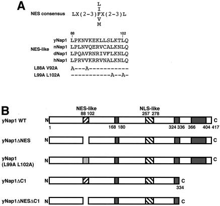
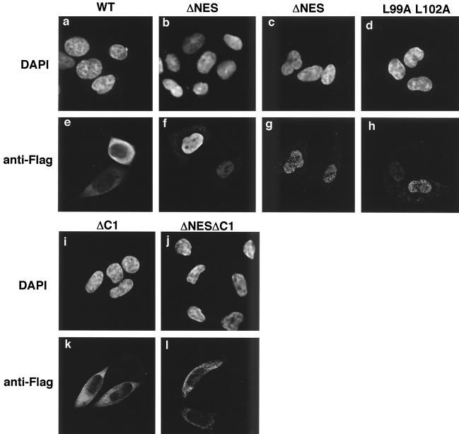
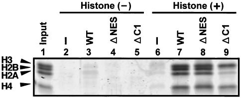
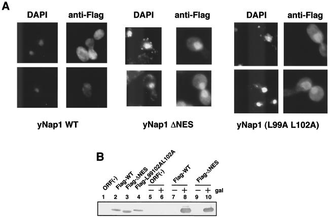
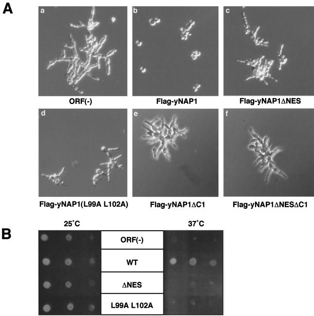
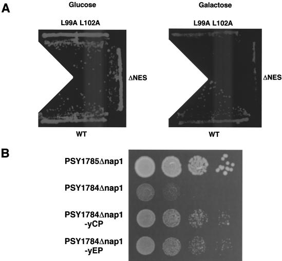
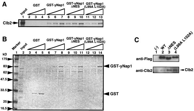
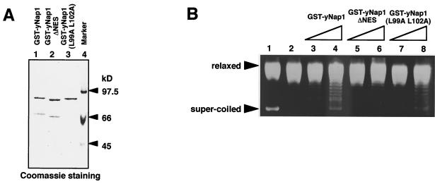
Nucleosome assembly protein 1 (Nap1) is widely conserved from yeasts to humans and facilitates nucleosome formation in vitro as a histone chaperone. Nap1 is generally localized in the cytoplasm, except that subcellular localization of Drosophila melanogaster Nap1 is dynamically regulated between the cytoplasm and nucleus during early development. The cytoplasmic localization of Nap1 is seemingly incompatible with the proposed role of Nap1 in nucleosome formation, which should occur in the nucleus. Here, we have examined the roles of a putative nuclear export signal (NES) sequence in yeast Nap1 (yNap1). yNap1 mutants lacking the NES-like sequence were localized predominantly in the nucleus. Deletion of NAP1 in cells harboring a single mitotic cyclin gene is known to cause mitotic delay and temperature-sensitive growth. A wild-type NAP1 complemented these phenotypes while nap1 mutant genes lacking the NES-like sequence or carboxy-terminal region did not. These and other results suggest that yNap1 is a nucleocytoplasmic shuttling protein and that its shuttling is important for yNap1 function during mitotic progression. This study also provides a possible explanation for Nap1's involvement in nucleosome assembly and/or remodeling in the nucleus.


Figures

FIG. 1.
FIG. 1.
(A) NES-like sequences of Nap1. The NES consensus sequence is taken from reference . The NES-like sequences of yNap1, nematode Nap1 (nNap1), Drosophila Nap1 (dNap1), and human Nap1 (hNap1) are aligned. Amino acid substitution mutants in yNap1 proteins used in this study are also indicated (L88A V92A and L99A L102A). Numbers indicate amino acid positions in the yNap1 protein. (B) yNap1 mutant proteins used this study are schematically summarized. An NES-like sequence (88 to 102), NLS-like sequence (257 to 278), and acidic regions (168 to 180, 324 to 336, and 404 to 417) are indicated.
FIG. 2.
FIG. 2.
Subcellular localization of Flag-yNap1 derivatives in mammalian cells. HeLa cells grown on coverslips were transfected with expression plasmids for Flag-yNap1 (a and e), Flag-yNap1ΔNES (b, c, f, and g), Flag-yNap1 (L99A L102A) (d and h), Flag-yNap1ΔC1 (i and k), or Flag-yNap1ΔNESΔC1 (j and l). For cells expressing Flag-yNap1ΔNES, two independent optical fields are shown. Cells were fixed, stained with DAPI (a to d, i, and j) or anti-Flag antibody (e to h, k, and l), and then examined under a fluorescence microscope and analyzed by a decombolution system (Zeiss).
FIG. 3.
FIG. 3.
Interactions between yNap1 derivatives and core histones. GST pull-down assays were performed with purified core histones and GST-tagged recombinant yNap1 proteins. Lysates prepared from E. coli cells expressing GST (lanes 2 and 6), WT GST-yNap1 (lanes 3 and 7), GST-yNap1ΔNES (lanes 4 and 8), and GST-yNAP-1ΔC1 (lanes 5 and 9) were incubated with (lanes 6 to 9) or without (lanes 2 to 5) 2 μg of core histones purified from HeLa cells as described previously (44) and then mixed with glutathione-Sepharose beads. Beads were washed with a buffer containing 150 mM NaCl and 0.5% NP-40 (lanes 2 to 9). Core histone (lane 1, 0.4 μg) and the bound proteins (lanes 2 to 9) eluted by an SDS-PAGE loading buffer were separated by SDS-15% PAGE and visualized by CYPRO Orange staining. The positions of each core histone component are indicated. The amounts of the bound histones were measured by National Institutes of Health imaging.
FIG. 4.
FIG. 4.
Subcellular localization of yNap1 derivatives in yeast cells. (A) Exponentially growing cells expressing WT Flag-yNap1, Flag-yNap1ΔNES, or Flag-yNap1 (L99A L102A) were fixed, and DNA and Flag-tagged proteins were stained with DAPI and anti-Flag antibody, respectively. (B) Amounts of Flag fusion proteins in transformed cells. DK168 cells were transformed with pRS316 (lane 1), pRS316-Flag-yNap1 (lane 2), pRS316-Flag-yNap1ΔNES (lane 3), or pRS316-Flag-yNap1 (L99A L102A) (lane 4). Exponentially growing transformants were lysed, and lysates were subjected to Western blot analysis with anti-Flag antibody. DK168 cells were separately transformed with pYES2 (lanes 5 and 6), pYES2-Flag-yNap1 (lanes 7 and 8), or pYES2-Flag-yNap1ΔNES (lanes 9 and 10). Lysates prepared from exponentially growing transformants in either SD (lanes 5, 7, and 9) or SG (lanes 6, 8, and 10) were subjected to Western blot analysis.
FIG. 5.
FIG. 5.
Complementation of a NAP1 disruptant with yNAP1 derivatives. The morphological change (A) and ts phenotype (B) of a NAP1-disrupted strain, DK168, were examined when DK168 was transformed with an empty vector [ORF(−)] (a) or expression vectors for Flag-yNap1 WT (WT) (b), Flag-yNap1ΔNES (ΔNES) (c), Flag-yNap1(L99A L102A) (L99A L102A) (d), Flag-yNap1ΔC1 (e), or Flag-yNap1ΔNESΔC1 (f). (A) The morphologies of exponentially growing transformants were observed under a phase microscope. (B) The ts phenotypes during growth were examined by incubation of serially diluted cell suspension spots at permissive (25°C) and nonpermissive (37°C) temperatures.
FIG. 6.
FIG. 6.
Genetic analyses of NAP1. (A) Effect of overexpression of NES-deficient yNap1 in a Clb2-dependent strain. DK168 cells were transformed with pYES2-Flag-yNap1 (WT), pYES2-Flag-yNap1ΔNES (ΔNES), or pYES2-Flag-yNap1 (L99A L102A) (L99A L102A). Single colonies on an SD (without Ura) plate were isolated, and well-grown cultures were streaked onto SD (without Ura) and SG (without Ura) plates and incubated at 25°C for 2 and 3 days, respectively. (B) Genetic interaction between NAP1 and KAP114. Yeasts (PSY1785Δnap1 = KAP114 nap1::LEU2; PSY1784Δnap1 = kap114::HIS3 nap1::LEU2; PSY1784Δnap1-yCP = kap114::HIS3 nap1::LEU2 carrying a low copy number of NAP1; PSY1784Δnap1-yEP = kap114::HIS3 nap1::LEU2 carrying a high copy number of NAP1) fully grown in liquid media were diluted to OD600s of 0.1 with distilled water. Then 1/10 serially diluted cultures were prepared from the 0.1-OD600 cell suspensions in distilled water, and 5 μl of each dilution was grown on YPD agar plates for 2 days at 30°C.
FIG. 7.
FIG. 7.
Interaction between yNap1 and Clb2. (A) Fifty nanograms (lanes 2, 5, and 8), 150 ng (lanes 3, 6, and 9), or 500 ng (lanes 4, 7, and 10) of GST (lanes 2 to 4), GST-yNap1 (lanes 5 to 7), GST-yNap1ΔNES (lanes 8 to 10), or GST-yNap1 (L99A L102A) (lanes 11 to 13) was mixed with glutathione beads and incubated with in vitro-translated [35S]methionine-labeled Clb2 (lane 1, input). Bound proteins were separated by SDS-10% PAGE and visualized by autoradiography. (B) The amount of loaded GST proteins used in panel A is shown by CBB R-250 staining. (C) Yeast cell lysates prepared from DK168::pYES2 (lane 1), DK168::pYES-Flag-yNap1 (lane 2), DK168::pYES-Flag-yNap1ΔNES (lane 3), or DK168::pYES-Flag-yNap1 (L99A L102A) (lane 4) were precipitated with anti-Flag antibody-conjugated agarose beads, and bound proteins were separated by SDS-7.5% PAGE and stained with anti-Flag and anti-Clb2 antibodies.
FIG. 8.
FIG. 8.
Nucleosome assembly activities of yNap1 mutants. (A) Purified GST-fused yNap1 proteins used for nucleosome assembly assays. Four hundred nanograms of each protein (lane 1, GST-yNap1; lane 2, GST-yNap1ΔNES; lane 3, GST-yNap1 [L99A L102A]) was separated by SDS-7.5% PAGE and stained with CBB R-250. (B) Supercoiling assays. Nucleosome assembly reactions (lanes 2 to 8) were carried out with relaxed circular DNA and purified core histones in the absence (lane 2) or presence of GST-yNap1 (lanes 3 and 4), GST-yNap1ΔNES (lanes 5 and 6), or GST-yNap1 (L99A, L102A) (lanes 7 and 8). After the reaction, DNA in each reaction mixture was deproteinized, separated by 1% agarose gel electrophoresis, and visualized by staining with Cyber gold (Molecular Probes). A mixture of fully supercoiled DNA and relaxed DNA is also shown in lane 1.

Similar articles

Cited by

References

    1. Aalfs, J. D., and Kingston, R. E. 2000. What does ‘chromatin remodeling’ mean? Trends Biochem. Sci. 25:548-555. - PubMed
    1. Adams, C. R., and R. T. Kamakaka. 1999. Chromatin assembly: biochemical identities and genetic redundancy. Curr. Opin. Genet. Dev. 9:185-190. - PubMed
    1. Altman, R., and D. Kellogg. 1997. Control of mitotic events by Nap1 and the Gin4 kinase. J. Cell Biol. 138:119-130. - PMC - PubMed
    1. Booher, R. N., R. J. Deshaies, and M. W. Kirschner. 1993. Properties of Saccharomyces cerevisiae wee1 and its differential regulation of p34CDC28 in response to G1 and G2 cyclins. EMBO J. 12:3417-3426. - PMC - PubMed
    1. Bortvin, A., and F. Winston. 1996. Evidence that Spt6p controls chromatin structure by a direct interaction with histones. Science 272:1473-1476. - PubMed

Publication types

MeSH terms