. 2003 Aug 27;23(21):7750-8.
doi: 10.1523/JNEUROSCI.23-21-07750.2003.

Submillisecond precision of the input-output transformation function mediated by fast sodium dendritic spikes in basal dendrites of CA1 pyramidal neurons

Affiliations

Submillisecond precision of the input-output transformation function mediated by fast sodium dendritic spikes in basal dendrites of CA1 pyramidal neurons

Gal Ariav et al. J Neurosci. .

Abstract

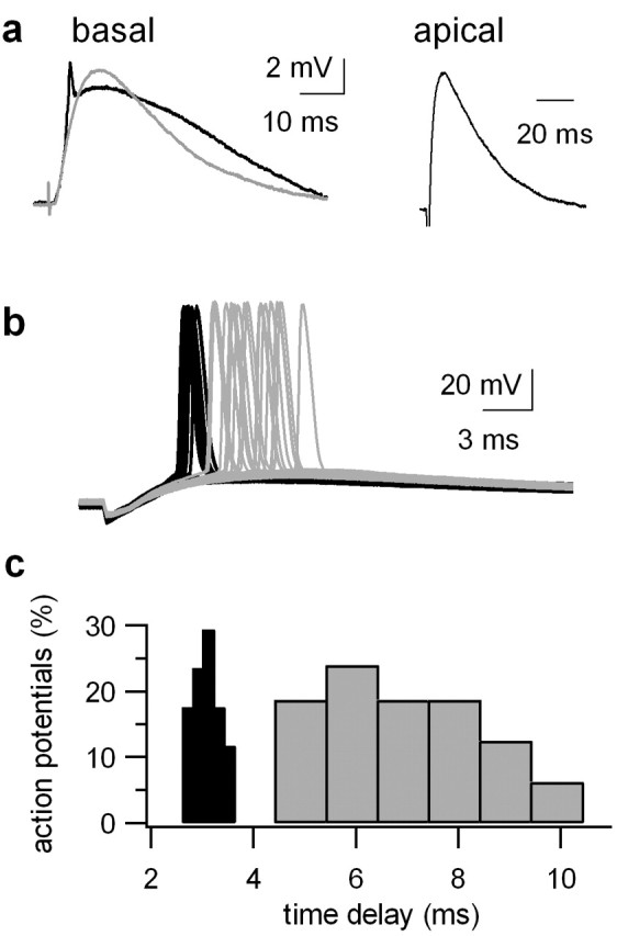
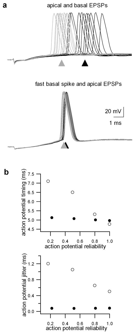
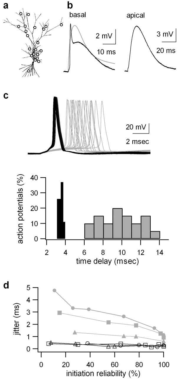
The ability of cortical neurons to perform temporally accurate computations has been shown to be important for encoding of information in the cortex; however, cortical neurons are expected to be imprecise temporal encoders because of the stochastic nature of synaptic transmission and ion channel gating, dendritic filtering, and background synaptic noise. Here we show for the first time that fast local spikes in basal dendrites can serve to improve the temporal precision of neuronal output. Integration of coactivated, spatially distributed synaptic inputs produces temporally imprecise output action potentials within a time window of several milliseconds. In contrast, integration of closely spaced basal inputs initiates local dendritic spikes that amplify and sharpen the summed somatic potential. In turn, these fast basal spikes allow precise timing of output action potentials with submillisecond temporal jitter over a wide range of activation intensities and background synaptic noise. Our findings indicate that fast spikes initiated in individual basal dendrites can serve as precise "timers" of output action potentials in various network activity states and thus may contribute to temporal coding in the cortex.


Figures

Figure 1.
Figure 1.
Rise time sharpening and supralinear amplitude amplification of the summed synaptic potential after coincident activation of synaptic inputs innervating the same dendritic branch. a, Fluorescent image of a CA1 pyramidal neuron loaded with OGB-1 (200 μm) via the somatic patch electrode. Two bipolar theta synaptic stimulating electrodes were placed in close proximity to a basal dendrite. Scale bar, 40 μm. b, Traces showing the individual EPSPs evoked by each of the synaptic stimulating electrodes, the summed synaptic potential during coincident activation of the two synaptic stimulating electrodes, and the arithmetic sum of the two individual responses (bold line). Note the large supralinear amplification and sharpening of the summed synaptic potential, as compared with the expected arithmetic sum response. c, Voltage traces obtained in response to coincident activation of two closely spaced electrodes at various time delays (0-20 msec). Black traces represent voltage responses to activation of the electrodes at time delays of 0 and 2 msec. Gray traces show the responses for activation at time delays of 3, 5, 10, 15, and 20 msec. Note that, in this experiment, the time window for coincident detection was <3 msec.
Figure 2.
Figure 2.
Synaptically evoked local spikes in basal dendrites of CA1 pyramidal neurons. a, Postsynaptic voltage responses were evoked by focal synaptic stimulation of a basal dendrite at increasing stimulus intensities. Note the sharp threshold of initiation of the local spike. b, Hyperpolarization of the membrane potential (from -64 to -80 mV) eliminated the fast and slow components of the spike. Two representative traces evoked by the same stimulus intensity (2 stimuli at 50 Hz) at a resting membrane potential of -64 and -80 mV (bold trace) are shown. c, The effect of the NMDA-receptor channel blocker APV (100 μm) on local dendritic spikes evoked by focal synaptic stimulation to a basal dendrite. Voltage traces in control conditions and after application of APV are shown for the same synaptic stimulus intensity (2 stimuli at 50 Hz). After significantly increasing the stimulus intensity (2-fold) in the presence of APV, local fast spikes could be reinitiated (APV-strong). Note that APV abolished the slow component of the spike. Consecutive addition of CNQX (10 μm) to the APV-containing bath solution eliminated altogether the voltage response (CNQX+APV). d, Top panel presents the peak amplitudes of the postsynaptic voltage responses plotted as a function of the synaptic stimulus intensities at resting membrane potential (•, -64 mV) and at hyperpolarized membrane potential (⋄, -80 mV). The data presented are from the same neuron as in a. Bottom panel shows the peak amplitudes of the postsynaptic responses plotted as a function of the synaptic stimulus intensities under control conditions (•) and in the presence of the NMDA-receptor blocker APV (⋄). The data presented are from the same neuron as in c. e, The effect of carbenoxolone (100 μm) on the local dendritic spike. Black trace = control; gray trace in the presence of carbenoxolone. f, The spatial spread of calcium transients evoked by a local basal spike. The cell was loaded with OGB-1 (200 μm) via the somatic patch electrode. Focal stimulation to a distal basal branch (marked by the electrode drawing; scale bar, 25 μm) evoked a local basal spike composed of early fast and later slow components. Calcium fluorescence imaging was performed in the line-scan mode. Slant line scans (512 Hz time resolution) covering at least 30 μm of a basal branch at a single scan were performed. Calcium transients were analyzed off-line and presented as ΔF/F in percentage values. The calcium transients were measured from all regions of the dendritic basal branches shown, but for simplicity, only calcium transients in representative locations are shown. Note the marked decline in calcium transients along the activated basal branch, and the very small calcium transients evoked by local basal spikes in the mother and sister dendritic branches.
Figure 3.
Figure 3.
The ionic mechanism of the fast and slow components of local spikes in basal dendrites. A CA1 pyramidal neuron was filled with CG-1, and glutamate was uncaged via a UV laser at distal basal dendrites. a, Single traces of excitatory, postsynaptic-like potentials (EPSLPs) evoked by UV-laser-induced glutamate uncaging at increasing UV-laser intensities (left panel). The inset in the frame represents the net spike calculated by subtraction of the just subthreshold response from the just suprathreshold response. The peak EPSLP responses were plotted as a function of the laser intensity (right panel). b, Single traces of EPSLPs recorded under control conditions (black) and after the addition of TTX (1 μm) and cadmium (100 μm) (gray traces). The black and smaller gray traces were evoked by similar laser intensity (12 mW), whereas the larger gray trace was evoked by larger laser intensity (25 mW). Addition of TTX and cadmium blocked at first the fast and slow components of the local basal spikes; however, after further increasing the laser intensity (from 12 to 25 mW), reinitiation of the slow NMDA spike occurred in the presence of TTX and cadmium. c, Single traces of EPSLPs recorded under control conditions (black trace) and after the addition of the NMDA-receptor blocker APV (100 μm; gray trace).
Figure 4.
Figure 4.
The effect of local fast basal spikes on the timing and temporal jitter of axonal action potentials. a, Single representative voltage traces of a basal EPSP (gray) and a local basal fast spike (black) that were coincidently activated with an apical EPSP. b, Single traces of 20 consecutive runs evoked by paired activation of a distributed apical EPSP with either a distributed basal EPSP (gray) or a local basal spike (black). The 20 consecutive stimulations were given every 20 sec. The stimulus intensity of the apical electrode was set to a level in which 10-20% of the trials failed to initiate action potentials. c, The timing of the axonal action potentials was monitored for each run and plotted in a time-delay histogram for the paired activation of distributed apical and basal EPSPs (gray) and distributed apical EPSPs with a local fast basal spike (black). The time delays were measured between the stimulation artifact and the peak of the action potential.
Figure 5.
Figure 5.
The impact of the activation intensity on the jitter and timing of output axonal action potentials. a, Pairing of basal with apical EPSPs (top traces) was compared with pairing of fast basal spikes with apical EPSPs (bottom traces). Pairing was performed at different stimulation intensities of the apical inputs, yielding different reliability values of axonal action potential initiation (reliability was defined as the fraction of traces resulting in action potential initiation). Consecutive successful runs resulting in action potential initiation are presented for two reliability values (100% gray and 20% black). Traces that failed to initiate action potentials were omitted. Arrowheads indicate the average timing of axonal action potentials in the two stimulation intensities. b, Action potential timing (top panel) and jitter (bottom panel) are plotted as a function of action potential reliability. The jitter was measured by the SD of action potential timing in consecutive runs. Black dots represent pairing with fast basal spikes, and open circles represent pairing with basal EPSPs. Note that the timing and jitter of axonal action potentials in the case of the fast spike was independent of the activation intensity.
Figure 6.
Figure 6.
The effect of local fast basal spikes on the temporal jitter of axonal action potentials: computer simulation data. a, A CA1 pyramidal neuron was reconstructed and the responses were simulated with the NEURON software. This panel shows the reconstructed neuron, 20 of 100-800 randomly distributed apical inputs (open circles), and the basal inputs that produced the fast spike (single open circle in basal region). The reconstructed neuron was kindly provided by Dr. Guy Major. b, Single representative voltage traces of a basal EPSP (gray, left panel) and a local basal fast spike (black, left panel) that were coincidently activated with an apical EPSP (right panel). c, Top panel, Consecutive activation of randomly distributed apical inputs paired with either randomly distributed basal inputs (gray) or a local basal spike (black) to produce a reliability value of 80% in action potential initiation. Bottom panel, A time-delay histogram is plotted for the paired activation of distributed apical and basal EPSPs (gray) and distributed apical EPSPs with a local fast basal spike (black). d, The action potential jitter is plotted as a function of action potential reliability values at different background synaptic noise in two conditions: paired activation of distributed apical and basal EPSPs (gray) and distributed apical EPSPs with a fast basal spike (black). To achieve different synaptic baseline noises, the number of arbitrarily activated synapses was changed (circles, 1500; squares, 1000; triangles, 0 synapses). Note the constant low jitter in the case of the fast basal spike activation.

Similar articles

Cited by

References

    1. Abeles M ( 1990) Corticonics. Cambridge, UK: Cambridge UP.
    1. Archie KA, Mel BW ( 2000) A model for intradendritic computation of binocular disparity. Nat Neurosci 3: 54-63. - PubMed
    1. Arieli A, Sterkin A, Grinvald A, Aertsen A ( 1996) Dynamics of ongoing activity: explanation of the large variability in evoked cortical responses. Science 273: 1868-1871. - PubMed
    1. Azouz R, Gray CM ( 1999) Cellular mechanisms contributing to response variability of cortical neurons in vivo J Neurosci 19: 2209-2223. - PMC - PubMed
    1. Azouz R, Gray CM ( 2000) Dynamic spike threshold reveals a mechanism for synaptic coincidence detection in cortical neurons in vivo. Proc Natl Acad Sci USA 97: 8110-8115. - PMC - PubMed

Publication types

Substances

LinkOut - more resources