. 2003 Aug 27;23(21):7759-66.
doi: 10.1523/JNEUROSCI.23-21-07759.2003.

Increased morphological diversity of microglia in the activated hypothalamic supraoptic nucleus

Affiliations

Increased morphological diversity of microglia in the activated hypothalamic supraoptic nucleus

Albert E Ayoub et al. J Neurosci. .

Abstract

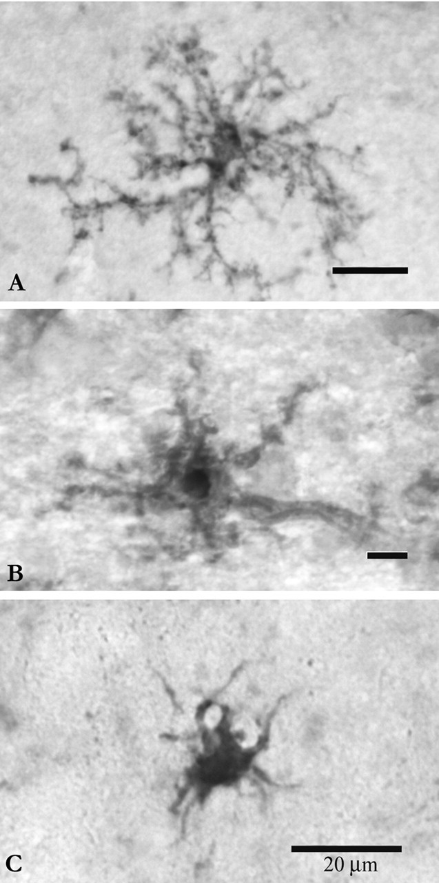
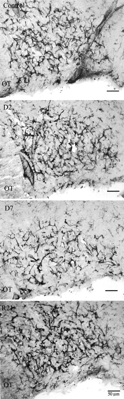
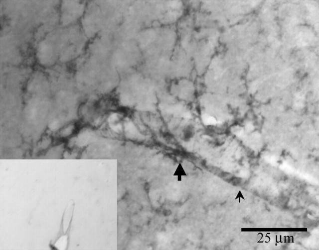
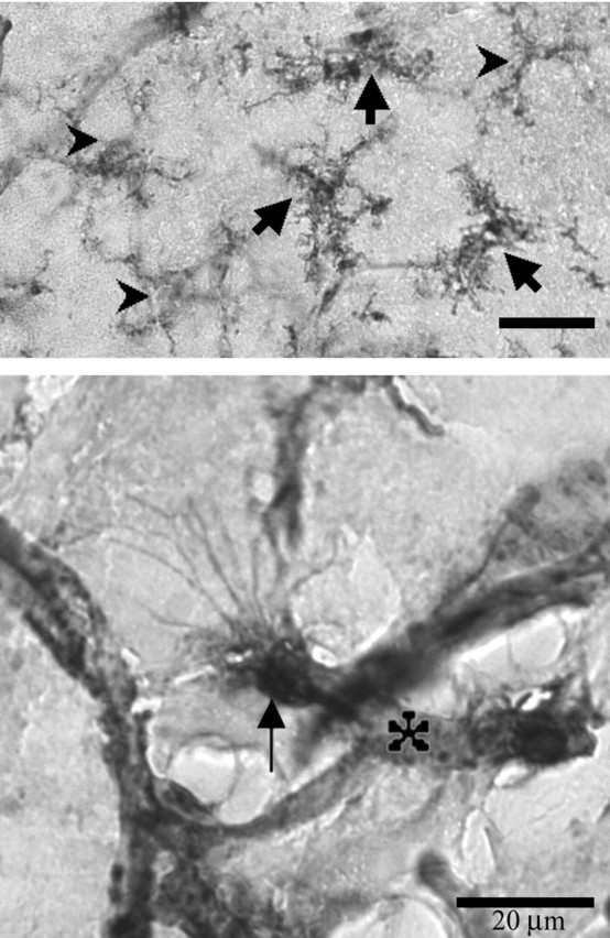
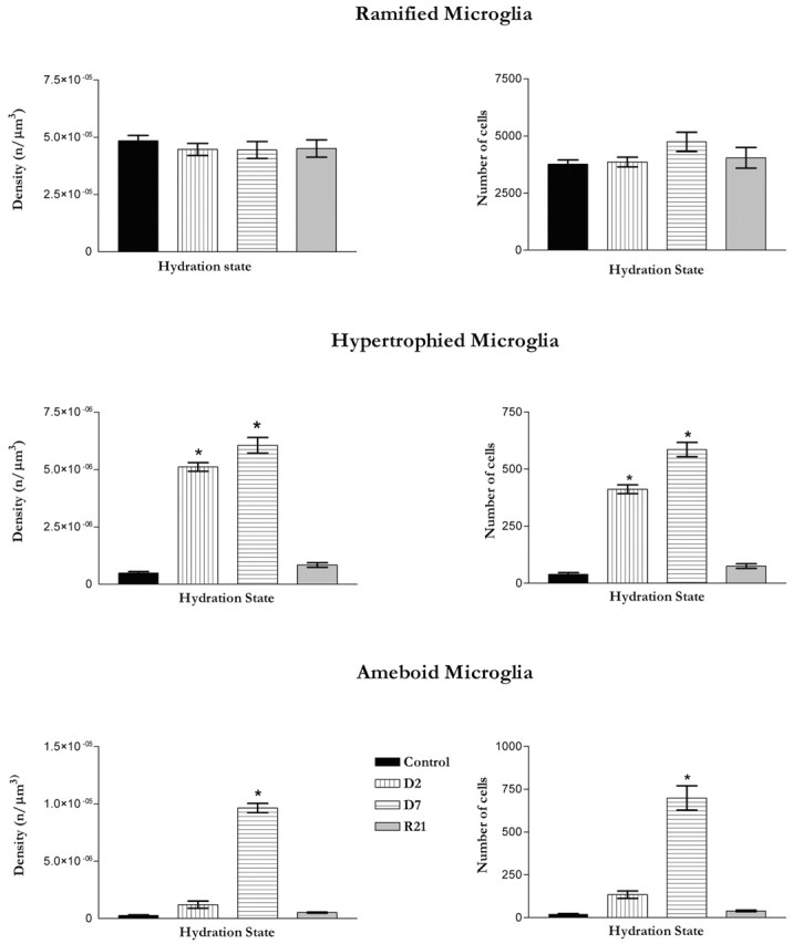
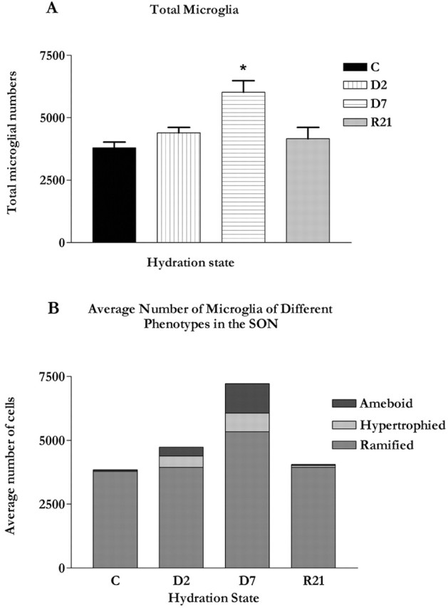
Microglia are the immune cells of the CNS. In the normal adult mammalian brain, the majority of these cells is quiescent and exhibits a ramified morphology. Microglia are perhaps best known for their swift transformation to an activated ameboid morphology in response to pathological insults. Here we have observed the responsiveness of these cells to events surrounding the normal activation of neurosecretory neurons in the hypothalamic supraoptic nucleus (SON), a well studied model of structural plasticity in the CNS. Neurons in the SON were activated by substituting 2% saline for drinking water. Brain sections were collected from four experimental groups [controls (C), 2 d-dehydrated (2D), 7 d-dehydrated (D7), and 7 d-dehydrated/21 d-rehydrated animals (R21)] and stained with Isolectin-B4-HRP to visualize microglial cells. Based on morphological criteria, we quantified ramified, hypertrophied, and ameboid microglia using unbiased stereological techniques. Statistical analyses showed significant increases in the number of hypertrophied microglia in the D2 and D7 groups. Moreover, there was a significant increase in the number of ameboid microglia in the D7 group. No changes were seen across conditions in the number of ramified cells, nor did we observe any significant phenotypic changes in a control area of the cingulate gyrus. Hence, increased morphological diversity of microglia was found specifically in the SON and was reversible with the cessation of stimulation. These results indicate that phenotypic plasticity of microglia may be a feature of the normal structural remodeling that accompanies neuronal activation in addition to the activation that accompanies brain pathology.


Figures

Figure 1.
Figure 1.
Microglial cells were classified as ramified (A), hypertrophied (B), or ameboid (C). Ramified microglia were defined as having a normal-appearing soma with thin, delicate, and radially projecting processes. Hypertrophied microglia were defined as a having an enlarged, darkened soma and shorter, thicker, and less branched processes. Ameboid microglia were defined as having densely stained, enlarged cell bodies, a few short processes, if any, and often several filopodia. Intermediate morphologies were rare, but excluded from the analysis when encountered. Scale bars, 20 μm.
Figure 2.
Figure 2.
Representative sections from the SON from control, D2, D7, and R21 experimental groups (top to bottom). At low magnification it can be appreciated that the majority of microglia are in the vicinity of SON capillaries. Microglia were easily distinguished from capillary endothelia at higher magnification (Fig. 4). OT, Optic tract.
Figure 3.
Figure 3.
Vascular endothelium in the SON stains slightly with isolectin-B4 (thin arrow). Clearly identifiable ramified microglia (thick arrow) are also visible. Inset, A negative control section pretreated with melibiose does not show stained microglia.
Figure 4.
Figure 4.
Example of microglia in the SON taken at high magnification and showing all three phenotypes: carets, ramified; thick arrows, hypertrophied; thin arrow, ameboid; asterisk, blood vessel (images edited with Adobe Photoshop; Adobe Systems Inc., San Jose, CA). Notice extensive filopodia protruding from the ameboid cell in the bottom panel.
Figure 5.
Figure 5.
Graphs depicting densities and numbers of microglial phenotypes in the SON across hydration states. n = 6 or 7 in each group. The number of ramified microglia did not show significant change with stimulation. In addition, the density of ramified microglia did not change across hydration states. The number of hypertrophied microglia significantly increased (p < 0.001) in the D2 and D7 groups compared with the control and R21 groups. The density of hypertrophied microglia showed a similar increase. The number of ameboid microglia increased significantly (p < 0.001) in the D7 group compared with the control, D2, and R21 groups. The density of ameboid microglia was also significantly higher in the D7 group, but not in any of the other experimental groups. Error bars indicate SEMs for ramified microglia and minimum-maximum values for hypertrophied and ameboid microglia.
Figure 6.
Figure 6.
A, Graphs depicting densities and numbers of microglial phenotypes in the control area of the cingulate cortex across hydration states. An area of the ipsilateral cingulate cortex, of dimensions equal to the SON of that section, was quantified as a per-section-control. As shown in this graph, no significant changes in the density of any phenotype was detected in the area of the cingulate cortex. Error bars indicate SEMs. B, A representative section from a D2 animal showing the control area of the cingulate cortex. The majority of microglia identified in this area had a ramified phenotype in each experimental group.
Figure 7.
Figure 7.
A, Graph showing the total numbers of microglia in the SON across hydration state. Significantly more microglia were observed in the SON at D7 (p < 0.01) relative to control and R21. Error bars indicate SEMs. B, Composite graph showing the relative numbers of each microglial phenotype in the SON across hydration states (graph generated using Microsoft Excel; Microsoft Corp., Seattle, WA).

Similar articles

Cited by

References

    1. Andrew RD, MacVicar BA, Dudek FE, Hatton GI ( 1981) Dye transfer through gap junctions between neuroendocrine cells of rat hypothalamus. Science 211: 1187-1189. - PubMed
    1. Ayoub A, Salm AK ( 2000) Morphological plasticity of microglia in the activated supraoptic nucleus. Soc Neurosci Abstr 26: 1943. 2000.
    1. Banati RB, Myers R, Kreutzberg GW ( 1997) PK (“peripheral benzodiazepine”)-binding sites in the CNS indicate early and discrete brain lesions: microautoradiographic detection of [3H]PK11195 binding to activated microglia. J Neurocytol 26: 77-82. - PubMed
    1. Basu A, Krady JK, O'Malley M, Styren SD, DeKosky ST, Levison SW ( 2002) The type 1 interleukin-1 receptor is essential for the efficient activation of microglia and the induction of multiple proinflammatory mediators in response to brain injury. J Neurosci 22: 6071-6082. - PMC - PubMed
    1. Bennett EL, Diamond MC, Krech D, Rosenzweig MR ( 1964) Chemical and anatomical elasticity of brain. Science 146: 610-619. - PubMed

Publication types

LinkOut - more resources