. 2003 Aug 27;23(21):7789-800.
doi: 10.1523/JNEUROSCI.23-21-07789.2003.

Ephrin-B2 and EphB2 regulation of astrocyte-meningeal fibroblast interactions in response to spinal cord lesions in adult rats

Affiliations

Ephrin-B2 and EphB2 regulation of astrocyte-meningeal fibroblast interactions in response to spinal cord lesions in adult rats

Liza Q Bundesen et al. J Neurosci. .

Abstract

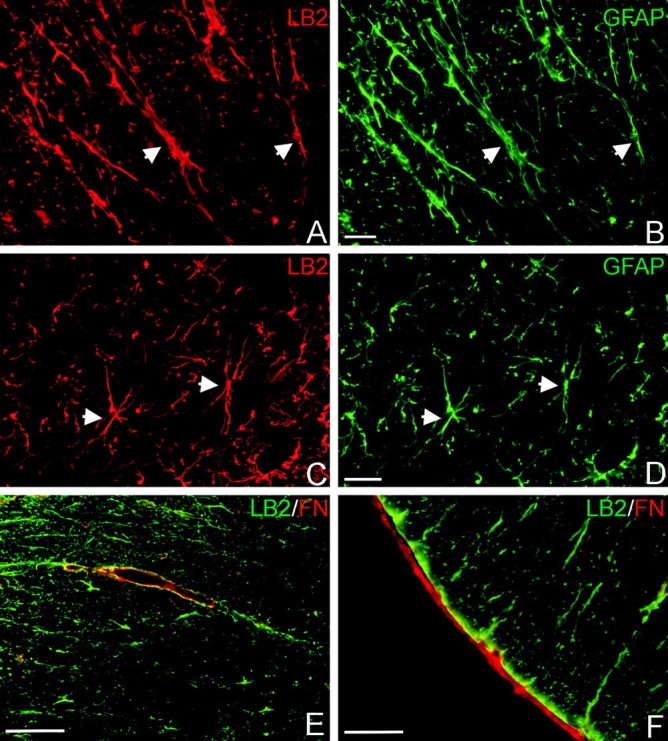
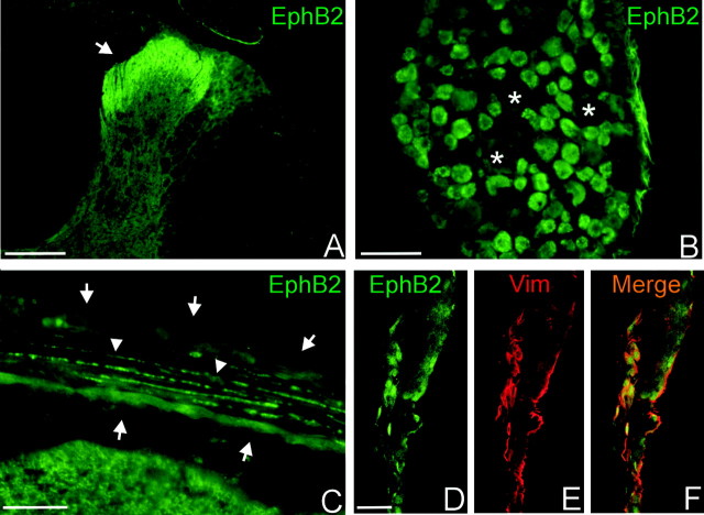
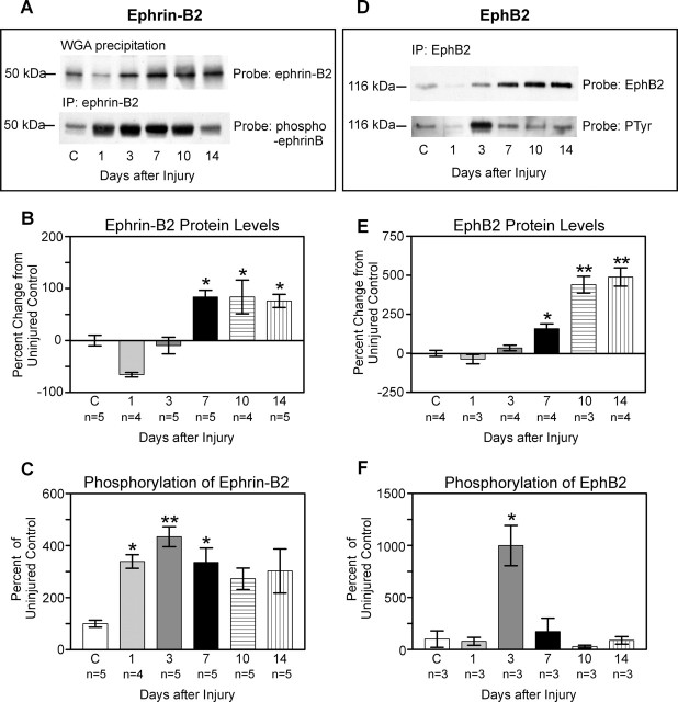
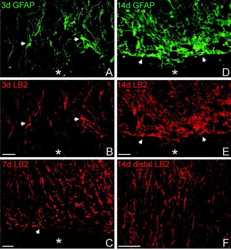
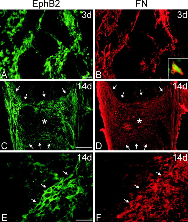
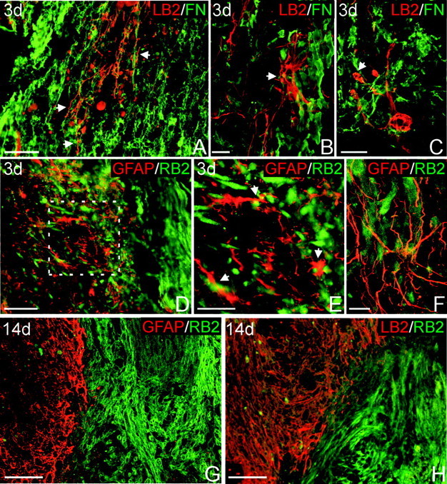
The present study provides the first evidence that signaling occurs between B-ephrins and EphB receptors in the adult CNS in response to injury. Specifically, our combined histological and biochemical data indicate that two members of the B-class of ephrins and Eph receptors, ephrin-B2 and EphB2, are expressed by astrocytes and meningeal fibroblasts, respectively, in the adult spinal cord. In response to thoracic spinal cord transection lesions, ephrin-B2 and EphB2 protein levels exhibit an initial decrease (1 d after lesion), followed by a significant increase by day 14. Immunohistochemical data indicate that ephrin-B2 is expressed by reactive CNS astrocytes, and EphB2 is present on fibroblasts invading the lesion site from the adjacent meninges. During the first 3 d after injury, there is intermingling of ephrin-B2-expressing reactive astrocytes at the lesion surface with EphB2-containing fibroblasts that is concurrent with bidirectional activation (phosphorylation) of ephrin-B2 and EphB2. By 7 d, both cell types are establishing restricted cellular domains containing dense networks of cells and interweaving processes. This astroglial-meningeal fibroblast scar is fully developed by day 14 when there is strict segregation of ephrin-B2-expressing astrocytes from EphB2-positive meningeal fibroblasts. These morphological changes are concomitant with a simultaneous decrease in ephrin-B2 and EphB2 activation. These observations provide strong evidence that cell contact-mediated bidirectional signaling between ephrin-B2 on reactive astrocytes and EphB2 on meningeal fibroblasts is an early event in the cellular cascades that result in the development of the glial scar and the exclusion of meningeal fibroblasts from the injured spinal cord.


Figures

Figure 1.
Figure 1.
Ephrin-B2 ligand immunohistochemistry in the uninjured adult spinal cord. Ephrin-B2 (LB2; red) is expressed by white matter (A) and gray matter (C) astrocytes, as confirmed by GFAP double labeling (green; B, D). Arrowheads mark some double-stained cells. E, F, Ephrin-B2-positive glial end feet (LB2; green) contact fibronectin-positive blood vessels (FN; red) in the white matter (E) and the fibronectin-positive pial fibroblasts (red) on the surface of the spinal cord (F). Scale bars: A-D, 20 μm; E, F, 50 μm.
Figure 2.
Figure 2.
EphB2 receptor immunohistochemistry in the uninjured adult spinal cord and DRG. EphB2 protein (green; A-D) is present in laminas I-III of the dorsal horn (A) and on small- and medium-diameter neurons of the DRG (B). Some EphB2-negative large-diameter DRG neurons are indicated with asterisks. C, The meningeal covering of the spinal cord (indicated by arrows) is highly immunoreactive for EphB2, and immunostaining appears to be preferentially localized along the interface between layers of fibroblasts (small arrowheads). D-F, Confocal images of the meninges double stained for EphB2 (green, D) and vimentin (Vim; red, E) confirms that EphB2 colocalizes with fibroblasts in the meninges (F, merged). Scale bars: A, 200 μm; B, 100 μm; C, 50 μm; D-F, 10 μm.
Figure 3.
Figure 3.
Western blots and quantification of protein and phosphorylation levels at progressive time points (1, 3, 7, 10, and 14 d) after complete T7 transections of the spinal cord. A, Representative Western blot illustrating temporal changes in ephrin-B2 protein and phosphorylation. Ephrin-B2 proteins were precipitated with WGA, and blots were probed with anti-ephrin-B2 (top). Ephrin-B2 protein was immunoprecipitated (IP) with anti-ephrin-B2, and resulting Western blots were probed with an antibody recognizing phosphorylated B-ephrins (bottom). B, Quantification of ephrin-B2 protein. At 1 d after lesion, there was a decrease in ephrin-B2 of ∼66% from control (C) levels. Ephrin-B2 protein quickly rebounded to near control levels by 3 d and exhibited a significant increase of 76-84% above uninjured levels from 7 to 14 d after lesion. Protein levels 7, 10, and 14 d after lesion were not significantly different from one another but were significantly different (*) from uninjured control (p < 0.05), from 1 d after lesion (p < 0.001), and 3 d after lesion (p < 0.01; Tukey's post hoc test). C, Quantification of ephrin-B2 phosphorylation. Low levels of ephrin-B2 phosphorylation were detected in uninjured tissue. At 1 d after lesion, there was a rapid and significant activation of ephrin-B2 compared with control (339%; p < 0.05), which additionally increased 3 d after lesion (435%; p < 0.01). Phosphorylation levels remained elevated at 7 d (335%; p < 0.05) before slowly decreasing. D, Representative Western blot illustrating temporal changes in EphB2 protein and phosphorylation. EphB2 protein was immunoprecipitated with anti-EphB2, and resulting Western blots were probed with anti-EphB2 (top). Activated EphB2 receptors were identified by immunoprecipitating EphB2 and probing the resulting Western blots with anti-phosphotyrosine (PTyr; bottom). E, Quantification of EphB2 protein. As for ephrin-B2, there was an initial drop in EphB2 protein at 1 d, which was followed by an increase that reached ∼160% of uninjured levels by day 7. At both 10 and 14 d, there was a highly significant increase in EphB2 protein of >400% from uninjured levels. Protein levels 10 and 14 d after lesion were significantly different (**) from uninjured, 1, 3 (p < 0.001), and 7 (p < 0.01) d. Protein levels 7 d after lesion were significantly different (*) from levels 1 d after lesion (p < 0.05). F, Quantification of EphB2 phosphorylation. A low level of EphB2 phosphorylation was detected in uninjured spinal cord tissue. At 3 d after injury, there was a highly significant and transient increase (1000%; p < 0.001) in EphB2 phosphorylation.
Figure 4.
Figure 4.
Reactive astrocytes at the lesion interface express ephrin-B2. Horizontal sections through lesioned tissue were double stained for GFAP (A, D; green) and ephrin-B2 (LB2; B, C, E, F; red) to demonstrate colocalization within astrocytes. Rostral is oriented toward the top of the page, and the lesion cavity is marked with an asterisk in each panel. The injured tissue immediately rostral to the lesion is depicted in A-E. A, B, At 3 d after injury, ephrin-B2-positive astrocytes were loosely distributed around the lesion area. A few hypertrophic astrocytes double stained for GFAP (arrows) had increased ephrin-B2 immunoreactivity. Astrocytic processes were disorganized and oriented radially toward the lesion but not parallel along the lesion surface. C, By 7 d after injury, many ephrin-B2-positive astrocytes had reoriented their processes parallel to the lesion interface (arrow). D, E, At 14 d after injury, astrocytes at the glial scar formed a dense cellular network with slender processes oriented parallel to the lesion surface. All astrocytes and astrocytic processes along the lesion surface possessed high levels of ephrin-B2 immunoreactivity compared with astrocytes in white matter several millimeters distal to the lesion site in the same tissue section (F). Scale bars, 50 μm.
Figure 5.
Figure 5.
Immunohistochemistry for EphB2 after injury. Horizontal sections through the spinal cord lesion were double stained for EphB2 (A, C, E; green) and fibronectin (FN; B, D, F; red) to detect fibroblasts in the lesion cavity at 3 and 14 d after injury. In all panels, rostral is oriented toward the top of the page. A, B, At 3 d after transection, there was generally weak EphB2 staining in the necrotic regions of the cord surrounding the lesion cavity. However, a loose meshwork of cells positive for EphB2 (A) and fibronectin (B) infiltrated the lesion cavity along the lesion interface. These cells appeared to originate from the meninges. B, Inset, Higher magnification of a cell double stained for EphB2 and fibronectin. C-F, By 14 d after injury, the lesion cavity was filled with a dense meshwork of EphB2-positive cells. C, D, Low-magnification micrographs of horizontal tissue sections through the entire lesion site. Asterisks mark the lesion epicenter, in which there is reduced EphB2 staining (C), and arrows demarcate the lesion interface with the spinal cord. Although fibronectin-positive fibroblasts have filled the entire lesion cavity (D), those that are the most immunoreactive for EphB2 are concentrated along the spinal cord border and meninges. E, F, Higher magnification of EphB2-positive meningeal fibroblasts within the lesion. Arrows mark cells double stained for EphB2 and fibronectin at the lesion interface. Scale bars: A, B, E, F, 50 μm; C, D, 500 μm.
Figure 6.
Figure 6.
EphB2-positive fibroblasts and ephrin-B2-positive astrocytes intermingle 3 d after injury but form restricted domains along the spinal cord lesion interface 14 d after injury. A-C, Double staining for ephrin-B2 (LB2; red) to detect astrocytes and fibronectin (FN; green) to detect fibroblasts 3 d after injury. A, Ephrin-B2-positive astrocytic processes (arrows) intermingle with fibroblasts at the lesion site. B, Confocal image of an ephrin-B2-positive stellate astrocyte (arrow) in direct contact with fibroblasts. C, Confocal image of an ephrin-B2-positive astrocyte with swollen processes (arrow), potentially attributable to retraction after contacting fibroblasts. D, F, Double staining for GFAP (red) and EphB2 (RB2; green) to confirm astrocyte-fibroblast intermingling 3 d after injury. E, Higher magnification of lesion interface outlined in D, demonstrating direct contact between GFAP-positive astrocytic processes (arrows) and EphB2-positive fibroblasts. F, Confocal image demonstrating astrocytic processes interwoven among EphB2-positive cells. G, Lesion interface at 14 d after injury double stained for GFAP (red) and EphB2 (green). EphB2-positive fibroblasts are strictly segregated from the spinal cord astrocytes at this survival time. H, Similar region of the spinal cord lesion double stained for ephrin-B2 (red) and EphB2 (green). Ephrin-B2-positive astrocytes and EphB2-positive fibroblasts are clearly segregated along the lesion border. Scale bars: A, D, 100 μm; B, C, F, 20 μm; E, 50 μm; G, H, 200 μm.
Figure 7.
Figure 7.
Schematic model for temporal interactions between reactive astrocytes and meningeal fibroblasts along the surface of the lesioned spinal cord. A, Three days after lesion, meningeal fibroblasts migrating into the lesion cavity begin making contact with astrocytes (yellow glow) along the lesioned surface of the spinal cord. In response to direct cell contact (inset), ephrin-B2 ligands on the surface of astrocytes bind to EphB2 receptors on invading fibroblasts, resulting in the phosphorylation of the tyrosine kinase domain on EphB2 (red receptors) and phosphorylation of conserved tyrosine residues on the cytoplasmic domain of ephrin-B2 (blue triangles). Receptor-ligand phosphorylation results in the initiation of bidirectional intracellular signaling cascades within the meningeal fibroblasts and reactive astrocytes. We propose that stimulation of EphB2 on the meningeal fibroblasts activates the Rho-GTPase pathway, producing local actin depolymerization that prevents additional infiltration of the fibroblasts into the spinal cord parenchyma. Activation of additional bidirectional signaling cascades in astrocytes and fibroblasts may initiate gene transcription for the deposition of ECM components by both cell types along their interface. B, Seven days after lesion, ECM components secreted by reactive astrocytes and meningeal fibroblasts form a basal lamina along areas of previous glial-meningeal contact. The deposition of this ECM prevents additional contact between ephrin-B2 and EphB2, resulting in a decrease in the endogenous phosphorylation and activation of EphB2 receptors on meningeal fibroblasts and ephrin-B2 on astrocytes. This decreased activation of the Ephephrin pathways may result in enhanced signaling through integrin receptor binding to the ECM components in the basal lamina, stabilizing the glial-meningeal boundary. C, Fourteen days after lesion. By this time, a basal lamina has completely formed along the glial-meningeal interface, and signaling between ephrin-B2 and EphB2 is terminated. Integrin-mediated binding of astrocytes and meningeal fibroblasts to the basal lamina now maintains the integrity of the glial-meningeal boundary.

Similar articles

Cited by

References

    1. Abnet K, Fawcett JW, Dunnett SB ( 1991) Interactions between meningeal cells and astrocytes in vivo and in vitro. Brain Res Dev Brain Res 59: 187-196. - PubMed
    1. Berry M, Maxwell WL, Logan A, Mathewson A, McConnell P, Ashhurst DE, Thomas GH ( 1983) Deposition of scar tissue in the central nervous system. Acta Neurochir (Wien) [Suppl] 32: 31-53. - PubMed
    1. Bradbury EJ, Moon LD, Popat RJ, King VR, Bennett GS, Patel PN, Fawcett JW, McMahon SB ( 2002) Chondroitinase ABC promotes functional recovery after spinal cord injury. Nature 416: 636-640. - PubMed
    1. Bruckner K, Pasquale EB, Klein R ( 1997) Tyrosine phosphorylation of transmembrane ligands for Eph receptors. Science 275: 1640-1643. - PubMed
    1. Bruckner K, Pablo Labrador J, Scheiffele P, Herb A, Seeburg PH, Klein R ( 1999) EphrinB ligands recruit GRIP family PDZ adaptor proteins into raft membrane microdomains. Neuron 22: 511-524. - PubMed

Publication types

MeSH terms

LinkOut - more resources