. 2003 Aug 27;23(21):7810-9.
doi: 10.1523/JNEUROSCI.23-21-07810.2003.

Novel insights into the regulation of the timeless protein

Affiliations

Novel insights into the regulation of the timeless protein

Lesley J Ashmore et al. J Neurosci. .

Abstract

In the Drosophila circadian clock, period (per) and its partner, timeless (tim), play a central role in the negative limb of an autoregulatory feedback loop. Unlike per, the dosage of which affects the frequency (tau) of the circadian cycle, we found that increasing copies of the tim gene has no effect on clock period length. The use of the tim promoter to express per results in a shortening of circadian period, also indicating that the regulation of tim is different from that of per. Drosophila TIM is similar to the mammalian circadian protein mPER2 in that it shuttles independently between the nucleus and cytoplasm both in vivo and in vitro. Contrary to the current model that PER and TIM heterodimerization is a prerequisite for their nuclear entry, PER is not required to transport TIM into nuclei, although it influences TIM localization and vice versa. Blocking nuclear export led to increased nuclear expression of TIM in S2 cells and in wild-type and per01 larvae, suggesting that PER may be required for nuclear retention of TIM. Unlike PER, nuclear TIM alone has no ability to repress transcription. We propose that TIM drives cycles of PER expression by regulating its stability, and in turn, PER retains TIM in the nucleus, either for the regulation of its own stability or for a novel nuclear role of TIM.


Figures

Figure 2.
Figure 2.
TIM alone cannot repress dCLK-CYC-mediated transcription of a circadian E-box reporter. An inducible tim gene was modified to permit nuclear entry of TIM in the absence of PER, as shown in A (see Results). Although the modifications still allowed TIM and PER together to repress transcription (asterisk indicates signal is significantly less than pAct-dClk alone; p < 0.01; t test), even high doses of nuclear TIM cannot repress transcription without PER. Surprisingly, TIM alone appeared to activate transcription (double asterisk indicates that signal is significantly greater than dCLK activation alone; p < 0.01; t test). Differences among hs-tim, tim+NLS, and timΔCLD are not significant. Drosophila S2 cells were transiently transfected with two reporter genes (E-box luc and i.e. β-gal), and a combination of pAct-dClk, full-length or mutated hs-tim, and pAct-per, as indicated by +. After 48 hr, cells were harvested and assayed for luciferase and β-gal activity. Data were normalized to β-gal expression to control for transfection efficiency. Results are presented as percentage of full activation seen in the dClk alone condition and expressed as averages (±SEM) of three triplicate measures. Two other identical independent experiments showed similar results.
Figure 4.
Figure 4.
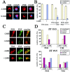
Nucleocytoplasmic shuttling of TIM in cell culture and per01 lateral neurons. Localization of TIM was measured in the presence of a nuclear export blocker (LMB) in transiently transfected S2 cells and larvae. In both cases, blocking nuclear export led to an increased accumulation of TIM trapped in the nucleus, indicating that even in the absence of PER, TIM is transported into the nucleus and actively exported. A shows representative examples of TIM subcellular localization in S2 cells. Five hundred nanograms of hs-tim (full-length only) were transfected, expressed, and scored as described in Figure 3, with one exception: LMB (10 ng/ml) was added to the medium before heat shock. Sample cells (A) were fixed 2 hr after induction, but we noted that TIM nuclear expression occurs extremely rapidly, even within 30 min after induction (data not shown). In B, TIM localization was quantified as described in Figure 3. LMB dosage was varied, and TIM was localized to the nucleus in a dose-dependent manner. C, Representative TIM (red) and PDF (green overlay) signals in per01 larval LNs. TIM is cytoplasmic in per01 flies (above), and LMB addition significantly increased the proportion of TIM found in the nucleus in per01 flies (below) in seven independent experiments, quantified in D (asterisk indicates that this group is significantly different from vehicle control; p < 0.01; t test). Whole brains from per01 third-instar larvae were dissected at ZT 20-21 and incubated for 3 hr in medium containing (bottom) or lacking (top) LMB (40 ng/ml) and supplemented with MG132 (a proteasomal inhibitor) to block TIM degradation. After the incubation period, the tissue was fixed and probed with antibodies to TIM and PDF. For the purpose of this Figure, the contrast of the TIM signal was adjusted slightly to enhance the signal over the background. As with the cell culture quantification, data are presented as percentages of LNs in which most of the TIM signal was nuclear, cytoplasmic, or uniformly distributed. Only LNs with clear cytoplasmic-nuclear boundaries (visualized by the PDF signal) were scored (blind) for TIM localization. LNs in which the TIM signal was too faint to score were discarded. A total of seven independent experiments were performed in which the incubation time (1.5-5 hr), LMB dosage (20-40 ng/ml), and DMSO (1-3%) concentration were varied, with similar results.
Figure 5.
Figure 5.
PER alters TIM localization in vitro and in vivo. PER sequesters TIM in the cytoplasm of S2 cells during LMB treatment, indicating that the presence of PER inhibits TIM from entering the nucleus when PER is cytoplasmic. In yw larvae, however, there is still a slight trend toward more nuclear expression of TIM and PER when nuclear export is blocked. The trend is more pronounced when PER is already more nuclear. A shows representative examples of S2 cells transiently transfected with 500 ng each of hs-tim and pAct-per. Quantification of TIM and PER localization is presented in B, scored as described in Figure 3. S2 cells were treated with LMB during heat shock as described in Figure 4, fixed 2 hr after induction, and stained for either TIM (left, red) or PER (right, red) and DAPI (blue overlay). Data in B were pooled from two independent experiments, and in each experiment 100-300 cells per group were scored. C presents examples of PER (top) and TIM (bottom) signals in yw larvae, collected at ZT 19.5, incubated with LMB or vehicle for 1.5 hr. At this time point, PER is more nuclear. The sample cells show an example of nuclear and uniform PER distribution without LMB and nuclear distribution with LMB. TIM is normally still cytoplasmic, as shown in the (-)LMB example, but there is a definite trend toward nuclear localization when export is blocked by LMB (bottom). PER localization is also affected, as seen in the quantifications of two independent experiments (D). Whole brains from yw third-instar larvae were dissected at ZT 20-21 and incubated for 3 hr in medium containing (bottom) or lacking (top) LMB (40 ng/ml) and supplemented with MG132 (a proteasomal inhibitor) to block TIM degradation. After the incubation period, the tissue was fixed and probed with antibodies to TIM or PER (visualized with Cy3-conjugated secondary antibodies, red) and compared with the cytoplasmic PDF signal detected with FITC (green). All LNs were scored blind as described in Figure 4.
Figure 1.
Figure 1.
TIM and PER levels in overexpression lines. Protein extracted from adult heads was separated on a 6% polyacrylamide gel. TIM levels are increased over wild-type (yw) levels in Tim 4 (A) and Tim 7 (B) lines collected at ZT 21. Spaces indicate noncontiguous lanes from the same blot. Asterisk indicates a common nonspecific band, used here as a loading control. All Tim 4 and 7 lines were tested, but only one representative line each is shown here for simplicity. In C, flies were collected at an earlier time point, ZT 16.5. PER levels are unaltered in Tim 4 (lanes 3, 5, and 6) and Tim 7 (lane 4) overexpression lines. PER is only mildly increased in Per 1-A and decreased in Per 1-B. Surprisingly, Per 1-B generally has a stronger behavioral phenotype than Per 1-A (Table 2), which probably indicates that Per 1-B has stronger negative feedback (see Discussion). HSP 70 (bottom blot) was used as a loading control. Another analysis of PER levels at ZT 21 showed similar results.
Figure 3.
Figure 3.
TIM localizes predominantly to the cytoplasm in transiently transfected S2 cells despite modifications to increase nuclear entry. Shown in A are representative samples of the subcellular distribution patterns of TIM with and without modifications to increase nuclear entry. Quantification of three independent experiments (B) revealed that although the modifications do tend to increase TIM distribution in the nucleus, the effect is not as robust as we expected, indicating that there may be alternative mechanisms acting on TIM localization. S2 cells transfected with 500 ng of full-length or modified hs-tim (in 6-well plates) were fixed 2 hr after heat shock induction of TIM expression. TIM localization was scored as mostly nuclear, mostly cytoplasmic, or uniformly distributed (left columns, in red), and nuclei were visualized with DAPI (right columns, overlaid against TIM signal). Data are presented as mean percentages of cells in each category (±SEM). Asterisk indicates that this group is significantly different from full-length TIM for that subcellular localization (p < 0.01; one-way ANOVA).
Figure 6.
Figure 6.
TIM alone does not repress dCLK-CYC-mediated transcription even when its nuclear export is blocked. LMB treatment had no significant effect on either TIM repression or TIM+PER repression. After transient transfection of S2 cells (described previously in Fig. 2), cells were heat shocked for 30 min every 12 hr, in medium supplemented with 5 ng/ml LMB. Four hours after induction, the medium was replaced with LMB-free medium. After 36 hr, cells were collected and analyzed for luciferase and β-gal expression as described in Figure 2. Results are expressed as averages (±SEM) of three triplicate measures. Two other identical independent experiments showed similar results. Asterisk indicates that the amount of activation is significantly less than activation by dCLK alone (p < 0.01; t test).

References

    1. Bae K, Lee C, Sidote D, Chuang KY, Edery I ( 1998) Circadian regulation of a Drosophila homolog of the mammalian Clock gene: PER and TIM function as positive regulators. Mol Cell Biol 18: 6142-6151. - PMC - PubMed
    1. Baylies MK, Bargielli TA, Jackson FR, Young MW ( 1987) Changes in abundance or structure of the per gene product can alter periodicity of the Drosophila clock. Nature 326: 390-392. - PubMed
    1. Ceriani MF, Hogenesch JB, Yanovsky M, Panda S, Straume M, Kay SA ( 2002) Genome-wide expression analysis in Drosophila reveals genes controlling circadian behavior. J Neurosci 22: 9305-9319. - PMC - PubMed
    1. Chen Y, Hunter-Ensor M, Schotland P, Sehgal A ( 1998) Alterations of per RNA in noncoding regions affect periodicity of circadian behavioral rhythms. J Biol Rhythms 13: 364-379. - PubMed
    1. Claridge-Chang A, Wijnen H, Naef F, Boothroyd C, Rajewsky N, Young M ( 2001) Circadian regulation of gene expression systems in the Drosophila head. Neuron 32: 657-671. - PubMed

Publication types

MeSH terms