. 2003 Sep 16;100(19):10989-94.
doi: 10.1073/pnas.1832776100. Epub 2003 Aug 28.

The EBV nuclear antigen 1 (EBNA1) enhances B cell immortalization several thousandfold

Affiliations

The EBV nuclear antigen 1 (EBNA1) enhances B cell immortalization several thousandfold

Sibille Humme et al. Proc Natl Acad Sci U S A. .

Abstract

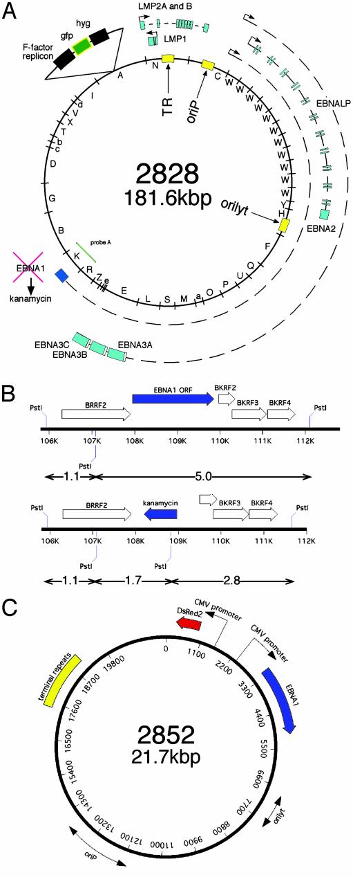
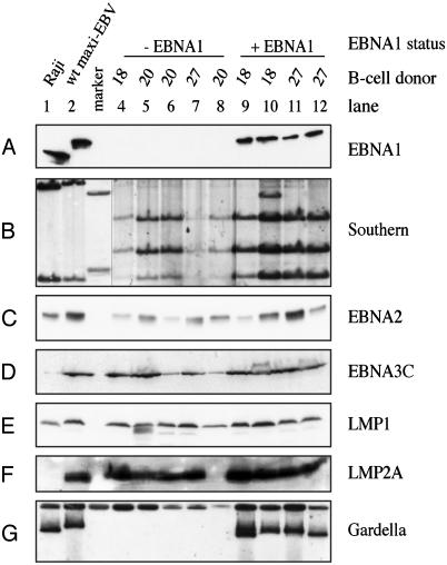
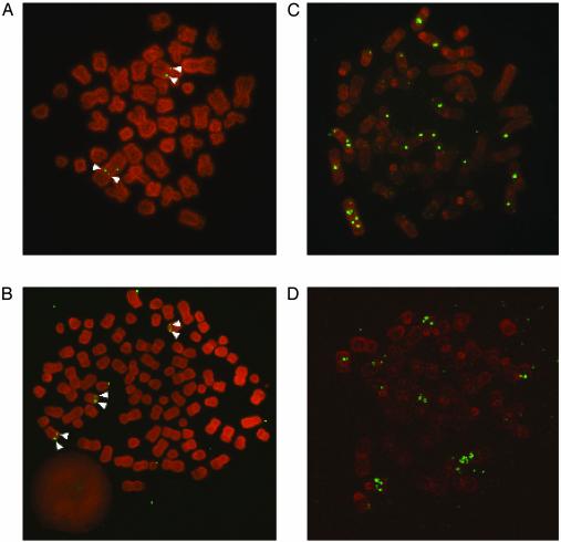
The Epstein-Barr virus (EBV) nuclear antigen 1 (EBNA1) is one of the earliest viral proteins expressed after infection and is the only latent protein consistently expressed in viral-associated tumors. EBNA1's crucial role in viral DNA replication, episomal maintenance, and partitioning is well examined whereas its importance for the immortalization process and the tumorgenicity of EBV is unclear. To address these open questions, we generated, based on the maxi-EBV system, an EBNA1-deficient EBV mutant and used this strain to infect primary human B cells. Surprisingly, lymphoblastoid cell lines (LCL) emerged from these experiments, although with very low frequency. These cell lines were indistinguishable from normal LCLs with respect to proliferation and growth conditions. A detailed analysis indicated that the entire viral DNA was integrated into the cellular genome. At least 5 of the 11 latent EBV proteins were expressed, indicating the integrity of the EBV genome. EBNA1-positive and DeltaEBNA1-EBV-LCLs were injected into severe combined immunodeficient (SCID) mice to examine their tumorgenicity in comparison. Both groups supported tumor growth, indicating that EBNA1 is not mandatory for EBV's oncogenic potential. The results shown provide genetic evidence that EBNA1 is not essential to establish LCLs but promotes the efficiency of this process significantly.


Figures

Fig. 1.
Fig. 1.
(A) Schematic picture of ΔEBNA1-genome p2828: The circular episome of the B95.8 strain is shown with its BamHI fragments (inner circle). Nine of the 11 latent viral genes (EBNA1, EBNA2, EBNA3A, -B, and -C, EBNA-LP; LMP1, LMP2A, and -2B) generally expressed in LCLs are depicted as blue boxes together with their primary transcripts and promoters. The EBNA1 gene was deleted by homologous recombination and replaced by the kanamycin gene. (B) Enlargement of the EBNA1-flanking regions used for homologous recombination to replace EBNA1 by kanamycin. The kanamycin resistance gene contains a diagnostic PstI restriction site used to distinguish the ΔEBNA1-genome p2828 (Lower) from the wild-type EBNA1 situation (Upper) by Southern blotting (Fig. 2B). (C) Composition of plasmid p2852 used for transcomplementation experiments in which both EBNA1 and the DsRed2 gene are expressed from the cytomegalovirus promoter. Both origins of DNA replication are indicated as double arrows, and the terminal repeats are shown as a yellow box.
Fig. 2.
Fig. 2.
Analysis of LCLs generated by different EBV strains. ΔEBNA1-LCLs from three different donors (donors 18, 20, and 27) were obtained from infections of human primary B cells without (lanes 4–8) or with the EBNA1 trans-complementing plasmid p2852 (lanes 9–12). Raji cells (lane 1) and a generic EBV-LCL (lane 2) were used as controls. Raji is negative for EBNA3C and LMP2A due to deletions in the genome. (A) Western blot analysis of EBNA1. (B) Southern blot of genomic DNA to distinguish generic EBV from ΔEBNA1-LCLs. The sizes of diagnostic DNA fragments are shown in Fig. 1B, and the location of probe A is depicted in Fig. 1 A. Lanes 4–8 are from the same Southern blot as the other lanes, but were exposed longer. (CF) Western blot analysis of different latent EBV proteins: EBNA2 (C), EBNA3C (D), LMP1 (E), and LMP2A (F) are expressed in all LCLs, with the exception of LMP2A (lane 8). (G) Gardella gel analysis used to determine the status of the viral DNA by using p135.16 as hybridization probe. EBV genomes are extrachromosomal when EBNA1 is present (lanes 1, 2, and 9–12) but integrated in the absence of the viral protein (lanes 4–8).
Fig. 3.
Fig. 3.
Metaphase chromosomes of LCLs were analyzed for the status of the viral genome by in situ hybridization. As representative examples, two different donors are shown: donor 27 (A and C) and donor 18 (B and D) (see Fig. 2). (A and B) ΔEBNA1-EBV 2828-derived LCLs show integration events of the viral genome into host chromosomes. Individual LCLs are of clonal nature as integration sites appear as doublets (white arrowheads). (C and D) ΔEBNA1-EBV-LCLs generated by coinfection with 2852 virus maintain the ΔEBNA1-EBV genome episomally with high copy number. Most viral episomes are randomly attached to metaphase chromosomes. A corresponds to lane 7 in Fig. 2, B to lane 4, C to lane 11, and D to lane 9.

References

    1. Rickinson, A. B. & Kieff, E. (2001) in Virology, eds. Knipe, D. M. & Howley, P. M. (Lippincott, Philadelphia), Vol. 2, pp. 2575–2627.
    1. Kieff, E. & Rickinson, A. B. (2001) in Virology, eds. Knipe, D. M. & Howley, P. M. (Lippincott, Philadelphia), Vol. 2, pp. 2511–2573.
    1. Leight, E. R. & Sugden, B. (2000) Rev. Med. Virol. 10, 83–100. - PubMed
    1. Aiyar, A., Tyree, C. & Sugden, B. (1998) EMBO J. 17, 6394–6403. - PMC - PubMed
    1. Sugden, B. (2002) Trends Biochem. Sci. 27, 1–3. - PubMed

Publication types

Substances

LinkOut - more resources