. 2003 Sep;4(9):877-82.
doi: 10.1038/sj.embor.embor917. Epub 2003 Aug 8.

Cdyl: a new transcriptional co-repressor

Affiliations

Cdyl: a new transcriptional co-repressor

Cécile Caron et al. EMBO Rep. 2003 Sep.

Abstract

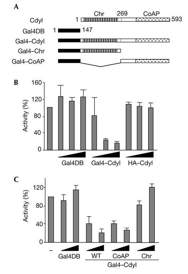
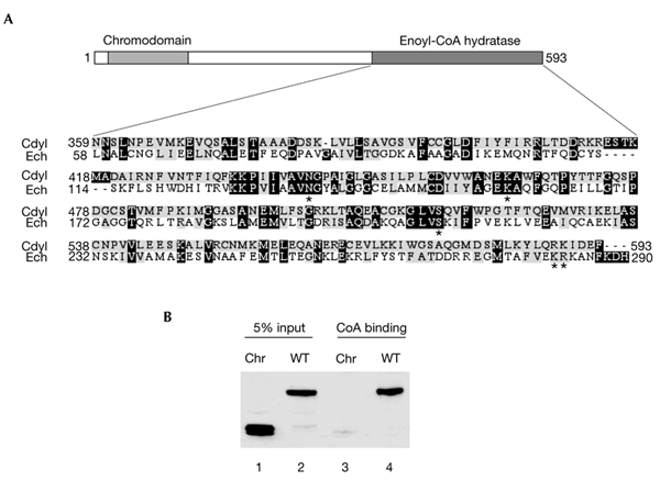
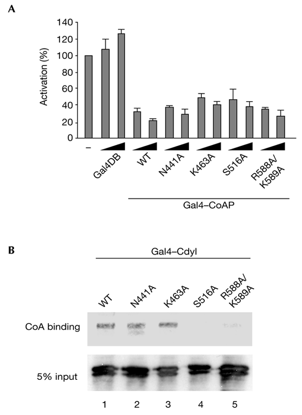
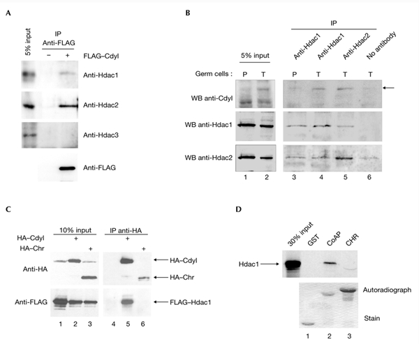
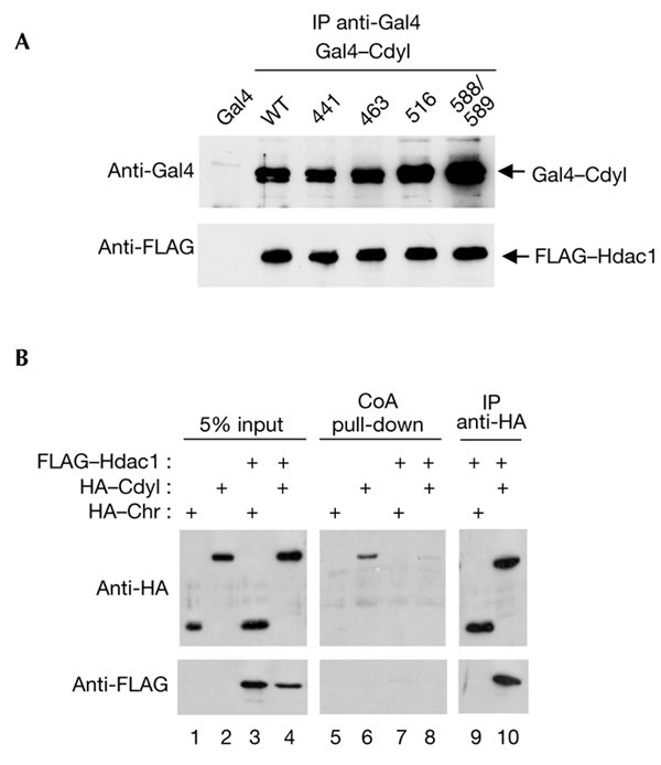
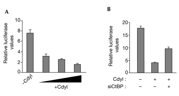
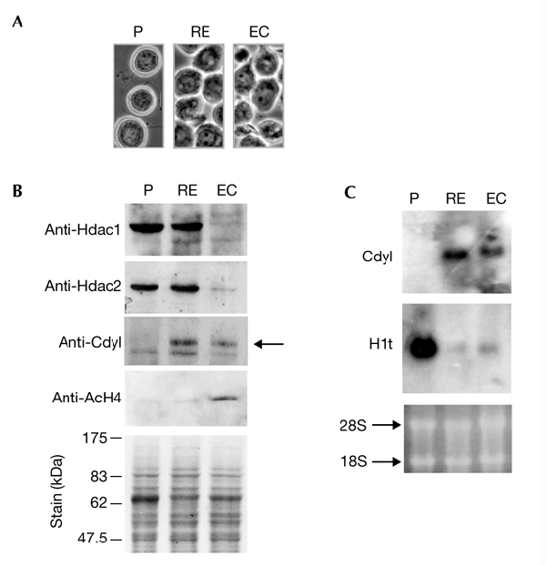
Cdyl (chromodomain-Y-like) is a chromodomain-containing protein that is predominantly expressed during mouse spermiogenesis. In its carboxy-terminal portion, there is a domain with homology to the coenzyme A (CoA) pocket of the enoyl-CoA hydratase/isomerase, which is shown here to be able to bind CoA and histone deacetylases (HDACs). It also efficiently represses transcription. Moreover, the binding of Hdac1 represses the ability of Cdyl to bind CoA, and a Cdyl-CoA interaction only occurs in the absence of HDACs. These data suggest that Cdyl is primarily a transcriptional co-repressor. However, the degradation of cellular Hdac1 and Hdac2, as observed here in the elongating spermatids, may provide an HDAC-free environment in which Cdyl could bind CoA and participate in the global chromatin remodelling that occurs in these cells.


Figures

Figure 1
Figure 1
Cdyl is a transcriptional repressor. (A) The Gal4 DNA-binding domain (Gal4DB) was fused to either full-length Cdyl (Gal4–Cdyl), the Cdyl chromodomain (Chr; the Gal4–Chr construct), or the Cdyl carboxy-terminal half, which is homologous to the enoyl-coenzyme A (CoA) hydratase (the Gal4–CoA-pocket (CoAP) construct). (B,C) HeLa cells were cotransfected with 200 ng of a reporter plasmid containing five Gal4-binding sites cloned upstream of the simian virus 40 early promoter, and increasing amounts (10, 30 or 100 ng in (B) and 30 or 100 ng in (C)) of the indicated expression constructs. Gal4DB corresponds to the Gal4 DNA-binding domain alone, and haemagglutinin (HA)–Cdyl is Cdyl fused to an HA-tag (and does not contain Gal4DB). Luciferase activity was measured and normalized with respect to that of β-galactosidase. Mean values of at least three independent assays are shown. Cdyl, chromodomain-Y-like; WT, wild type.
Figure 2
Figure 2
The domain of Cdyl that is homologous to enoyl-coenzyme-A hydratase binds coenzyme A. (A) Sequence homology between the coenzyme A (CoA) pocket of mouse enoyl-CoA hydratase (Ech) and mouse Cdyl. The asterisks indicate the amino acids that are replaced by alanine in the mutants shown in Fig. 3. (B) Whole-cell extracts from U2OS cells that express wild-type (WT) Cdyl and a Cdyl fragment that contains the chromodomain (Chr) were incubated with CoA–agarose. The beads were washed and the bound proteins were detected by western blotting using an anti-Cdyl antibody. Cdyl, chromodomain-Y-like.
Figure 3
Figure 3
Cdyl mutated in its coenzyme A pocket is also a transcriptional repressor. (A) Amino acids N441, K463, S516, R588 and K589 of Cdyl, which are conserved between enoyl-coenzyme A (CoA) hydratase and Cdyl (see Fig. 2A), were replaced by alanine. The mutated proteins were fused to the Gal4 DNA-binding domain (Gal4DB), and their ability to repress transcription was monitored, as described in Fig. 1. Mean values from at least three independent assays are shown. (B) U2OS cells were transfected with vectors that express either wild-type (WT) Cdyl or one of the four mutants of Cdyl. Extracts from transfected cells were used to perform a CoA pull-down assay on CoA beads as described in Fig. 2. Beads were washed and the bound proteins were detected by western blotting using an anti-Cdyl antibody. Cdyl, chromodomain-Y-like.
Figure 4
Figure 4
Cdyl interacts with histone deacetylases 1 and 2 through its coenzyme-A-pocket domain. (A) Cdyl recruits endogenous histone deacetylase 1 (Hdac1) and Hdac2. COS7 cells were transfected with 5 μg of expression vector for FLAG–Cdyl (+) or an empty vector (−), and Cdyl-containing complexes were immunoprecipitated from whole-cell extracts with an anti-FLAG antibody, followed by elution with a 3X FLAG peptide (Sigma). A western blot probed with an anti-FLAG antibody showed the efficiency of the immunoprecipitations (anti-FLAG panel). A western blot probed with anti-Hdac1, anti-Hdac2 and anti-Hdac3 antibodies showed the association of endogenous Hdac1 and Hdac2, but not Hdac3, with Cdyl. (B) Immunoprecipitations (IPs) were performed on extracts prepared from fractionated spermatogenic cells enriched in pachytene spermatocytes (P) and spermatids (T) with anti-Hdac1 (lanes 3 and 4), anti-Hdac2 (lane 5) covalently linked to protein-G–sepharose, or with no antibody as a negative control (lane 6). Cdyl (upper panels), Hdac1 (middle panels) and Hdac2 (lower panels) were detected using the corresponding antibodies. (C) COS7 cells were transfected with 5 μg of expression vectors for haemagglutinin (HA)–Cdyl (lane 2), HA–chromodomain (Chr; lane 3) or with an empty vector as a control (lane 1), and with 5 μg of expression vectors for FLAG-tagged Hdac1. IPs were performed on whole-cell extracts using an anti-HA antibody and were analysed by SDS–polyacrylamide gel electrophoresis (PAGE) on an 8% gel using anti-HA and anti-FLAG antibodies (lanes 4–6). (D) The CoA-pocket domain (CoAP) and the Chr of Cdyl fused to glutatione-S-transferase (GST), and GST alone as a control, were immobilized on gluthatione beads and incubated with 35S-radiolabelled Hdac1. Bound proteins were eluted and loaded onto a 10% SDS–PAGE gel, which was stained (lower panel) and used to expose an autoradiograph (upper panel). Cdyl, chromodomain-Y-like; WB, western blot.
Figure 5
Figure 5
Coenzyme A and histone deacetylase binding by Cdyl. (A) Although they are unable to bind coenzyme A (CoA), Cdyl mutants can recruit histone deacetylase 1 (Hdac1). Wild-type (WT) Gal4–Cdyl and each of the four Gal4–Cdyl mutants described in the legend of Fig. 3 were co-expressed with FLAG–Hdac1. Twenty-four hours post-transfection, extracts were prepared and Gal4 fusion proteins were immunoprecipitated (IP) using an anti-Gal4 antibody. The association of the co-expressed Hdac1 was then monitored using an anti-FLAG antibody. (B) Hdac1 interaction inhibits CoA binding by Cdyl. COS7 cells were transfected with expression vectors for haemagglutinin (HA)–Cdyl full-length (lanes 2 and 4) or chromodomain (Chr; lanes 1 and 3; as negative control), with (lanes 3 and 4) or without (lanes 1 and 2) the expression vector for FLAG–Hdac1. Whole-cell extracts were prepared and subjected to a CoA pull-down (lanes 5–8; corresponding to inputs 1–4, respectively) and (on extracts corresponding to inputs 3 and 4 only) to an anti-HA immunoprecipitation (lanes 9 and 10). The beads were then washed and the bound proteins were analysed by western blotting using anti-HA (upper panels) or anti-FLAG (lower panels) antibodies. Cdyl, chromodomain-Y-like.
Figure 6
Figure 6
Cdyl is involved in the repression of the E-cadherin gene. (A) 293T cells were co-transfected with 20 ng of a reporter plasmid containing the E-cadherin promoter (positions −308 to +40) and increasing amounts (8, 16 or 50 ng) of a haemagglutinin (HA)–Cdyl expression vector. (B) 293T cells were co-transfected with 20 ng of a reporter plasmid that contains the E-cadherin promoter in the presence (+) or absence (−) of 12.5 ng of an HA–Cdyl expression vector and 40 ng of a C-terminal binding protein small interfering RNA plasmid (siCtBP; Shi et al., 2003). Luciferase activity was measured and normalized as described in the legend to Fig. 1. Cdyl, chromodomain-Y-like.
Figure 7
Figure 7
Histone hyperacetylation during spermiogenesis is associated with the disappearance of endogenous histone deacetylases 1 and 2. (A,B) Equivalent amounts of extracts were prepared from fractionated spermatogenic cells enriched in pachytene spermatocytes (P), round and elongating spermatids (RE) and elongating and condensing spermatids (EC; corresponding cell fractions shown in (A)). These were used to prepare western blots (B), which were successively probed with the following antibodies: anti-histone deacetylase 1 (Hdac1), anti-Hdac2, anti-Cdyl (chromodomain-Y-like) and anti-acetylated histone H4 (AcH4). The arrowhead indicates the Cdyl protein. (C) RNA prepared from the indicated fractions was used to obtain a northern blot, which was successively probed with Cdyl and testis H1 (H1t) probes. The ethidium-bromide-stained gel before transfer is shown (lower panel).

References

    1. Bellve A.R. ( 1993) Purification, culture, and fractionation of spermatogenic cells. Methods Enzymol., 225, 84–113. - PubMed
    1. Comijn J., Berx G., Vermassen P., Verschueren K., van Grunsven L.A., Bruyneel E., Mareel M., Huylebroek D. & van Roy F. ( 2001) The two handed E-box binding zinc finger protein SIP1 downregulates E-cadherin and induces invasion. Mol. Cell, 7, 1267–1278. - PubMed
    1. Engel C.K., Mathieu M., Zeelen J.P., Hiltunen J.K. & Wierenga R.K. ( 1996) Crystal structure of enoyl-coenzyme A (CoA) hydratase at 2.5 angstroms resolution: a spiral fold defines the CoA-binding pocket. EMBO J., 15, 5135–5145. - PMC - PubMed
    1. Lahn B.T. & Page D.C. ( 1999) Retroposition of autosomal mRNA yielded testis-specific gene family on human Y chromosome. Nature Genet., 21, 429–433. - PubMed
    1. Lahn B.T., Tang Z.L., Zhou J., Barndt R.J., Parvinen M., Allis C.D. & Page D.C. ( 2002) Previously uncharacterized histone acetyltransferases implicated in mammalian spermatogenesis. Proc. Natl Acad. Sci. USA, 99, 8707–8712. - PMC - PubMed

Publication types