. 2003 Sep;4(9):861-6.
doi: 10.1038/sj.embor.embor922. Epub 2003 Aug 22.

Condensed chromatin domains in the mammalian nucleus are accessible to large macromolecules

Affiliations

Condensed chromatin domains in the mammalian nucleus are accessible to large macromolecules

Pernette J Verschure et al. EMBO Rep. 2003 Sep.

Abstract

Most chromatin in interphase nuclei is part of condensed chromatin domains. Previous work has indicated that transcription takes place primarily at the surface of chromatin domains, that is, in the perichromatin region. It is possible that genes inside chromatin domains are silenced due to inaccessibility to macromolecular components of the transcription machinery. We have tested the accessibility of chromatin domains in nuclei of living cells with proteins and dextrans of different molecular sizes. Our results show that chromatin domains are readily accessible to large macromolecules, including proteins with a molecular weight of several hundred kilodaltons. Therefore, the silencing of genes that are incorporated into such domains is not due to the physical inaccessibility of condensed chromatin domains to transcription factors.


Figures

Figure 1
Figure 1
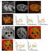
Spatial distribution of 3- and 10-kDa dextrans in relation to chromatin domains. Fluorescently labelled dextrans with molecular weights of 3 and 10 kDa were microinjected into nuclei of living HeLa cells. (A,B) Typical nuclei microinjected with 3-kDa dextran. (C,D) Nuclei microinjected with 10-kDa dextran. (Aa,Ba,Ca,Da) Distribution of microinjected fluorescently labelled dextrans. (Ab,Bb,Cb,Db) Histone-H2B–green-fluorescent-protein (GFP) signal. (Ac,Bc,Cc,Dc) Overlay of dextrans in red and chromatin in green. (Ad,Bd,Cd,Dd) Line scans showing the local intensity distribution of fluorescently labelled dextrans (red) and GFP-tagged chromatin (green). In the overlay images, the positions and directions of the line scans are indicated by long arrows. Small arrows point to several positions on the lines that correspond to asterisks in the line scans. The positions of the nucleoli are shown by double-headed black arrows below the line scans. Three-dimensional images were recorded; images shown are individual mid-nuclear optical sections. Scale bars in (Ac,Bc,Cc), 2 μm; scale bar in (Dc), 1.5 μm.
Figure 2
Figure 2
Spatial distribution of 70-kDa dextran in relation to chromatin domains. Fluorescently labelled dextran with a molecular weight of 70 kDa was microinjected into nuclei of living HeLa cells. (A,B) Typical nuclei, showing (Aa,Ba) the distribution of microinjected fluorescently labelled dextrans, (Ab,Bb) the histone-H2B–green-fluorescent-protein (GFP) signal, (Ac,Ad,Bc,Bd) overlays of the dextran signal in red and GFP-tagged chromatin in green, (Ae,Be) line scans through the two nuclei shown in (Ac,Ad,Bc,Bd). In the overlay pictures in (Ad,Bd), the line-scan positions are indicated by long arrows. Small arrows point to several positions on the line that are represented by asterisks in the line scans. The positions of the nucleoli are represented by black double-headed arrows in the line scans (Ae,Be). Three-dimensional images were recorded; images show individual mid-nuclear optical sections. Scale bars, 2 μm.
Figure 3
Figure 3
Spatial distribution of GFP-tagged TFIIH and RNA polymerase II in relation to chromatin domains. Chromatin from human SV40 (simian virus 40)-transformed fibroblasts that express GFP-tagged TFIIH, and from CHO-K1 cells that express GFP-tagged RNA polymerase II, were stained with Hoechst. (A,B) Typical nuclei; TFIIH–GFP distribution in relation to condensed chromatin is shown. (C,D) Typical nuclei; RNA-polymerase–II-GFP distribution in relation to condensed chromatin is shown. (Aa,Ba,Ca,Da) Distribution of GFP-tagged protein. (Ab,Bb,Cb,Db) Hoechst-labelled condensed chromatin. (Ac,Bc,Cc,Dc) Overlay of the GFP-tagged protein signal in red and the Hoechst-labelled chromatin in green. (Ad,Bd,Cd,Dd) Line scans through the nuclei shown in Ac,Bc,Cc and Dc, respectively. In the overlay images, the positions and directions of the line scans are indicated by long arrows. Small arrows point to several positions on the lines that correspond to asterisks in the line scans. The positions of the nucleoli are shown by double-headed black arrows below the line scans. Three-dimensional images were recorded; images shown are individual, mid-nuclear optical sections. Scale bars, 1.5 mm. GFP, green fluorescent protein.

Similar articles

Cited by

References

    1. Brown K.E., Baxter J., Graf D., Merkenschlager M. & Fisher A.G. ( 1999) Dynamic repositioning of genes in the nucleus of lymphocytes preparing for cell division. Mol. Cell, 3, 207–217. - PubMed
    1. Chubb J.R. & Bickmore W.A. ( 2003) Considering nuclear comparmentalization in the light of nuclear compartments. Cell, 112, 403–406. - PubMed
    1. Chubb J.R., Boyle S., Perry P. & Bickmore W.A. ( 2002) Chromatin motion is constrained by association with nuclear compartments in human cells. Curr. Biol., 12, 439–445. - PubMed
    1. Cmarko D., Verschure P.J., Martin T.E., Dahmus M.E., Krause S., Fu X.D., Van Driel R. & Fakan S. ( 1999) Ultrastructural analysis of transcription and splicing in the cell nucleus after bromo-UTP microinjection. Mol. Biol. Cell, 10, 211–223. - PMC - PubMed
    1. Cockell M. & Gasser S.M. ( 1999) Nuclear compartments and gene regulation. Curr. Opin. Genet. Dev., 9, 199–205. - PubMed

Publication types