. 2003 Sep;4(9):889-93.
doi: 10.1038/sj.embor.embor929. Epub 2003 Aug 29.

An exosome-like complex in Sulfolobus solfataricus

Affiliations

An exosome-like complex in Sulfolobus solfataricus

Elena Evguenieva-Hackenberg et al. EMBO Rep. 2003 Sep.

Abstract

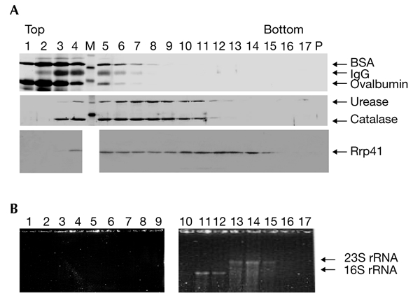
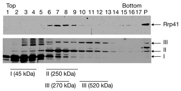
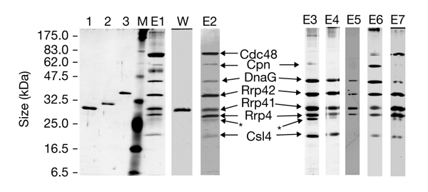
We present the first experimental evidence for the existence of an exosome-like protein complex in Archaea. In Eukarya, the exosome is essential for many pathways of RNA processing and degradation. Co-immunoprecipitation with antibodies directed against the previously predicted Sulfolobus solfataricus orthologue of the exosome subunit ribosomal-RNA-processing protein 41 (Rrp41) led to the purification of a 250-kDa protein complex from S. solfataricus. Approximately half of the complex cosediments with ribosomal subunits. It comprises four previously predicted orthologues of the core exosome subunits from yeast (Rrp41, Rrp42, Rrp4 and Csl4 (cepl synthetic lethality 4; an RNA-binding protein and exosome sub-unit)), whereas other predicted subunits were not found. Surprisingly, the archaeal homologue of the bacterial DNA primase DnaG was tightly associated with the complex. This suggests an RNA-related function for the archaeal DnaG-like proteins. Comparison of experimental data from different organisms shows that the minimal core of the exosome consists of at least one phosphate-dependent ribonuclease PH homologue, and of Rrp4 and Csl4. Such a protein complex was probably present in the last common ancestor of Archaea and Eukarya.


Figures

Figure 1
Figure 1
Sulfolobus solfataricus Rrp41 is a complex-bound protein. (A) Sulfolobus solfataricus lysate and marker proteins were fractionated in parallel through 10–60% glycerol gradients at 150 mM NaCl. Aliquots of each fraction were resolved by SDS–polyacrylamide gel electrophoresis. Rrp41 was detected by western blot analysis and marker proteins were detected by silver staining. Lanes 1–17 contain glycerol-gradient fractions. The proteins detected are indicated by arrows. (B) Analysis of the RNA content of the phenol/chloroform-extracted glycerol-gradient fractions in an ethidium-bromide-stained formaldehyde–agarose gel. The two panels are from the same gel. M, protein marker; P, pellet; Rrp41, ribosomal-RNA-processing protein 41.
Figure 2
Figure 2
Sulfolobus solfataricus Rrp41 cosediments with ribosomal subunits under high-salt conditions. Sulfolobus solfataricus lysate and marker proteins were fractionated in parallel through 5–20% glycerol gradients in the presence of 500 mM NaCl. Aliquots of each fraction were resolved by SDS–polyacrylamide gel electrophoresis. Rrp41 was detected by western blot analysis and marker proteins were detected by silver staining. Lanes 1–17 contain glycerol-gradient fractions. The proteins detected are indicated by arrows. The peak fractions of each marker are indicated under the lower panel. P, pellet; Rrp41, ribosomal-RNA-processing protein 41. I, Ovalbumin; II, catalase; III, urease.
Figure 3
Figure 3
Composition of the Sulfolobus solfataricus exosome. Gels from SDS–polyacrylamide gel electrophoresis and a western blot, showing the proteins that copurify with Sulfolobus solfataricus ribosomal-RNA-processing protein 41 (Rrp41) during immunoprecipitation. At the left side of the panel, the size of the marker polypeptides is shown. The coprecipitated proteins are indicated by arrows. The asterisks indicate the Rrp4 degradation product. Lane 1, His6-tagged Rrp4; lane 2, His6-tagged Rrp41; lane 3, His6-tagged Rrp42; lane M, protein marker; lane W, western hybridization of lane E1 with antibodies against Rrp41; lanes E1–E7, independent purifications of the exosome (first elution fractions); lanes E1 and E2, coprecipitation from 10–60% glycerol-gradient fractions; lanes E3 and E4, coprecipitation directly from cell-free extract; lane E4, treatment with ribonucleases was performed before elution; E5, coprecipitation after partial purification on anion- and cation-exchange columns; lane E6, coprecipitation after anion-exchange and size-exclusion chromatography steps; lane E7, coprecipitation from 5–20% glycerol-gradient fractions 6–9 (see Fig. 2). Lane E2 shows Coomassie-blue-stained proteins. The proteins in lanes 1–3, M, E1 and E3–E7 are silver stained.

References

    1. Allmang C., Petfalski E., Podtelejnikov A., Mann M., Tollervey D. & Mitchell P. ( 1999a) The yeast exosome and human PM–Scl are related complexes of 3′ to 5′ exonucleases. Genes Dev., 13, 2148–2158. - PMC - PubMed
    1. Allmang C., Kufel J., Chanfreau G., Mitchell P., Petfalski E. & Tollervey D. ( 1999b) Functions of the exosome in rRNA, snoRNA and snRNA synthesis. EMBO J., 18, 5399–5410. - PMC - PubMed
    1. Aloy P., Ciccarelli F.D., Leutwein C., Gavin A.C., Superti-Furga G., Bork P., Bottcher B. & Russell R.B. ( 2002) A complex prediction: three-dimensional model of the yeast exosome. EMBO Rep., 3, 628–635. - PMC - PubMed
    1. Anantharaman V., Koonin E.V. & Aravind L. ( 2002) Comparative genomics and evolution of proteins involved in RNA metabolism. Nucleic Acids Res., 30, 1427–1464. - PMC - PubMed
    1. Aravind L., Leipe D.D. & Koonin E.V. ( 1998) Toprim—a conserved catalytic domain in type IA and II topoisomerases, DnaG-type primases, OLD family nucleases and RecR proteins. Nucleic Acids Res., 26, 4205–4213. - PMC - PubMed

Publication types

MeSH terms