. 2003 Sep;185(18):5342-8.
doi: 10.1128/JB.185.18.5342-5348.2003.

Overproduction of inactive variants of the murein synthase PBP1B causes lysis in Escherichia coli

Affiliations

Overproduction of inactive variants of the murein synthase PBP1B causes lysis in Escherichia coli

Ute Meisel et al. J Bacteriol. 2003 Sep.

Abstract

Penicillin-binding protein 1B (PBP1B) of Escherichia coli is a bifunctional murein synthase containing both a transpeptidase domain and a transglycosylase domain. The protein is present in three forms (alpha, beta, and gamma) which differ in the length of their N-terminal cytoplasmic region. Expression plasmids allowing the production of native PBP1B or of PBP1B variants with an inactive transpeptidase or transglycosylase domain or both were constructed. The inactive domains contained a single amino acid exchange in an essential active-site residue. Overproduction of the inactive PBP1B variants, but not of the active proteins, caused lysis of wild-type cells. The cells became tolerant to lysis by inactive PBP1B at a pH of 5.0, which is similar to the known tolerance for penicillin-induced lysis under acid pH conditions. Lysis was also reduced in mutant strains lacking several murein hydrolases. In particular, a strain devoid of activity of all known lytic transglycosylases was virtually tolerant, indicating that mainly the lytic transglycosylases are responsible for the observed lysis effect. A possible structural interaction between PBP1B and murein hydrolases in vivo by the formation of a multienzyme complex is discussed.


Figures
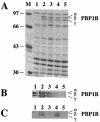
FIG. 1.
FIG. 1.
Overproduction of PBP1B variants in MC1061. Different variants of PBP1B were overproduced by the induction with 1 mM IPTG for 1 h at 37°C. (A and B) Membranes were prepared, and either the proteins were separated by SDS-8% PAGE and stained with Coomassie blue (A) or the proteins were transferred to nitrocellulose and PBP1B was detected with a polyclonal antiserum (B). In order to detect active penicillin-binding protein, the samples were incubated with biotinylated ampicillin prior to SDS-8% PAGE and transfer to nitrocellulose. (C) The biotin label was recognized by streptavidin-horseradish peroxidase and visualized with chloronaphthol. Lane M, low-molecular-weight protein marker. Lanes 1, MC1061(pJFK118EH) (control). Lanes 2, MC1061(pUM1Bα) (overproduction of native PBP1B forms). Lanes 3, MC1061(pUM1Bα*) (overproduction of PBP1B forms with inactive transpeptidase domain). Lanes 4, MC1061(pUM1BTG*α) (overproduction of PBP1B forms with inactive transglycosylase domain). Lanes 5, MC1061(pUM1BTG*α*) (overproduction of PBP1B forms with inactive transpeptidase and transglycosylase domains). Numbers on the left are molecular weights in thousands.
FIG. 2.
FIG. 2.
Growth of MC1061 with overproduction of inactive PBP1B variants. MC1061 harboring either empty expression plasmid pJFK118EH (open squares) or the PBP1B expression plasmid pUM1Bα (closed squares), pUM1Bα* (diamonds), pUM1BTG*α (triangles), or pUM1BTG*α* (circles) was grown at 37°C in LB medium containing 50 μg of kanamycin per ml. At an OD of 0.1, the production of PBP1B variants was induced by the addition of 1 mM IPTG to the cultures.
FIG. 3.
FIG. 3.
Overproduction of variants of PBP1Bαβγ and PBP1Bγ with different inducer concentrations. (A) Cultures of MC1061(pJFK118EH) (open diamonds) (control), MC1061(pUM1Bα*) (squares, triangles, and closed diamonds), or MC1061(pUM1Bγ*) (circles) were grown in LB medium with 50 μg of kanamycin per ml. At an OD of 0.1, the MC1061(pJFK118EH) and MC1061(pUM1Bγ*) cells received 1 mM IPTG, whereas the culture of MC1061(pUM1Bα*) was divided into three aliquots which received 0.01 (squares), 0.03 (triangles) or 0.05 mM (closed diamonds) IPTG. The growth was monitored by OD measurement. (B) One hour after the addition of IPTG, one aliquot was removed from each culture and membranes were isolated as described in Materials and Methods. The membrane proteins were separated by SDS-8% PAGE, and after blotting on nitrocellulose, PBP1B was detected with an antiserum. Lane 1, MC1061(pJFK118EH); lane 2, MC1061(pUM1Bα*) with 0.01 mM IPTG; lane 3, MC1061(pUM1Bα*) with 0.03 mM IPTG; lane 4, MC1061(pUM1Bα*) with 0.05 mM IPTG; lane 5, MC1061(pUM1Bγ*) with 1 mM IPTG.
FIG. 4.
FIG. 4.
Light microscopy of cells at the onset of lysis. MC1061(pUM1Bα*) was grown in LB medium with 50 μg of kanamycin per ml. Wild-type MC1061 was grown in LB medium. At an OD of 0.1, the cultures were divided. In MC1061(pUM1Bα*) the overproduction of inactive PBP1B was induced by the addition of 1 mM IPTG (B), whereas MC1061 received 100 μg of penicillin G per ml (D). After 70 min, the cells were fixed for light microscopy as described in Materials and Methods. Control cultures of MC1061(pUM1Bα*) (A) and MC1061 (C) received neither IPTG nor penicillin G. Bars, 10 μm.
FIG. 5.
FIG. 5.
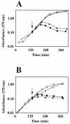
Penicillin-induced lysis and lysis upon overproduction of inactive PBP1B at pH 7.2 and 5.0. MC1061(pUM1Bα*) (squares) or MC1061 (triangles) was grown at 37°C in Bacto antibiotic medium 3 at a pH of 7.2 (A) or 5.0 (B). At an OD of 0.1 (arrow), the cultures were divided and one part (closed symbols) received either 1 mM IPTG to induce the overproduction of inactive PBP1B [MC1061(pUM1Bα*)] or 100 μg penicillin G per ml (MC1061).
FIG. 6.
FIG. 6.
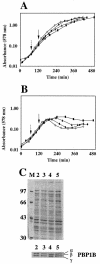
Overproduction of active or inactive PBP1B in murein hydrolase mutants. (A and B) Wild-type MC1061 (open circles), MHD61 (closed circles), or MHD82 (triangles and squares) harboring either pUM1Bα (A) (control, overproduction of active PBP1B) or pUM1Bα* (B) (overproduction of inactive PBP1B) was grown at 37°C in LB medium. At an OD of 0.1 (arrow), the cultures received 1 mM IPTG. One culture of MHD82 (squares) also received 20 μg of bulgecin per ml at the indicated time (dashed arrow) to inhibit the soluble lytic transglycosylase. (C) To compare the amounts of overproduced PBP1B variants in MHD82 grown in the presence of bulgecin and in MC1061, cells were harvested 1 h after induction with 1 mM IPTG and boiled in SDS sample buffer. The total protein was analyzed by SDS-8% PAGE following Coomassie blue staining (upper panel). PBP1B variants were detected with antiserum after blotting on nitrocellulose (lower panel). Lane M, molecular weight marker; lane 2, MC1061(pUM1Bα) (overproduction of active PBP1B); lane 3, MC1061(pUM1Bα*) (overproduction of inactive PBP1B); lane 4, MHD82(pUM1Bα) (overproduction of active PBP1B); lane 5, MHD82(pUM1Bα*) (overproduction of inactive PBP1B). Numbers on the left are molecular weights in thousands.

Similar articles

Cited by

References

    1. Bishop, R. E., and J. H. Weiner. 1993. Complementation of growth defect in an ampC deletion mutant of Escherichia coli. FEMS Microbiol. Lett. 114:349-354. - PubMed
    1. Casabadan, M. J., and S. N. Cohen. 1980. Analysis of gene control signals by DNA fusion and cloning in Escherichia coli. J. Mol. Biol. 138:179-207. - PubMed
    1. Chalut, C., X. Charpentier, M. H. Remy, and J. M. Masson. 2001. Differential responses of Escherichia coli cells expressing cytoplasmic domain mutants of penicillin-binding protein 1b after impairment of penicillin-binding proteins 1a and 3. J. Bacteriol. 183:200-206. - PMC - PubMed
    1. Chalut, C., M. H. Remy, and J. M. Masson. 1999. Disulfide bridges are not involved in penicillin-binding protein 1b dimerization in Escherichia coli. J. Bacteriol. 181:2970-2972. - PMC - PubMed
    1. Charpentier, X., C. Chalut, M. H. Remy, and J. M. Masson. 2002. Penicillin-binding proteins 1a and 1b form independent dimers in Escherichia coli. J. Bacteriol. 184:3749-3752. - PMC - PubMed

MeSH terms

Substances

LinkOut - more resources