. 2003 Sep;185(18):5357-62.
doi: 10.1128/JB.185.18.5357-5362.2003.

Mycobacterium tuberculosis FurA autoregulates its own expression

Affiliations

Mycobacterium tuberculosis FurA autoregulates its own expression

Claudia Sala et al. J Bacteriol. 2003 Sep.

Abstract

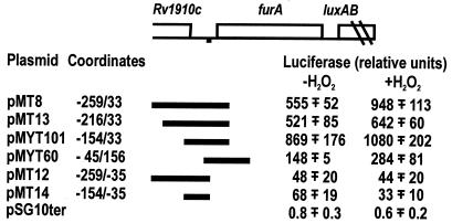
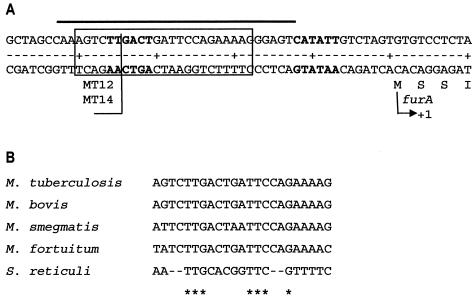
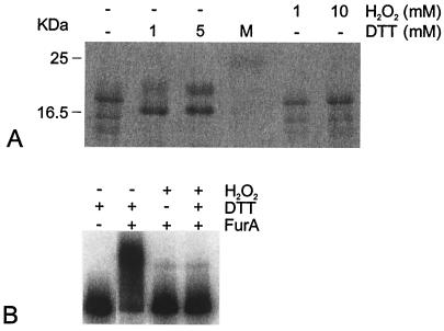
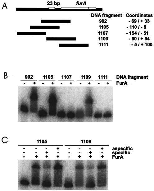
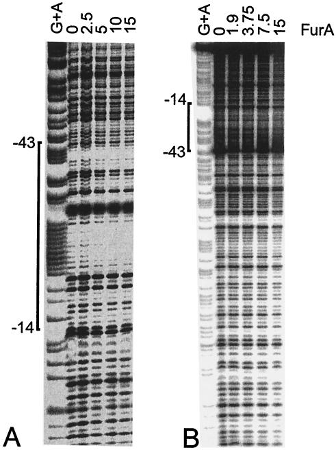
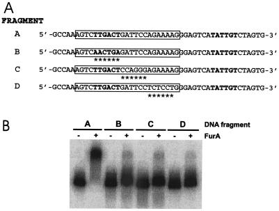
The furA-katG region of Mycobacterium tuberculosis, encoding a Fur-like protein and the catalase-peroxidase, is highly conserved among mycobacteria. Both genes are induced upon oxidative stress. In this work we analyzed the M. tuberculosis furA promoter region. DNA fragments were cloned upstream of the luciferase reporter gene, and promoter activity in Mycobacterium smegmatis was measured in both the presence and absence of oxidative stress. The shortest fragment containing an inducible promoter extends 45 bp upstream of furA. In this region, -35 and -10 promoter consensus sequences can be identified, as well as a 23-bp AT-rich sequence that is conserved in the nonpathogenic but closely related M. smegmatis. M. tuberculosis FurA was purified and found to bind upstream of furA by gel shift analysis. A ca. 30-bp DNA sequence, centered on the AT-rich region, was essential for FurA binding and protected by FurA in footprinting analysis. Peroxide treatment of FurA abolished DNA binding. Three different AT-rich sequences mutagenized by site-directed mutagenesis were constructed. In each mutant, both M. tuberculosis FurA binding in vitro and pfurA regulation upon oxidative-stress in M. smegmatis were abolished. Thus, pfurA is an oxidative stress-responsive promoter controlled by the FurA protein.


Figures

FIG. 1.
FIG. 1.
Oxidative stress induction of luciferase by plasmids carrying M. tuberculosis DNA regions. (Top) Schematic representation of the M. tuberculosis furA region. The AT-rich sequence upstream of furA is indicated by a black square. The DNA fragments cloned in pSG10ter are indicated, and the coordinates of the fragments are reported. The M. tuberculosis coordinates are arbitrary, with coordinate +1 corresponding to the first nucleotide of the furA gene. (Bottom) Luciferase activity expressed in M. smegmatis mc2155 transformed by the plasmids was measured, as described in Materials and Methods, both in the absence of (−H2O2) and after (+H2O2) oxidative stress. The mean values ± standard deviations for two to four independent clones tested are reported.
FIG. 2.
FIG. 2.
Sequence of the M. tuberculosis DNA fragment containing the oxidative stress-inducible promoter. (A) Sequence of the −50/+10 M. tuberculosis region. The coordinates are arbitrary, with coordinate +1 corresponding to the first nucleotide of the furA gene. The −35 and −10 sequences are in boldface. The 23-bp AT-rich region is boxed. The ends of the fragments cloned in MT12 and MT14 are indicated. The region protected by FurA is indicated by a line above the sequence. (B) Alignment of the AT-rich DNA sequences upstream of furA in M. tuberculosis, M. bovis, M. smegmatis, and M. fortuitum and the sequence upstream of furS in S. reticuli. The conserved bases are indicated by asterisks.
FIG. 3.
FIG. 3.
Electrophoretic mobility of the FurA protein and binding of FurA to DNA under different redox conditions. (A) Purified M. tuberculosis FurA (1.7 μg) expressed in E. coli was run in the first lane of an SDS-15% polyacrylamide gel. Equivalent samples were mixed with either DTT or H2O2 at the concentrations indicated above the lanes before being loaded on the gel. Lane M, molecular mass markers, with sizes indicated on the left. (B) Gel shift caused by FurA. The DNA probe covers the −154/+33 M. tuberculosis region. FurA (10 μM) was added, as indicated above the lanes, to 32P-labeled DNA probe in the presence of 200 μM Ni2+, and complexes were resolved on a Tris-acetate-8% polyacrylamide gel containing 200 μM Ni2+. DTT (1 mM) and H2O2 (10 mM) were added to the incubation buffer as indicated.
FIG. 4.
FIG. 4.
FurA binds DNA fragments covering the M. tuberculosis furA upstream region. (A) Map of the DNA fragments. (B) Gel shift caused by FurA. FurA binding was performed as described in the legend to Fig. 3, except that 12 μM FurA was added. (C) Specificity of FurA binding. Specific (same DNA fragment) and nonspecific (1111 DNA fragment) competitor DNA was added to each binding reaction mixture at 3,000 M.
FIG. 5.
FIG. 5.
Footprint analysis of the FurA binding site upstream of M. tuberculosis furA. The purified −154/+33 DNA region, labeled at either the +33 end (A) or the −154 end (B), was incubated in the presence of the amount of FurA protein (in micrograms) indicated above the lanes for 20 min at room temperature and digested with DNase I as described in Materials and Methods. Maxam-Gilbert A+G sequences of the same fragments were loaded in the first lanes.
FIG. 6.
FIG. 6.
Effect of mutations in the furA operator sequence on FurA binding. (A) Sequences of the mutations. Fragment A, wild type. Fragment B, C, and D carry different 6-bp substitutions within the AT-rich region (boxed). The mutant bases are indicated by asterisks. The −10 and −35 sequences are in boldface. (B) Gel shift of the mutant fragments caused by FurA. FurA binding was performed as described in the legend to Fig. 4.

Similar articles

Cited by

References

    1. Althaus, E. W., C. E. Outten, K. E. Olson, H. Cao, and T. V. Halloran. 1999. The ferric uptake regulation (Fur) repressor is a zinc metalloprotein. Biochemistry 38:6559-6569. - PubMed
    1. Bagg, A., and J. B. Neilands. 1987. Ferric uptake regulation protein acts as a repressor, employing iron (II) as a cofactor to bind the operator of an iron transport operon in Escherichia coli. Biochemistry 26:5471-5477. - PubMed
    1. Bashyam, M. D., D. Kaushal, S. K. Dasgupta, and A. K. Tyagi. 1996. A study of mycobacterial transcriptional apparatus: identification of novel features in promoter elements. J. Bacteriol. 178:4847-4853. - PMC - PubMed
    1. Bsat, N., A. Herbig, L. Casillas-Martinez, P. Setlow, and J. D. Helmann. 1998. Bacillus subtilis contains multiple Fur homologues: identification of the iron uptake (Fur) and peroxide regulon (PerR) repressors. Mol. Microbiol. 29:189-198. - PubMed
    1. Bullock, W., J. Fernandez, and J. Short. 1987. XLI-Blue: a high efficiency plasmid transforming recA E. coli strain with β-galactosidase selection. BioTechniques 5:376-379.

Publication types

MeSH terms

LinkOut - more resources