. 2003 Sep;185(18):5398-407.
doi: 10.1128/JB.185.18.5398-5407.2003.

Extracellular polysaccharides associated with thin aggregative fimbriae of Salmonella enterica serovar enteritidis

Affiliations

Extracellular polysaccharides associated with thin aggregative fimbriae of Salmonella enterica serovar enteritidis

A P White et al. J Bacteriol. 2003 Sep.

Abstract

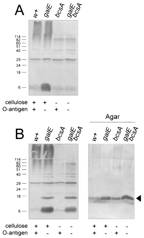
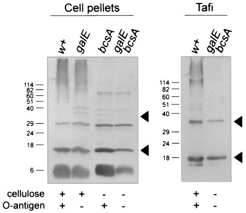
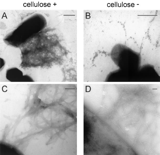
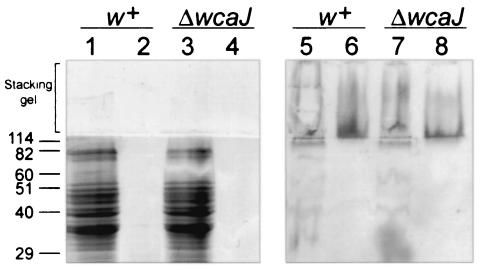
Lipopolysaccharide (LPS) O polysaccharide was identified as the principle factor impeding intercellular formation of intact thin aggregative fimbriae (Tafi) in Salmonella enterica serovar Enteritidis. The extracellular nucleation-precipitation assembly pathway for these organelles was investigated by quantifying fimbrial formation between deltaagfA (AgfA recipient) and deltaagfB (AgfA donor) cells harboring mutations in LPS (galE::Tn10) and/or cellulose (deltabcsA) synthesis. Intercellular complementation could be detected between deltaagfA and deltaagfB strains only when both possessed the galE mutation. LPS O polysaccharide appears to be an impenetrable barrier to AgfA assembly between cells but not within individual cells. The presence of cellulose did not restrict Tafi formation between cells. Transmission electron microscopy of w+ S. enterica serovar Enteritidis 3b cells revealed diffuse Tafi networks without discernible fine structure. In the absence of cellulose, however, individual Tafi fibers were clearly visible, appeared to be occasionally branched, and showed the generally distinctive appearance described for Escherichia coli K-12 curli. A third extracellular matrix component closely associated with cellulose and Tafi was detected on Western blots by using immune serum raised to whole, purified Tafi aggregates. Cellulose was required to tightly link this material to cells. Antigenically similar material was also detected in S. enterica serovar Typhimurium and one diarrheagenic E. coli isolate. Preliminary analysis indicated that this material represented an anionic, extracellular polysaccharide that was distinct from colanic acid. Therefore, Tafi in their native state appear to exist as a complex with cellulose and at least one other component.


Figures

FIG. 1.
FIG. 1.
Intercellular complementation of Tafi between S. enterica serovar Enteritidis 3b ΔagfB donor and ΔagfA recipient strains. CR binding of different ΔagfA recipient strains grown together with ΔagfB donor strains is shown. Recipient strain genotypes are indicated above each graph, while ΔagfB donor strain phenotypes are listed at the bottom. CR binding values for each combination of mutants were normalized by subtracting values for the corresponding ΔagfB or ΔagfA strains grown individually. Each bar shows the averages and standard errors from four separate experiments, and asterisks indicate significant differences within each group (*, P < 0.05; **, P < 0.01; ***, P < 0.001 [Tukey-Kramer multiple comparisons test]). The panels below the bar graphs show immunoblot analysis performed with immune serum raised to purified Tafi. The amount of AgfA fimbrial material associated with recipient and donor cells scraped off the TCR agar is noted for each recipient-donor combination.
FIG. 2.
FIG. 2.
Western blot analysis of S. enterica serovar Enteritidis 3b ΔagfB donor and ΔagfA recipient strains for the production of AgfA. S. enterica serovar Enteritidis 3b ΔagfA (A) and ΔagfB (B) strains were analyzed for production of AgfA after growth on T agar. Samples from strains were loaded as indicated, and strain phenotypes are listed below each lane. Panel A and the first four lanes of panel B represent proteins in the debris left over after boiling cells in SDS-PAGE sample buffer, whereas agar samples (B) represent proteins present in agar plugs after cells were removed. All samples were treated with formic acid prior to loading on SDS-PAGE. AgfA and associated material were detected by using immune serum raised to purified Tafi; the arrowhead in panel B indicates monomeric AgfA. Molecular mass markers (in kilodaltons) are indicated on the left.
FIG. 3.
FIG. 3.
Immunoblot analysis of AgfA and associated material produced by S. enterica serovar Enteritidis 3b and related strains. Proteins in the debris left over after boiling cells in SDS-PAGE sample buffer (cell pellets) or purified fimbrial material (Tafi) from S. enterica serovar Enteritidis 3b (w+) the galE::Tn10 strain (galE), the ΔbcsA strain (bcsA), or the galE::Tn10 ΔbcsA strain (galE bcsA) were loaded as indicated. Strain phenotypes are listed below each immunoblot. All samples were treated with 90% formic acid prior to loading on SDS-PAGE. AgfA and associated material were detected by using immune serum raised to whole Tafi; the lower and upper arrowheads indicate monomeric and dimeric AgfA, respectively. Molecular mass markers (in kilodaltons) are indicated on the left..
FIG. 4.
FIG. 4.
Transmission electron microscopy of Tafi fimbriae. Fimbriae produced by S. enterica serovar Enteritidis strain 3b (cellulose positive) (A and C) or the ΔbcsA mutant (cellulose negative) (B and D) were immunogold labeled with AgfA-specific monoclonal antibody 3A-12 ascites followed by goat anti-mouse immunoglobulin-10-nm-diameter gold (A and B) or simply negatively stained with uranyl acetate (C and D). Bars, 500 nm (A and B) or 100 nm (C and D).
FIG. 5.
FIG. 5.
Detection of immunoreactive Tafi-associated material in different enterobacterial species. Proteins in the debris left over after boiling cells in SDS-PAGE sample buffer (cell pellets) or acetone-precipitated proteins from a 10 mM Tris (pH 8) wash of whole cells of S. enterica serovar Enteritidis 3b, S. enterica serovar Typhimurium SL7207 or E. coli Vietnam I/1 after growth on T agar at 28 or 37°C were loaded as indicated. The brackets show the regions of immunoblots corresponding to the stacking gel from SDS-PAGE. AgfA and associated material were detected by using immune serum raised to whole Tafi; arrowheads indicate monomeric AgfA (Salmonella) or CsgA (E. coli). Molecular mass markers (in kilodaltons) are indicated on the left.
FIG. 6.
FIG. 6.
Detection of high-MW immunoreactive material in S. enterica serovar Enteritidis 3b and ΔwcaJ strains. SDS-PAGE (lanes 1 to 4) or immunoblot analysis (lanes 5 to 8) of whole cells of the S. enterica serovar Enteritidis 3b (w+) and ΔwcaJ strains boiled in SDS-PAGE sample buffer (lanes 1, 3, 5, and 7) and digested with 0.5 mg of proteinase K per ml for 1 h at 65°C (lanes 2, 4, 6, and 8) is shown. Proteins were detected with GelCode staining (Pierce) (lanes 1 to 4). High-MW material was detected by using immune serum raised to purified Tafi (lanes 5 to 8). The bracket shows the stacking gel region of SDS-PAGE and the corresponding immunoblot. Molecular mass markers (in kilodaltons) are indicated on the left.

Similar articles

Cited by

References

    1. Aguilar, A., S. Merino, M. M. Nogueras, M. Regue, and J. M. Tomas. 1999. Two genes from the capsule of Aeromonas hydrophila (serogroup O:34) confer serum resistance to Escherichia coli K12 strains. Res. Microbiol. 150:395-402. - PubMed
    1. Anriany, Y. A., R. M. Weiner, J. A. Johnson, C. E. De Rezende, and S. W. Joseph. 2001. Salmonella enterica serovar Typhimurium DT104 displays a rugose phenotype. Appl. Environ. Microbiol. 67:4048-4056. - PMC - PubMed
    1. Arnqvist, A., A. Olsen, J. Pfeifer, D. G. Russell, and S. Normark. 1992. The Crl protein activates cryptic genes for curli formation and fibronectin binding in Escherichia coli HB101. Mol. Microbiol. 6:2443-2452. - PubMed
    1. Austin, J. W., G. Sanders, W. W. Kay, and S. K. Collinson. 1998. Thin aggregative fimbriae enhance Salmonella enteritidis biofilm formation. FEMS Microbiol. Lett. 162:295-301. - PubMed
    1. Baumler, A. J., A. J. Gilde, R. M. Tsolis, A. W. van der Velden, B. M. Ahmer, and F. Heffron. 1997. Contribution of horizontal gene transfer and deletion events to development of distinctive patterns of fimbrial operons during evolution of Salmonella serotypes. J. Bacteriol. 179:317-322. - PMC - PubMed

Publication types

MeSH terms

LinkOut - more resources