. 2003 Sep;185(18):5508-18.
doi: 10.1128/JB.185.18.5508-5518.2003.

In vivo reconstitution of the FhuA transport protein of Escherichia coli K-12

Affiliations

In vivo reconstitution of the FhuA transport protein of Escherichia coli K-12

Michael Braun et al. J Bacteriol. 2003 Sep.

Abstract

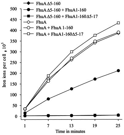
The FhuA protein in the outer membrane of Escherichia coli actively transports ferrichrome and the antibiotics albomycin and rifamycin CGP 4832 and serves as a receptor for the phages T1, T5, and phi80 and for colicin M and microcin J25. The crystal structure reveals a beta-barrel with a globular domain, the cork, which closes the channel formed by the barrel. Genetic deletion of the cork resulted in a beta-barrel that displays no FhuA activity. A functional FhuA was obtained by cosynthesis of separately encoded cork and the beta-barrel domain, each endowed with a signal sequence, which showed that complementation occurs after secretion of the fragments across the cytoplasmic membrane. Inactive complete mutant FhuA and an FhuA fragment containing 357 N-proximal amino acid residues complemented the separately synthesized wild-type beta-barrel to form an active FhuA. Previous claims that the beta-barrel is functional as transporter and receptor resulted from complementation by inactive complete FhuA and the 357-residue fragment. No complementation was observed between the wild-type cork and complete but inactive FhuA carrying cork mutations that excluded the exchange of cork domains. The data indicate that active FhuA is reconstituted extracytoplasmically by insertion of separately synthesized cork or cork from complete FhuA into the beta-barrel, and they suggest that in wild-type FhuA the beta-barrel is formed prior to the insertion of the cork.


Figures

FIG. 1.
FIG. 1.
Time-dependent transport of [55Fe3+]ferrichrome (1 μM) into E. coli MB97 ΔfhuA aroB expressing plasmid-encoded FhuA or FhuA β-barrel of E. coli together with FhuA cork domain fragments as indicated.
FIG. 2.
FIG. 2.
(A) Comparison of [35S]methionine-labeled cork domains of FhuA after transformation of E. coli BL21 fhuA with plasmids pT7-7 (lane 1), pCEc773 (lane 2), pCEc771 (lane 3), and pCEc772 (lane 4). The proteins were separated by SDS-PAGE of whole-cell lysates. No other bands were seen outside the gel section presented here. Arrows indicate the mature cork fragments. CP, cytoplasmic (without signal sequence). (B) Autoradiograph after SDS-PAGE with [35S]methionine-labeled proteins of E. coli BL21 fhuA whole-cell lysates after transformation with plasmids pCEc762 and pBK570 (lane 1), pCEc761 and pBK570 (lane 2), pCEc762 and pHSG576 (lane 3), and pCEc761 and pHSG576 (lane 4). Arrows indicate the mature FhuA cork domain proteins. (C) Autoradiograph after SDS-PAGE of different cell fractions of E. coli BL21 fhuA expressing [35S]methionine-labeled FhuA1-160 cork domain of E. coli encoded on plasmid pCEc761. Cytoplasmic fraction (lane 1), total membrane fraction (lane 2), periplasmic fraction (lane 3), and whole-cell lysates (lane 4) were applied. The control strain BL21 fhuA transformed with the pT7-6 vector showed no bands of [35S]methionine-labeled proteins. Arrows indicate the mature cork domains. Numbers on the right of each panel indicate molecular masses in kilodaltons.
FIG. 3.
FIG. 3.
Comparison of [35S]methionine-labeled FhuA proteins after cross-linking with 1% formaldehyde (+ [samples without formaldehyde are marked −]) of FhuA1-160 and FhuAΔ5-160. Cross-linking experiments were performed with E. coli strain BL21 fhuA carrying plasmid pT7-6 (lanes 2 and 3), pHK763 (lanes 4 and 5), pBK7 (lanes 6 and 7), pCEc761 (lanes 8 and 9), pMBlc3 (lanes 10 and 11), or pMBlc1 (lanes 12 and 13). Samples were heated at 65°C for 5 min prior to gel loading. Whole-cell lysate of E. coli BL21 fhuA expressing FhuA1-357 encoded on plasmid pMBlc7 was also applied on the gel (lane 1). The mature protein of the truncated FhuA is marked by an asterisk. The arrow in lane 13 indicates the FhuA1-160 FhuAΔ5-160 cross-link product; arrows with broken lines indicate possible cork dimer products in lanes 9, 11, and 13. Numbers on the right indicate molecular masses in kilodaltons.
FIG. 4.
FIG. 4.
Comparison of [35S]methionine-labeled FhuA proteins after in vivo cysteine cross-linking of FhuA T27C P533C and FhuA1-160 T27C cork mutant with the FhuAΔ5-160 P533C β-barrel mutant. Cross-linking experiments were performed with E. coli strain BL21 fhuA carrying plasmid pMBFE1 (lanes 1 and 2), pMBFE2 (lanes 3 and 4), pMBlc5 (lanes 5 and 6), or pMBlc6 (lanes 7 and 8). Samples were suspended in nonreducing sample buffer (−) or in sample buffer containing 1% β-mercaptoethanol (+) and heated at 90°C for 5 min prior to gel loading. Arrows in lanes 6 and 8 indicate the cross-linked complex between FhuA1-160 T27C and FhuAΔ5-160 P533C. The broken arrow in lane 6 indicates the possible cross-linked FhuA1-160 T27C dimer. Asterisks in lanes 2 and 4 indicate the intraprotein cross-linked complexes of FhuA T27C P533C and FhuAΔ5-17 T27C P533C showing lower electrophoretic mobilities than the un-cross-linked proteins (lanes 1 and 3). Numbers on the right indicate molecular masses in kilodaltons.

Similar articles

Cited by

References

    1. Bayer, M. H., G. P. Costello, and M. E. Bayer. 1982. Isolation and partial characterization of membrane vesicles carrying markers of the membrane adhesion sites. J. Bacteriol. 149:758-767. - PMC - PubMed
    1. Bös, C., D. Lorenzen, and V. Braun. 1998. Specific in vivo labeling of cell surface-exposed protein loops: reactive cysteines in the predicted gating loop mark a ferrichrome binding site and a ligand-induced conformational change of the Escherichia coli FhuA protein. J. Bacteriol. 180:605-613. - PMC - PubMed
    1. Braun, M., H. Killmann, and V. Braun. 1999. The β-barrel domain of FhuAΔ5-160 is sufficient for TonB-dependent FhuA activities of Escherichia coli. Mol. Microbiol. 33:1037-1049. - PubMed
    1. Braun, M., H. Killmann, E. Maier, R. Benz, and V. Braun. 2002. Diffusion through channel derivatives of the Escherichia coli FhuA transport protein. Eur. J. Biochem. 269:4948-4959. - PubMed
    1. Braun, V. 1995. Energy-coupled transport and signal transduction through the gram-negative outer membrane via TonB-ExbB-ExbD-dependent receptor proteins. FEMS Microbiol. Rev. 16:295-307. - PubMed

Publication types

MeSH terms

LinkOut - more resources