. 2003 Sep;185(18):5573-84.
doi: 10.1128/JB.185.18.5573-5584.2003.

Genome diversification in phylogenetic lineages I and II of Listeria monocytogenes: identification of segments unique to lineage II populations

Affiliations

Genome diversification in phylogenetic lineages I and II of Listeria monocytogenes: identification of segments unique to lineage II populations

Chaomei Zhang et al. J Bacteriol. 2003 Sep.

Abstract

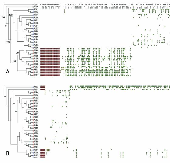
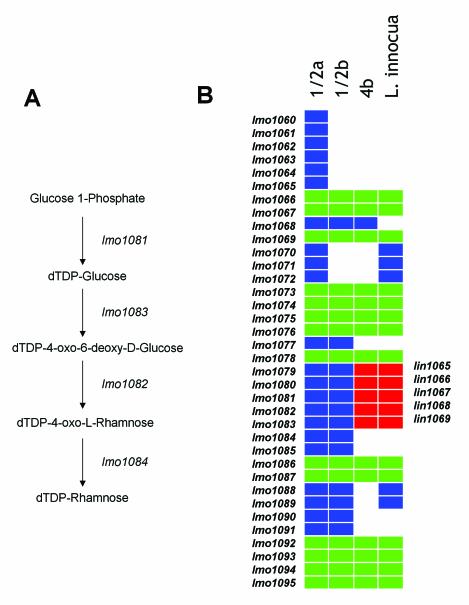
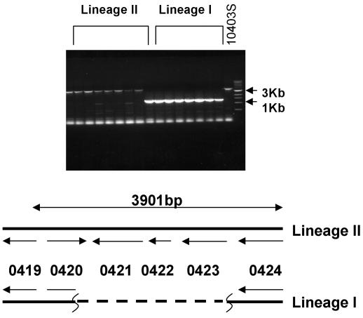
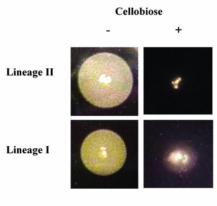
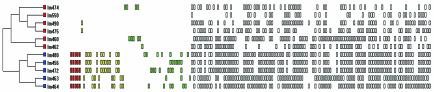
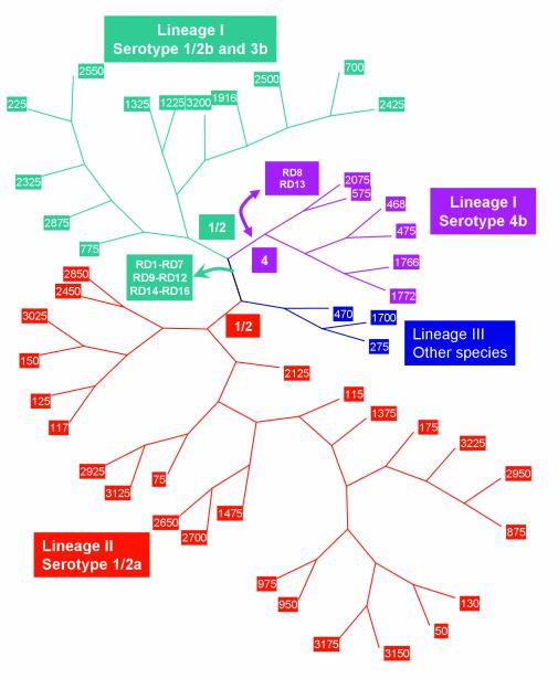
Thirteen different serotypes of Listeria monocytogenes can be distinguished on the basis of variation in somatic and flagellar antigens. Although the known virulence genes are present in all serotypes, greater than 90% of human cases of listeriosis are caused by serotypes 1/2a, 1/2b, and 4b and nearly all outbreaks of food-borne listeriosis have been caused by serotype 4b strains. Phylogenetic analysis of these three common clinical serotypes places them into two different lineages, with serotypes 1/2b and 4b belonging to lineage I and 1/2a belonging to lineage II. To begin examining evolution of the genome in these serotypes, DNA microarray analysis was used to identify lineage-specific and serotype-specific differences in genome content. A set of 44 strains representing serotypes 1/2a, 1/2b, and 4b was probed with a shotgun DNA microarray constructed from the serotype 1/2a strain 10403s. Clones spanning 47 different genes in 16 different contiguous segments relative to the lineage II 1/2a genome were found to be absent in all lineage I strains tested (serotype 4b and 1/2b) and an additional nine were altered exclusively in 4b strains. Southern hybridization confirmed that conserved alterations were, in all but two loci, due to absence of the segments from the genome. Genes within these contiguous segments comprise five functional categories, including genes involved in synthesis of cell surface molecules and regulation of virulence gene expression. Phylogenetic reconstruction and examination of compositional bias in the regions of difference are consistent with a model in which the ancestor of the two lineages had the 1/2 somatic serotype and the regions absent in the lineage I genome arose by loss of ancestral sequences.


Figures

FIG. 1.
FIG. 1.
Analysis of genome content by reference microarray. (A)Dendrogram derived from UPGMA analysis of binary data from the L. monocytogenes serotype 1/2a array. UPGMA was performed on binary data generated from 44 strains of Listeria monocytogenes and one strain each of L. welshimeri and L. innocua. Tree length, using 730 polymorphic characters, was 1,941 steps with a consistency index of 0.3509 and homoplasy index of 0.6491. Bootstrap scores, using 1,000 repetitions of a UPGMA search, are shown on nodes scoring > 70%. Leaves of the tree from lineage I strains are colored red, those from lineage II are colored blue, and those from lineage III or other Listeria species are uncolored. Rectangles to the right of the strain names depict polymorphisms present in a given strain sorted by the MARKFIND program. Those polymorphisms conserved within all members of a lineage are colored red, those that are lineage-specific but nonconserved are colored green (polyphyletic) or yellow (monophyletic). The 489 polymorphisms distributed among strains in both lineages are not shown. (B) The same data as those in panel A were analyzed to identify polymorphisms exclusive to and conserved within serotype 4b L. monocytogenes strains and L. welshimeri and L. innocua. Red rectangles depict altered loci in serotype 4b strains and L. welshimeri and L. innocua, green rectangles indicate those polymorphisms shared among polyphyletic groups of strains within the serotype 4b cluster and among the serotype 4b and 1/2a clusters.
FIG. 2.
FIG. 2.
Polymorphism in putative teichoic acid biosynthetic genes between serotype 1/2 and 4b populations. (A) The putative four-gene biosynthetic pathway for dTDP-l-rhamnose encoded by lmo1081-lmo1084 is shown. (B) The map shows the alignment of genes in the region relative to the L. monocytogenes serotype 1/2a, 4b, and L. innocua genome sequences and the inferred presence and absence of genes in serotype 1/2b relative to the microarray data. The rectangles represent individual genes. White space indicates absence of a gene at that relative position in the genome and the colored squares indicate orthologous genes at the respective positions. Gene indices relative to the L. monocytogenes EGD genome are indicated to the left of the illustration (lmo1060-lmo1094) and genes from the L. innocua genome (lin1065-lin1069) are on the right. Genes shared by L. monocytogenes serotype 4b and L. innocua are in red.
FIG. 3.
FIG. 3.
PCR analysis of the lmo0421-lmo0423 region. Agarose gel electrophoresis of PCR amplification products derived from PCR amplification of the lmo0420-lmo0424 region. Products from lineage I and lineage II strains are indicated. The control strain, lineage II strain 10403s, is indicated on the right end of the gel along with the 3- and 1-kb markers from the size standards. The map underneath depicts the region in lineage II and lineage I strains. The lineage I map is based on sequence analysis of PCR amplification products from two independent lineage I strains.
FIG. 4.
FIG. 4.
Phospholipase activity of L. monocytogenes strains. Strains from lineage I and lineage II were spotted onto egg yolk agar to measure phospholipase activity. Colonies on the left hand panels were grown on agar with no cellobiose and those on the right hand panel were grown on agar with cellobiose. The opaque zones around the colonies grown on plates without cellobiose are derived from the activity of the PrfA-dependent phospholipase B.
FIG. 5.
FIG. 5.
Analysis of genome content among strains derived from a single outbreak. DNA microarray analysis was performed on 11 different L. monocytogenes strains that were isolated from independent patients of a single outbreak. Each strain shared identical PFGE profiles and ribotypes. The dendrogram was rendered by UPGMA analysis and the data sorted based on the two groups (blue and red leaves) using the MARKFIND program.
FIG. 6.
FIG. 6.
Model for evolution of the L. monocytogenes genome. The unrooted dendrogram was rendered from UPGMA analysis of the microarray data. Branches corresponding to lineage I serotype 1/2b and 3b strains are colored green, lineage I serotype 4b colored purple, and lineage II serotype 1/2a colored red. The branch containing the lineage III strain and other species is colored blue. Branch points where genome deletions and acquisitions are postulated to have arisen are indicated with boxes colored according to the affected lineage.

Similar articles

Cited by

References

    1. Aarts, H. J., L. E. Hakemulder, and A. M. Van Hoef. 1999. Genomic typing of Listeria monocytogenes strains by automated laser fluorescence analysis of amplified fragment length polymorphism fingerprint patterns. Int. J. Food Microbiol. 49:95-102. - PubMed
    1. Begg, K. J., and W. D. Donachie. 1985. Cell shape and division in Escherichia coli: experiments with shape and division mutants. J. Bacteriol. 163:615-622. - PMC - PubMed
    1. Bibb, W. F., B. Schwartz, B. G. Gellin, B. D. Plikaytis, and R. E. Weaver. 1989. Analysis of Listeria monocytogenes by multilocus enzyme electrophoresis and application of the method to epidemiologic investigations. Int. J. Food Microbiol. 8:233-239. - PubMed
    1. Bishop, D. K., and D. J. Hinrichs. 1987. Adoptive transfer of immunity to Listeria monocytogenes: the influence of in vitro stimulation on lymphocyte subset requirements. J. Immunol. 139:2005-2009. - PubMed
    1. Botta, G. A., and J. T. Park. 1981. Evidence for involvement of penicillin-binding protein 3 in murein synthesis during septation but not during cell elongation. J. Bacteriol. 145:333-340. - PMC - PubMed

Publication types

LinkOut - more resources