. 2003 Nov 15;553(Pt 1):83-94.
doi: 10.1113/jphysiol.2003.048298. Epub 2003 Aug 29.

GABA mediates autoreceptor feedback inhibition in the rat carotid body via presynaptic GABAB receptors and TASK-1

Affiliations

GABA mediates autoreceptor feedback inhibition in the rat carotid body via presynaptic GABAB receptors and TASK-1

Ian M Fearon et al. J Physiol. .

Abstract

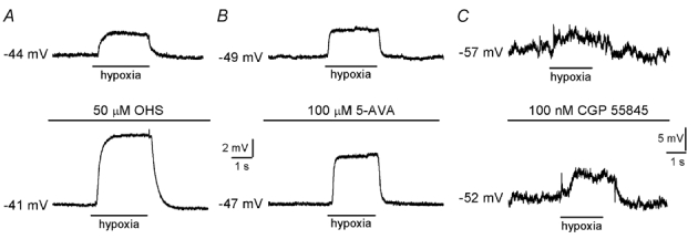
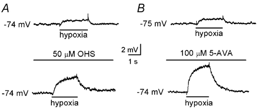
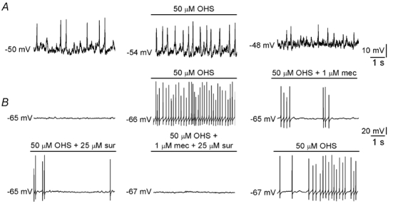
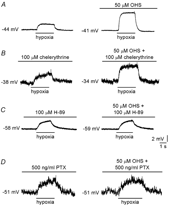
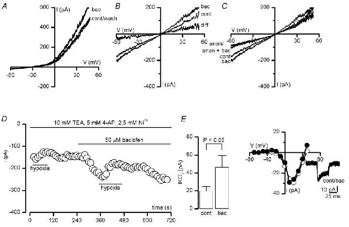
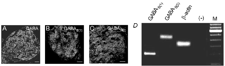
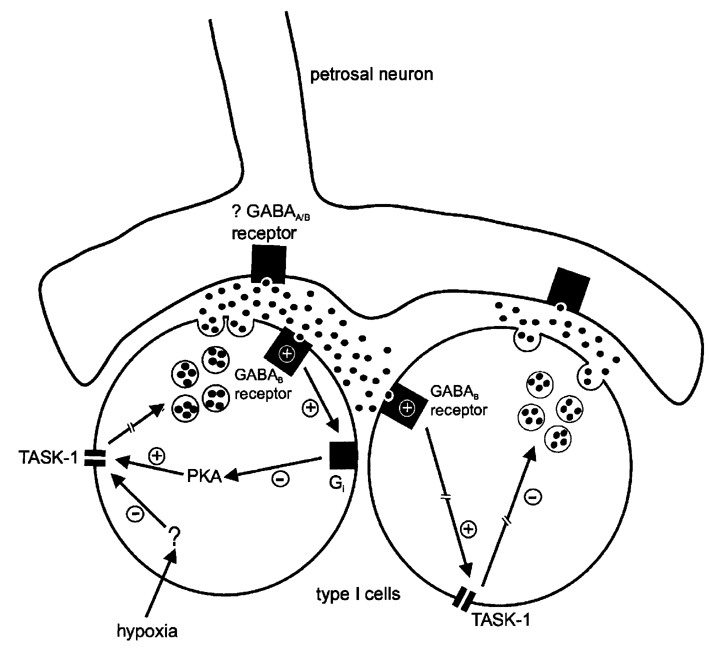
Background K+ channels exert control over neuronal excitability by regulating resting potential and input resistance. Here, we show that GABAB receptor-mediated activation of a background K+ conductance modulates transmission at rat carotid body chemosensory synapses in vitro. Carotid body chemoreceptor (type I) cells expressed GABAB(1) and GABAB(2) subunits as well as endogenous GABA. The GABAB receptor agonist baclofen activated an anandamide- and Ba2+-sensitive TASK-1-like background K+ conductance in chemoreceptor cell clusters, but was without effect on voltage-gated Ca2+ channels. Hydroxysaclofen (50 microM), 5-aminovaleric acid (100 microM) and CGP 55845 (100 nM), selective GABAB receptor blockers, potentiated the hypoxia-induced receptor potential; this effect was abolished by pre-treatment with pertussis toxin (PTX; 500 ng ml-1), an inhibitor of Gi, or by H-89 (50 microM), a selective inhibitor of protein kinase A. The protein kinase C inhibitor chelerythrine chloride (100 microM) was without effect on this potentiation. GABAB receptor blockers also caused depolarisation of type I cells in clusters, and enhanced spike discharge in spontaneously firing cells. In functional co-cultures of type I clusters and petrosal sensory neurones, GABAB receptor blockers potentiated hypoxia-induced postsynaptic chemosensory responses mediated by the fast-acting transmitters ACh and ATP. Thus GABAB receptor-mediated activation of TASK-1 or a related channel provides a presynaptic autoregulatory feedback mechanism that modulates fast synaptic transmission in the rat carotid body.


Figures

Figure 1
Figure 1. GABAB receptor blockade enhances receptor potential in chemoreceptor cells
A, typical current-clamp recordings made from a type I cell cluster. Application of hypoxia (PO2, 5 mmHg) is indicated by the horizontal bar below each trace. Under control conditions (upper), hypoxia induced a depolarisation which was increased in the presence of 50 µm hydroxysaclofen (OHS; lower). B and C, as in A, except the effects of further specific blockers of GABAB receptors, 5-aminovaleric acid (5-AVA; 100 µm) and CGP 55845 (100 nm), were examined. Note the different scale bars in C. In all cases, effects of GABAB receptor blockers were fully reversed on washout of the drug (not shown).
Figure 2
Figure 2. Postsynaptic depolarisation due to hypoxia is modulated by GABAB receptor inhibition
A, typical current-clamp recordings made from a petrosal neurone juxtaposed to a type I cluster in co-culture. Application of hypoxia (PO2, 5 mmHg) is indicated by the horizontal bar below each trace. Under control conditions (upper), hypoxia induced a postsynaptic depolarisation which was increased in the presence of the GABAB receptor antagonist, OHS (50 µm; lower). B, as in A, except the effect of a further specific GABAB receptor antagonist, 5-aminovaleric acid (5-AVA; 100 µm), was examined.
Figure 3
Figure 3. GABAB receptor blockers enhance excitability and release of ACh and ATP from type I cells
A, effect of 50 µm hydroxysaclofen (OHS) on membrane potential in a presynaptic type I cell. This cell exhibited spontaneous activity, which was reversibly enhanced by OHS. B, effect of 50 µm OHS on membrane potential in a petrosal neurone juxtaposed to a type I cluster in co-culture. Application of this GABAB receptor antagonist induced spiking in this cell, representative of 6 cells that behaved in this way. This effect is due to disinhibition of ACh and ATP release since responses were reduced in the presence of 1 µm mecamylamine (mec) or 25 µm suramin (sur), blockers of nicotinic and purinergic receptors, respectively. In the presence of both blockers OHS-induced spike activity in the neurone was abolished. Scale bars apply to all traces. Together with the data in A, this demonstrates a GABAB receptor-mediated ‘braking’ mechanism which is constitutively active and reduces presynaptic excitability and the release of fast excitatory neurotransmitters onto postsynaptic neurones.
Figure 4
Figure 4. Mechanism of GABA-mediated regulation of synaptic transmission
A, current-clamp recordings made from a type I cell in a cluster. Application of hypoxia (PO2, 5 mmHg) is indicated by the horizontal bars below each trace. Under control conditions (left), hypoxia induced a depolarisation which was increased in the presence of 50 µm OHS (right). B, enhanced depolarisation still occurred in the presence of the selective PKC blocker chelerythrine chloride (100 µm, right). In contrast, enhancement was abolished in the presence of the selective PKA blocker H-89 (50 µm; C) or following pretreatment for 24 h with PTX (500 ng ml−1; D). In B and C, the effects of chelerythrine and H-89 were fully reversible on washout of the kinase inhibitors (not shown).
Figure 5
Figure 5. Baclofen activates a TASK-1-like conductance in type I cells
A, voltage-clamp recordings obtained from a type I cell in a cluster. Currents were evoked by ramp depolarisations between -60 and +50 mV under asymmetrical K+ conditions, in the absence (cont) and presence of 50 µm baclofen (bac) and following washout (wash), as indicated. B, as in A, except currents were recorded under symmetrical K+ conditions. Subtraction of the current obtained in the presence of baclofen from that seen under control conditions (to give the indicated difference current) shows that baclofen activates a linear K+ conductance. C, the selective TASK-1 blocker anandamide (anan; 5 µm) ablated the response to baclofen. D, time-series recording demonstrating the enhancement of the oxygen-sensitive background K+ current (IKO2) by 50 µm baclofen. Typical of 4 such recordings, which were made under Ca2+-free, symmetrical K+ conditions and in the presence of 2.5 mm Ni2+, 10 mm TEA and 5 mm 4-AP to block voltage- and Ca2+-dependent K+ channels (Buckler et al. 2000). Periods of application of baclofen and hypoxia (PO2, 5 mmHg) are indicated by the horizontal bars. Currents were measured at a test potential of -60 mV. Inset shows the magnitude of the hypoxic response, obtained by subtracting the current evoked at -60 mV under normoxic conditions from that obtained in hypoxia. Plotted are mean (± s.e.m.) data obtained in 4 cells, under control conditions (cont) and in the presence of 50 µm baclofen (bac), as indicated. E, Ca2+ channel current-voltage relationships obtained from a type I cell under control conditions (○) and in the presence of 50 µm baclofen (•). Each point shows the peak current amplitude evoked by a 50 ms step depolarisation to the indicated test potential, from a holding potential of -80 mV. Ca2+ (5 mm) was used as charge carrier. Inset, individual current traces obtained by step-depolarising to +10 mV for 50 ms, under control conditions and in the presence of 50 µm baclofen (bac).
Figure 6
Figure 6. Presence of GABA and GABAB receptors in type I cells
Confocal images showing carotid body sections which were immunostained with a specific antibodies raised against GABA (A), GABAB(1) (B) and GABAB(2) (C) receptor subunits and visualised by secondary FITC fluorescence. Positive immunostaining of type I clusters is seen in each case. Scale bars represent 20 µm. In all cases, staining was abolished either when sections were exposed to the secondary antibody without prior exposure to the primary antibody, or in the case of GABA the primary antibody was pre-adsorbed with excess antigen (not shown). D, micrograph of a 2 % agarose gel stained with ethidium bromide and viewed under UV illumination. RT-PCR was carried out on isolated type I clusters following extraction of mRNA, and using gene-specific primers for the GABAB(1) and GABAB(2) subunits, and β-actin. Marker lane (M) shows bands at 100 bp increments with the 600 bp fragment at increased intensity. In negative control reactions without RT (−) no PCR products were observed.
Figure 7
Figure 7. Schematic representation of the autoregulatory pathways involved in the GABA-mediated regulation of neurotransmitter release from type I cells during hypoxia
Via an as yet uncharacterised intracellular pathway, hypoxia inhibits TASK-1-like background channels in type I cells, leading to membrane depolarisation and ultimately (broken arrow) neurotransmitter release. In this process GABA (black circles) is released from type I cells, and acts at presynaptic GABAB receptors on either the same type I cell (autocrine) or on an adjacent type I cell (paracrine) in the cluster. This causes stimulation of the pertussis toxin-sensitive inhibitory G protein Gi, causing inhibition of protein kinase A (PKA) and subsequently activation of TASK-1. This would serve to hyperpolarise the type I cell and limit the degree of depolarisation during exposure to hypoxia, regulating the further release of transmitters. GABA may also act at postsynaptic ionotropic or metabotropic GABA receptors to modulate chemoreceptor output. For clarity, the involvement of other K+ channels and neurotransmitters in chemotransmission, and the intracellular events leading to transmitter release, have been omitted.

Similar articles

Cited by

References

    1. Bairam A, Neji H, De Grandpre P, Carroll JL. Autoreceptor mechanism regulating carotid body dopamine release from adult and 10-day-old rabbits. Resp Physiol. 2000;120:27–34. - PubMed
    1. Bayliss DA, Talley EM, Sirois JE, Lei Q. TASK-1 is a highly modulated pH-sensitive ‘leak’ K+ channel expressed in brainstem respiratory neurones. Resp Physiol. 2001;129:159–174. - PubMed
    1. Benot AR, Lopez-Barneo J. Feedback inhibition of Ca2+ currents by dopamine in glomus cells of the carotid body. Eur J Neurosci. 1990;2:809–812. - PubMed
    1. Bowery N, Enna SJ. γ-Aminobutyric acidB receptors: first of the functional metabotropic heterodimers. J Pharmacol Exp Ther. 2000;2:2–7. - PubMed
    1. Buckler KJ, Vaughan-Jones RD. Effects of hypoxia on membrane potential and intracellular calcium in rat neonatal carotid body type I cells. J Physiol. 1994;476:423–428. - PMC - PubMed

Publication types

MeSH terms

LinkOut - more resources