Comparative Study
. 2003 Aug 15;298(1):109-22.
doi: 10.1002/jez.b.29.

Molecular biology of feather morphogenesis: a testable model for evo-devo research

Affiliations
Comparative Study

Molecular biology of feather morphogenesis: a testable model for evo-devo research

Randall B Widelitz et al. J Exp Zool B Mol Dev Evol. .

Abstract

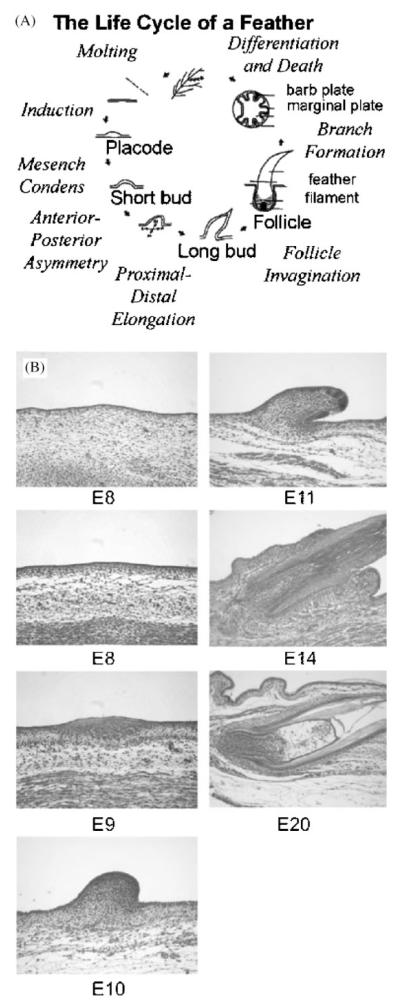
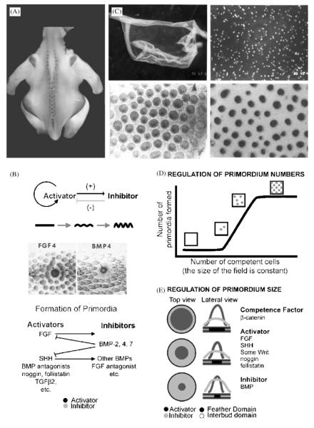
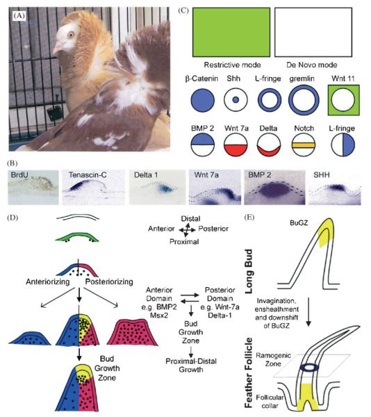
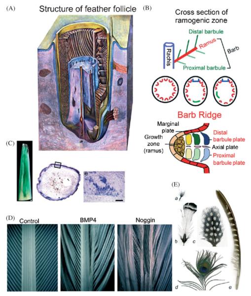
Darwin's theory describes the principles that are responsible for evolutionary change of organisms and their attributes. The actual mechanisms, however, need to be studied for each species and each organ separately. Here we have investigated the mechanisms underlying these principles in the avian feather. Feathers comprise one of the most complex and diverse epidermal organs as demonstrated by their shape, size, patterned arrangement and pigmentation. Variations can occur at several steps along each level of organization, leading to highly diverse forms and functions. Feathers develop gradually during ontogeny through a series of steps that may correspond to the evolutionary steps that were taken during the phylogeny from a reptilian ancestor to birds. These developmental steps include 1) the formation of feather tract fields on the skin surfaces; 2) periodic patterning of the individual feather primordia within the feather tract fields; 3) feather bud morphogenesis establishing anterio-posterior (along the cranio-caudal axis) and proximo-distal axes; 4) branching morphogenesis to create the rachis, barbs and barbules within a feather bud; and 5) gradual modulations of these basic morphological parameters within a single feather or across a feather tract. Thus, possibilities for variation in form and function of feathers occur at every developmental step. In this paper, principles guiding feather tract formation, distributions of individual feathers within the tracts and variations in feather forms are discussed at a cellular and molecular level.


Figures

Fig. 1
Fig. 1
The process of feather formation involves temporal and spatial regulation of cellular processes, including localized cell proliferation, migration, adhesion, death and differentiation. A. The life cycle of a feather: the stages and major morphogenetic processes in feather development are listed. B. Haemotoxylin and eosin stained tissue sections of developing feathers from dorsal skin: at E7, it is flat. At E 8, dense dermis starts to form. At E9, feather primordia form. At E10, feather buds form. At E11, buds have elongated. At E14, buds invaginate to form follicles. At E20, a new feather is forming.
Fig. 2
Fig. 2
Periodic patterning of feather primordia within the feather tract field. It is a self-organizing process, based on the interplay of molecules with positive or negative roles on feather primordia formation. It results in individual feather buds of certain size and numbers. A. β-catenin marks the initial appearance of feather primordia: during development, the β-catenin transcript is first expressed in the whole tract field, then becomes restricted to individual feather primordia with a lateral inhibitory zone (Widelitz et al., 2000). B. Feather periodic patterning involving reaction-diffusion and competitive equilibrium: through experimentation, some molecules are found to enhance feather formation (activators), and some suppress feather formation (inhibitors). An exemplary activator (FGF 4) and inhibitor (BMP 4) are shown. Data show that both activators and inhibitors are located in the primordial region, not one in the primordia and one in the inter-primordia, respectively. The activators induce both activators and inhibitors, while the inhibitors suppress the activators. These results favor the involvement of a reaction-diffusion mechanism (Turing, ’52; Nagorcka and Mooney, ’85; Moore et al., ’98; Jiang et al., ’99). C. A novel reconstitution assay that allows the study of periodic pattern formation from a zero state: Upper panels. Embryonic day 6 chicken skin was dissected, and epidermis (upper left panel) and mesenchyme were separated. Mesenchyme was dissociated into single cells (upper right panel). Lower panels. Cells are recombined with epithelium and cultured. In 24 hours, they self-organize into many feather buds simultaneously (lower left panel, Jiang et al., ’99). These buds start to express bud specific genes such as SHH (lower right panel, Ting-Berreth and Chuong, ’96a). D. Regulation of the number of feather primordia: Using the reconstitution assay, the effect of using a fixed size of epidermis while increasing the number of competent mesenchymal cells was tested. Logically, either the number of buds or the size of the feather primordia could increase. Using skin from a certain region, it was found that the size of the feather primordia is a constant. At low cell density, buds did not form. At higher cell density, feather primordia started to appear randomly. The density of feather primordia gradually increased until they reached the highest packing density, which yields the hexagonal patterning (Jiang et al., ’99). E. Regulation of the size of feather primordia: Competent cells are first distributed homogeneously in the field. These cells adhere randomly and this adhesion is reversible. When these small unstable aggregates surpass a threshold density, they become stable dermal condensations. The size of each dermal condensation is dependent on the ratio of activator molecules (noggin, FGF, Shh, etc) to inhibitor molecules (BMPs). A higher activator to inhibitor ratio allows the formation of larger sized feather buds while a higher inhibitor to activator ratio favors the formation of the interbud region (Jiang et al., ’99).
Fig. 3
Fig. 3
Further regionalization within feather buds: It involves setting up anterior - posterior asymmetric long feather buds and a proximal - distal axis. A. Orientation of the feather: when feather primordia first form, they have no orientation. Soon, they acquire anterior-posterior asymmetry from the epithelium (Novel, ’73; Chuong et al., ’96). The anterior end is the side of the future rachis. Feather orientation is essential to feather function in view of the need for flight. However, the orientation signal can go wrong as seen in these two genetic variants of the pigeon. Feathers in the scapular feather tract are inversely oriented. Individual feathers are otherwise normal. B. Molecular asymmetry of the feather buds: cell proliferation is enriched in the posterior buds. Tenascin-C is in the anterior mesenchyme (Jiang and Chuong, ’92). Delta-1 is in the posterior bud mesenchyme (Chen et al., ’97), Wnt 7a in the posterior epithelium (Chuong et al., ’96), BMP 2 in the mesenchyme (Jung et al., ’98) and SHH in the distal epithelium (Ting-Berreth and Chuong, ’96a). Notice, while BMP 2 is a feather formation inhibitor, it is expressed in the feather primordia region. C. Schematic drawing of representative molecular expression modes: two general expression profiles for all molecules studied to date have been observed. In the restrictive mode, the genes are initially expressed throughout a feather tract field before the buds are formed, and later become restricted to specific regions of the buds or interbuds. In the de novo mode, they appear in specific regions of the buds or interbuds after the buds form. D. Coupling of anterior - posterior asymmetry and proximal - distal growth: experimental data suggest that the co-existence of anterior and posterior bud domains is critical for the proximal - distal elongation of buds (Noveen et al., ’95a; Widelitz et al., ’99). A bud growth zone (yellow) appears at the distal interface of the anterior (blue) and posterior (red) domains. At this stage, proliferation is highest within this bud growth zone. Some possible molecular relationships are listed. E. Growth zones in buds and follicle: at the time of invagination and ensheathment to form the feather follicle, the growth zone gradually shifts from the distal to the proximal end of the follicle, and becomes the collar (Chodankar et al., 2003).
Fig. 4
Fig. 4
Branching morphogenesis of the feather: forming rachis, barbs and barbules. A new level of periodic patterning process within the cylindrical feather collar epithelium leads to the three-leveled hierarchal branches. Modulation of their size, angle and symmetry through molecular pathways can generate complex and diverse feather forms. A. A schematic drawing showing the inside of a forming feather filament (from Lucas and Stettenheim, ’72): The ramogenic zone is the region in which the barb ridge starts to form. Its cross section is shown in panel B. B. Formation of rachis, barbs and barbule: from the cylinder shaped collar, feather barb ridges (Red) start to form in the ramogenic zone. This can occur in a radially symmetric fashion (middle left) and form downy feathers. Or, a rachis (blue) and a barb forming zone (green) are formed in a bilaterally symmetric (middle center) or asymmetric (middle right) fashion, leading to bilaterally symmetric or asymmetric vanes. Proximal and distal barbules (top, here as the proximal and distal barbule plates) can be the same or different, leading to plumulaceous or pennaceous vanes. C. RCAS mediated gene transduction of feathers: To generate feathers with mis-expressed genes, regenerating feathers are infected with RCAS chicken retrovirus. Ectopic expression of β galactosidase and Noggin are shown. D. Alteration of feather branching by perturbation of the BMP pathway: Left, normal. Middle, Over-expression of BMP4 causes enlargement of rachis. Right, Over-expression of noggin causes splitting of the rachis. E. Examples of feathers showing different feather forms: a. Downy feather shows radially arranged barbs inserted on a short stalk. b. A contour feather with a bilaterally symmetric vane. c. A feather with a bilaterally symmetric vane. d. A bilaterally symmetric peacock retrice (tail feather). e. Highly asymmetric remige (flight feather) of a pheasant. Pigmentation patterns can be either symmetric stripes (b), spots, note the half spots surrounding the rachis (c), one “eye spot” (concentric rings) (d) or stripes on only one of the vanes (e) Model on how these patterns may form based on reaction diffusion was proposed by Prum and Williamson, 2002. Plumulaceous barbs like those shown in a and d are due to symmetric barbules. Pennaceous barbs like those found in b, c and e are the results of asymmetric barbule formation.
Fig. 5
Fig. 5
Different feather growth rate. The length of feathers from different skin regions from a budgie was measured.

References

    1. Bartels T, Meyer W, Neurand K. Skin and plumage changes in domestic birds. II. Plumage changes Tierarztl Prax. 1992;20:275–281. - PubMed
    1. Brantjes H, Barker N, van Es J, Clevers H. TCF: Lady Justice casting the final verdict on the outcome of Wnt signalling. Biol Chem. 2002;383:255–261. - PubMed
    1. Chen CWJ, Jung HS, Jiang TX, Chuong CM. Asymmetric expression of Notch, Serrate and Delta is associated with the anterior posterior axis of feather buds. Dev Biol. 1997;188:181–187. - PubMed
    1. Chen CW, Chuong CM. Avian integument provides multiple possibilities to analyse different phases of skin appendage morphogenesis. J Investig Dermato. Symp Proc. 1999;4:333–337. - PubMed
    1. Chen CWJ, Chuong CM. Dynamic expression of lunatic fringe during feather morphogenesis: a switch from medial-lateral to anterior-posterior asymmetry. Mech Dev. 2000;91:351–354. - PubMed

Publication types

LinkOut - more resources