. 2003 Sep;49(6):1699-713.
doi: 10.1046/j.1365-2958.2003.03669.x.

VirE2, a type IV secretion substrate, interacts with the VirD4 transfer protein at cell poles of Agrobacterium tumefaciens

Affiliations

VirE2, a type IV secretion substrate, interacts with the VirD4 transfer protein at cell poles of Agrobacterium tumefaciens

Krishnamohan Atmakuri et al. Mol Microbiol. 2003 Sep.

Abstract

Agrobacterium tumefaciens transfers oncogenic DNA and effector proteins to plant cells during the course of infection. Substrate translocation across the bacterial cell envelope is mediated by a type IV secretion (TFS) system composed of the VirB proteins, as well as VirD4, a member of a large family of inner membrane proteins implicated in the coupling of DNA transfer intermediates to the secretion machine. In this study, we demonstrate with novel cytological screens - a two-hybrid (C2H) assay and bimolecular fluorescence complementation (BiFC) - and by immunoprecipitation of chemically cross-linked protein complexes that the VirE2 effector protein interacts directly with the VirD4 coupling protein at cell poles of A. tumefaciens. Analyses of truncation derivatives showed that VirE2 interacts via its C terminus with VirD4, and, further, an NH2-terminal membrane-spanning domain of VirD4 is dispensable for complex formation. VirE2 interacts with VirD4 independently of the virB-encoded transfer machine and T pilus, the putative periplasmic chaperones AcvB and VirJ, and the T-DNA transfer intermediate. Finally, VirE2 is recruited to polar-localized VirD4 as a complex with its stabilizing secretion chaperone VirE1, yet the effector-coupling protein interaction is not dependent on chaperone binding. Together, our findings establish for the first time that a protein substrate of a type IV secretion system is recruited to a member of the coupling protein superfamily.


Figures
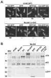
Fig. 1
Fig. 1
VirD4-dependent localization of GFP-VirE2 to A. tumefaciens cell poles. A. A348 (WT) and Mx355 (virD4 null mutant) cells producing proteins indicated above each panel photographed 10 h after induction with 200 μM AS by fluorescence microscopy. The proteins indicated were synthesized from the following IncP plasmids: D4-GFP (pKA62); GFP (pZDB69); GFP-E2 (pZDB73) and D4 + GFP-E2 (pKA77). The number below each panel represents the percentage of cells with polar fluorescence out of a total of at least 1000 cells examined; the ‘−’ denotes no detectable polar fluorescence. B. Immunodetection of fusion proteins produced in Mx355 derivatives at 10 h post induction. The proteins listed above each lane were synthesized from the IncP plasmids listed in (A); for D4 (pKA21). Blots were developed with the antisera listed at the right. The reactive species (~60-kDa) in all lanes detected by anti-VirE2 antisera is native VirE2 produced from pTi.
Fig. 2
Fig. 2
Cytology-based dihybrid screens for demonstrating VirE2–VirD4 complex formation. A. C2H: A348 (WT) cells producing the proteins indicated above each panel were examined 4 h post induction by fluorescence microscopy. The proteins indicated were synthesized from the following plasmids: DivIVA-D4 + GFP-E2 (pZD76, pZDB73); DivIVA + GFP-E2 (pKA76, pZDB73); DivIVA-D4 + D4-GFP (pZD76, pKA62); DivIVA + D4-GFP (pKA76, pKA62). The number below each panel represents the percentage of cells with polar fluorescence. Typically, ~ 15% of cells exhibiting DivIVA-dependent targeting in the C2H screen show fluorescence at the midcell. B. BiFC: KE1 (ΔvirE) and Mx355 cells producing the proteins indicated adjacent to each panel were examined 4 and 10 h postinduction, respectively, by fluorescence microscopy. The proteins indicated were synthesized from the following plasmids introduced into the strains singly or in combination: E1-N’GFP (pZDB88); GFP’C-E2 (pZDB89); D4-N’GFP (pKAB64); N’GFP + GFP’C (pKVB35, pKVB39); D4-N’GFP (pKAB64); D4-N’GFP + GFPC’ (pKAB64, pKVB39). C. Immunodetection of fusion proteins produced in KE1 (top panel) and Mx355 (bottom panel) at 10 h post induction. The proteins listed above each lane were synthesized from the following plasmids introduced into the strains singly or in combination: E1-N’GFP (pZDB88); GFP’C-E2 (pZDB89); D4-N’GFP (pKAB64); D4-N’GFP + GFPC’-E2 (pKAB64, pZDB89); and DivIVA-D4 + GFP-E2 (pKV42). The dark arrowheads point to distinct proteins that exhibit similar mobilities, e.g., VirD4-N’GFP (~ 92-kDa) and GFP-VirE2 (~ 90-kDa) (immunoreactive with anti-GFP antiserum), and VirD4-N’GFP and DivIVA-VirD4 (~ 93 kDa) (immunoreactive with anti-VirD4 antiserum).
Fig. 3
Fig. 3
A. Co-immunoprecipitation of VirD4 and VirD4ΔN87 with VirE2. Isolated membranes were cross-linked with DSP and detergent-solubilized complexes were immunoprecipitated (IP) with preimmune (−) or immune (+) antisera listed at the left of the immunoblots. Blots were developed (BD) with antisera listed at the right. Strains: A348 (WT), At12516 (virE2), Mx355 (virD4); Mx355(D4) and Mx355(D4ΔN87) are the virD4 null mutant producing VirD4 and VirD4ΔN87 from plasmids pKA21 and pKA43, respectively. Lanes headed with slanted strain names, A348 and Mx355(D4), were loaded with total solubilized membrane protein extracts to show presence of VirE2 (~ 60-kDa), VirD4 (~ 76-kDa), and VirD4ΔN87 (~ 65-kDa). Immunoreactive bands at the bottoms of all blots correspond to the IgG heavy chain. B. C2H screen for a VirD4ΔN87 interaction with GFP-VirE2. Strain Mx355 producing proteins indicated adjacent to each panel were examined 4 h post induction by fluorescence microscopy. Proteins were produced from the following plasmids: DivIVA-D4ΔN87 + GFP-E2 (pKV43); D4ΔN87-GFP (pKAB75); DivIVA-D4ΔN87 + GFP (pKA43, pZDB69); D4ΔN87 + GFP-E2 (pKA43, pZDB72).
Fig. 4
Fig. 4
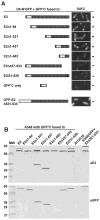
Localization of a VirD4 interaction domain to the C terminus of VirE2. A. A348 producing VirD4-N’GFP and GFP’C fused to the VirE2 derivatives listed at the left and schematically represented were examined at 10 h (top two panels) or 18 h (bottom six panels) post induction by fluorescence microscopy. The proteins indicated were produced from the following plasmids: D4-N’GFP (pKAB64); GFPC’-E2 (pZDB89); GFPC’-E2Δ1–84 (pZDB838); GFPC’-E2Δ1–331 (pZDB834); GFPC’-E2Δ1–437 (pZDB836); GFPC’-E2Δ1–483 (pZDB122); GFPC’-E2Δ437–533 (pZDB846); GFPC’-E2i31–529 (pZDB827); GFPC’ (pKVB39); GFP-VirE2Δ524–533 (pZDB120). ‘ +’, fluorescent foci. ‘−’, no detectable foci, although these cells exhibited weak uniform fluorescence at t = 18. B. Immunodetection of fusion proteins produced in A348 derivatives at 10 h postinduction. The proteins listed above each lane were synthesized from the IncP plasmids listed in (A). Blots were developed with the antisera listed at the right. The reactive species (~60-kDa) in all lanes, except At12516 (virE2), reactive with the anti-VirE2 antisera is native VirE2 produced from pTi.
Fig. 5
Fig. 5
VirE2 targeting to VirD4 independently of T-strand-VirD2, Mpf proteins and VirE1 secretion chaperone. A. Complex formation assessed with the C2H and BiFC screens. The A348 mutants listed at the left co-produced VirD4 and GFP-E2 from pKA77 [C2H], or D4-N’GFP and GFPC’-E2 from pKAB64 and pZDB89). Strains were examined 10 h post induction by fluorescence microscopy. The numbers to the right of panels depicting the C2H results correspond to the percentage of cells with polar fluorescence. For BiFC, ‘+’, fluorescent foci; ‘−’, no detectable foci. B. VirD4 recruitment of VirE2 as a complex with VirE1. KE1 and PC3001 (ΔvirE1) producing E1-GFP from pZDB12, or DivIVA-D4 and E1-GFP from pZD76 + pZDB12 were examined 4 h after induction with 200 μM AS by fluorescence microscopy.

Similar articles

Cited by

References

    1. Backert S, Ziska E, Brinkmann V, Zimny-Arndt U, Fauconnier A, Jungblut PR, et al. Translocation of the Helicobacter pylori CagA protein in gastric epithelial cells by a type IV secretion apparatus. Cell Microbiol. 2000;2:155–164. - PubMed
    1. Baron C, O’Callaghan D, Lanka E. Bacterial secrets of secretion: Euro Conference on the biology of type IV secretion processes. Mol Microbiol. 2002;43:1359–1365. - PubMed
    1. Berger BR, Christie PJ. Genetic complementation analysis of the Agrobacterium tumefaciens virB operon: virB2 through virB11 are essential virulence genes. J Bacteriol. 1994;176:3646–3660. - PMC - PubMed
    1. Binns A, Beaupre C, Dale E. Inhibition of VirB-mediated transfer of diverse substrates from Agrobacterium tumefaciens by the IncQ plasmid RSF1010. J Bacteriol. 1995;177:4890–4899. - PMC - PubMed
    1. Burgess RR, Arthur TM, Pietz BC. Mapping protein–protein interaction domains using ordered fragment ladder far-western analysis of hexahistidine-tagged fusion proteins. Methods Enzymol. 2000;328:141–157. - PubMed

Publication types

MeSH terms