. 2003 Sep 1;17(17):2123-37.
doi: 10.1101/gad.1117903.

A transcriptional regulatory cascade that controls left/right asymmetry in chemosensory neurons of C. elegans

Affiliations

A transcriptional regulatory cascade that controls left/right asymmetry in chemosensory neurons of C. elegans

Sarah Chang et al. Genes Dev. .

Abstract

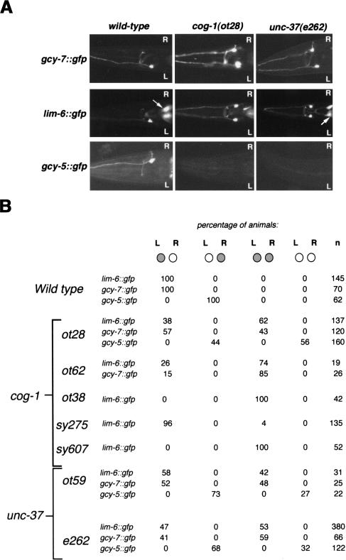
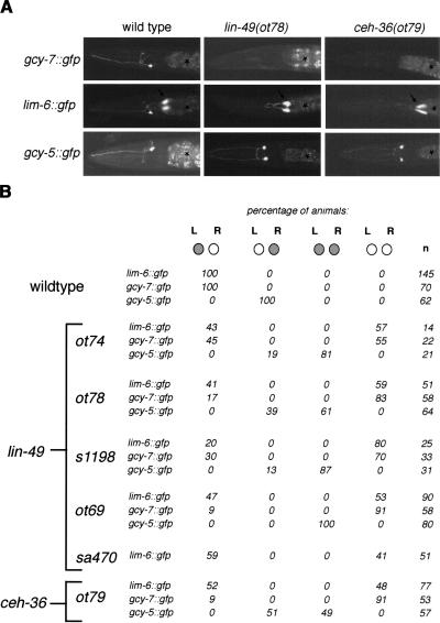
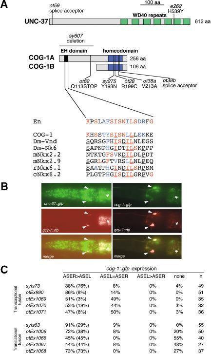
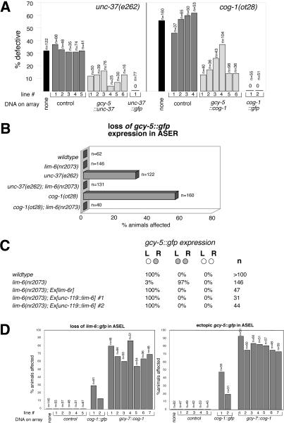
The molecular mechanisms of differential pattern formation along the left/right (L/R) axis in the nervous system are poorly understood. The nervous system of the nematode Caenorhabditis elegans displays several examples of L/R asymmetry, including the directional asymmetry displayed by the two ASE taste receptor neurons, ASE left (ASEL) and ASE right (ASER). Although bilaterally symmetric in regard to all known morphological criteria, these two neurons display distinct chemosensory capacities that correlate with the L/R asymmetric expression of three putative sensory receptor genes, gcy-5, expressed only in ASER, and gcy-6 and gcy-7, expressed only in ASEL. In order to understand the genetic basis of L/R asymmetry establishment, we screened for mutants in which patterns of asymmetric gcy gene expression are disrupted, and we identified a cascade of several symmetrically and asymmetrically expressed transcription factors that are sequentially required to restrict gcy gene expression to either the left or right ASE cell. These factors include the zinc finger transcription factor che-1; the homeobox genes cog-1, ceh-36, and lim-6; and the transcriptional cofactors unc-37/Groucho and lin-49. Specific features of this regulatory hierarchy are sequentially acting repressive interactions and the finely balanced activity of antagonizing positive and negative regulatory factors. A key trigger for asymmetry is the L/R differential expression of the Nkx6-type COG-1 homeodomain protein. Our studies have thus identified transcriptional mediators of a putative L/R-asymmetric signaling event and suggest that vertebrate homologs of these proteins may have similar functions in regulating vertebrate brain asymmetries.


Figures

Figure 1.
Figure 1.
Neural asymmetry in the AWC(L/R) and ASE(L/R) sensory neurons is genetically separable. The top panel is a schematic depiction of the anterior third of a worm, showing the anatomy and gene expression profiles of the AWC(L/R) odorsensory neurons (top left panels) and the ASE(L/R) gustatory neurons (top right panel). The bottom panel shows a list of mutants that were tested for asymmetry defects in each neuron class. Results with mutants that affect asymmetric str-2 expression in AWC(L/R), monitored using the kyIs140 transgene, are taken from Troemel et al. (1999), with the exception of the cog-1, unc-37, and lin-49 results (our own results; the ASE defects are described in more detail in other figures). ASE(L/R) asymmetry was monitored using the lim-6 reporter transgene otIs6 or otIs114. A complete list of mutants tested for effect on asymmetric ASE(L/R) expression patterns is shown in Supplementary Table 1. References for the individual genes and alleles can be found in Troemel et al. (1999) and at http://www.wormbase.org. Notes: 1Ectopic lim-6::gfp expression is observed in a set of neurons other than ASE(L/R). 2The effect of the lin-49(ot74) null allele on str-2 expression (which in wild-type animals is 100% “one AWC on”; Troemel et al. 1999) is as follows: 38% “one AWC on,” 38% “no AWC on,” 14% “two AWC on,” 10% “more than two cells on” (n = 42). In contrast to these pleiotropic effects on AWC(L/R), the effect of lin-49 on lim-6 is qualitatively different; there is a stereotyped gain of ASER fate at the expense of the ASEL fate (see Fig. 5B). Given this qualitative difference and also given the molecular identity of LIN-49 as a broadly expressed transcriptional cofactor with roles in multiple tissue types (see text), we do not consider the effect of lin-49 on AWC and ASE as specific evidence that the AWC and ASE asymmetries are mechanistically related.
Figure 2.
Figure 2.
Mutations resulting in a symmetric, “two ASEL” phenotype (class I phenotype). (A) Expression patterns of gcy-7::gfp, lim-6::gfp, and gcy-5::gfp in wild-type, cog-1(ot28), and unc-37(e262) mutant backgrounds. Adult animals are shown. Both gcy-7::gfp and lim-6::gfp are derepressed in ASER in unc-37 and cog-1 backgrounds, whereas gcy-5::gfp expression is lost in ASER. Arrows denote the excretory gland cell that expresses lim-6::gfp. (B) Quantification of the effects of cog-1 and unc-37 on ASE asymmetry. Animals were scored as adults (with the exception of ot59, which, because of their lethality, were scored as L1s). Note that derepression of lim-6 in ASER is always followed by a concomitant loss of gcy-5 expression.
Figure 5.
Figure 5.
Mutations resulting in a symmetric, “two ASER” phenotype (class II phenotype). (A) Expression patterns of gcy-7::gfp, lim-6::gfp, and gcy-5::gfp in wild-type, lin-49(ot78), and ceh-36(ot79) adult animals. Arrows denote the excretory gland cell that expresses lim-6::gfp. Asterisks denote gut autofluorescence. (B) Quantification of the effects of lin-49 and ceh-36 on ASE asymmetry. Animals were scored as adults (with the exception of s1198 and ot74 animals which, because of their late L1 lethality, were scored as viable L1s). The lackof a perfect correlation between loss of lim-6 expression and gain of gcy-5 expression [e.g., in lin-49(ot69), 100% of animals gain gcy-5 expression, but only 53% lose lim-6 expression] is likely a levels issue; that is, animals whose lim-6 expression levels appear unaffected by reporter gene analysis may have experienced a drop of lim-6 levels below a critical threshold required for gcy-5 repression.
Figure 8.
Figure 8.
Mutations in the che-1 locus eliminate adoption of the ASE(L/R) fate (class I phenotype). (A) ASE(L/R) expression of gcy-7::gfp (otIs3), lim-6::gfp (otIs114), gcy-5::gfp (ntIs1), and flp-6::gfp (otIs125) in adult wild-type and adult che-1 (ot27) mutant animals. ASE-specific expression of all reporters is lost in che-1 mutants. Arrows in the “lim-6::gfp” panel denote the excretory gland cell that expresses lim-6::gfp and the ADF or AFD neuron class in the flp-6::gfp panel; note that gfp expression in neither of these cell types is affected in che-1 mutants. Asterisks denote gut autofluorescence. (B) Quantification of che-1 effects on ASE gene expression patterns. (C) Schematic CHE-1 protein structure (drawn to scale), denoting the position of mutant alleles retrieved from our screen. The deletion breakpoints in ot66 have not been sequenced but are inferred by PCR.
Figure 9.
Figure 9.
Transcriptional cascade regulating ASE(L/R) asymmetry. (A) Summary of expression of transcription factors and their effectors in ASE(L/R). Circles indicate gfp expressing (green)/nonexpressing (not filled) ASEL and ASER cells. The ASE(L/R) patterns of expression of ubiquitously expressed genes (unc-37 and lin-49) are not shown. Notes: 1Low levels of cog-1 activity in ASEL are inferred from reporter gene assays as well as the observation that cog-1 is required in ASEL in a ceh-36 mutant to repress lim-6 expression. 2lim-6 is not required to initiate its own expression but to maintain it (data not shown); the empty circles refer to expression in the adult, after autoregulation has been established. 3flp-6::gfp was used to assess bilaterally symmetric ASE fate. 4See Figure 7C for an explanation of the reappearance of ASE asymmetry. (B) A molecular model for the establishment of asymmetric gcy-5 and gcy-7 expression. Arrows reflect genetic pathway interactions, which subsume either a direct interaction of a protein with the respective transcriptional regulatory elements or the presence of intermediary factors. Different sizes of the COG-1 protein in ASEL versus ASER are meant to reflect different protein levels brought about by differential activation or repression of cog-1 transcription (model #1 or model #2, which are not mutually exclusive). Note that low levels of cog-1 must be present in ASEL because, in a ceh-36 and lin-49 mutant background, a role for cog-1 is revealed in ASEL (Fig. 7). Differential expression of COG-1 in ASEL and ASER is either achieved through differential transcriptional repression in ASEL (model #1) or differential transcriptional activation in ASER (model #2). Because the loss of lim-6 results in activation of gcy-5 expression, we invoke CHE-1 as a potential direct positive regulator of gcy-5 expression, a notion supported by ectopically expressed CHE-1 being able to induce gcy-5 expression (data not shown; Uchida et al. 2003). We and others have also shown that CHE-1 regulates bilaterally symmetric features of ASE fate (Uchida et al. 2003; this paper). CEH-36 and LIN-49 do not regulate these features (data not shown).
Figure 3.
Figure 3.
Mutations in cog-1 and unc-37, two transcription factors expressed in ASE(L/R), cause ASE asymmetry defects. (A) Schematic depiction of the UNC-37 and COG-1 protein structure (drawn to scale) with mutant alleles noted. The blowup of the COG-1 sequence depicts a motif similar to the eh1 domain [alignment extended from Muhr et al. (2001)]. Colored residues indicate conservation to the Engrailed protein (red, identity; blue, conservative substitution), underlining indicates identity (in >50% of aligned sequences) among the NK-type protein that cannot be observed in the Engrailed protein. In contrast to the previously characterized cog-1 alleles sy607 and sy275, which still produce progeny, the ot38 and ot62 alleles, the latter of which only affects the cog-1a splice form (A), are completely sterile. ot28 animals are fertile. The lsy phenotype of ot62, but not any other cog-1 allele, is dominant (23% of ot62/+ animals are lsy; n = 22). cog-1(ot62) animals may produce a truncated COG-1A protein that contains only its transcriptional repressor domain, the eh1 domain, but not its DNA-binding domain. In heterozygous animals, this truncated protein may interfere in a dominant-negative manner with the activity of the wild-type copy of the COG-1A protein. (B) Expression of a translational UNC-37::GFP fusion protein (left panel; Kelly et al. 1997) and a translational COG-1::GFP fusion protein (right panel; transgenic line, syIs63; Palmer et al. 2002) in the ASE neurons in midlarval stage animals (white arrowheads). ASEL and ASER are marked with the ASE(L/R)-expressed reporter rfp transgene otIs131 (Is[gcy-7::rfp]) or otEx445 (Ex[gcy-7::rfp]). Asterisks denote gut autofluorescence. (C) Quantification of cog-1 reporter gene expression in different transgenic lines in ASEL and ASER. The top five lines carry transcriptional reporter fusions that contain the promoter of the cog-1 gene. syIs73 is a chromosomally integrated array (Palmer et al. 2002). The bottom five lines carry translational reporter fusions that contain the promoter as well as all coding sequences of cog-1. otEx1006 rescues the cog-1 mutant phenotype (Fig. 4A; other lines were not tested for rescue). ASER > ASEL indicates stronger gfp fluorescence in ASER compared with ASEL. Numbers in parentheses indicate complete absence of fluorescence in ASEL. In theory, the occasional, less consistent expression of cog-1 in ASEL could be a reporter gene artifact, explained through the titration of an ASEL-specific negative regulator of cog-1 expression. However, genetic evidence described in the text indicates that, in specific mutant backgrounds (ceh-36, lin-49), a function for cog-1 in ASEL is revealed, demonstrating that low levels of cog-1 activity are indeed present in ASEL. All animals contained gcy-7::rfp reporters in the background (otIs131 or otEx445), were photographed at midlarval stages in which gcy-7::rfp reporter is expressed in ASEL and ASER, and were scored for the gfp phenotype as gravid adults.
Figure 4.
Figure 4.
cog-1 and unc-37 act autonomously to repress ASEL fate via repression of lim-6. (A) cog-1 and unc-37 act cell autonomously. Transformation rescue data of the unc-37 and cog-1 mutant defects are shown. Numbers below bars indicate independent transgenic strains. “% defective” refers to the absence of gcy-5::gfp expression in ASER, assessed with the ntIs1 (Is[gcy-5::gfp]) integrated transgene. “cog-1::gfp” (otEx1006, otEx1007; see Materials and Methods) and “unc-37::gfp” (Kelly et al. 1997) are translational gfp fusions in which the respective genomic locus is fused to gfp. cog-1::gfp was injected at 10 ng/μL and gcy-5::cog-1 at 2 ng/μL (lines 1 and 2), 5 ng/μL (lines 3 and 4), and 50 ng/μL (lines 5 and 6). gcy-5::unc-37 was injected at 50 ng/μL. rol-6 was the injection marker. Control lines have the cog-1 or unc-37 coding region replaced with gfp and were generated at 50 ng/μL injected DNA. We explain the ability to rescue gcy-5 expression through supplying cog-1 and unc-37 under control of the gcy-5 promoter by the gcy-5 promoter not being entirely shut off in the respective mutants. (B) cog-1 and unc-37 act through lim-6. Loss of gcy-5::gfp expression (monitored with ntIs1) in ASER in cog-1 and unc-37 is suppressed by removing lim-6 activity. (C) lim-6 is not sufficient to repress gcy-5 expression. gcy-5::gfp (ntIs1) is in the background of all strains. “lim-6r” is a rescuing lim-6 genomic fragment previously described to rescue other lim-6 mutant defects (Hobert et al. 1999) and was injected at 20 ng/μL. unc-119::lim-6 was injected at 20 ng/μL (#1) or 5 ng/μL (#2) and resulting F1s were scored. (D) Converting ASEL to ASER fate through raising the activity of cog-1. cog-1 activity was raised by generating multicopy arrays of a gfp-tagged cog-1 genomic clone (cog-1::gfp) or of a cog-1 cDNA driven by the gcy-7 promoter (gcy-7::cog-1). Arrays were expressed in wild-type animals, except in the right panel, where cog-1::gfp was expressed in a cog-1(ot28) mutant background to assess its rescuing capacity. The left panel shows the repression of lim-6 in ASEL and the right panel shows the concomitant gain of ASER features in ASEL (assessed with ntIs1). Note that cog-1::gfp is a rescuing gfp construct that, as shown in A, rescues the gcy-5 expression in ASER, but, as shown here, also causes ectopic gcy-5 expression in ASEL—both effects that can be attributed to the repression of the lim-6 repressor. Thus, gcy-5 expression is observed in ASEL + ASER in cog-1-overexpressing animals, only in ASER in wild-type animals, and neither in ASEL nor in ASER in cog-1 mutants. cog-1::gfp was injected at 5 ng/μL and gcy-7::cog-1 was injected at 50 ng/μL. As a control, gcy-7::rfp was injected at 50 ng/μL. Animals were scored as adults.
Figure 6.
Figure 6.
Mutations in lin-49 and ceh-36, two transcription factors expressed in ASE(L/R), cause ASE asymmetry defects. (A) The protein structure of LIN-49 and CEH-36 is schematically depicted with mutant alleles noted. The SMART domain search tool (http://smart.embl-heidelberg.de) was used to predict all domains shown here. ceh-36(ot79) is a likely hypomorph. Like the previously characterized s1198 allele (which also displays a lsy phenotype; Fig. 5B), the ot74 allele is an early stop codon in the lin-49 gene that also causes larval lethality; both alleles are likely null alleles (A). ot78 and ot69 are viable, yet ot69 is completely vulvaless and of low brood size. All alleles are recessive for their lsy phenotype. (B) Dendrogram showing the relationship of the homeodomain of CEH-36 to other OTX-type homeodomains (created with ClustalX and NJPlot). The homeodomain of the TTX-3 LIM-type homeodomain protein was used as an outlier. (C) Rescue of ceh-36(ot79) with a genomic ceh-36 fragment and an rfp-tagged ceh-36 construct and of lin-49(ot78) with a gfp-tagged lin-49 genomic fragment. “Control” indicates injection marker (rol-6) alone. Numbers refer to number of transgenic lines. Rescuing constructs were injected at 2.5 ng/μL. Animals were scored as adults. (D) Expression of a ceh-36 transcriptional gfp reporter fusion construct (promoter only) in ASEL and ASER in midlarval stage animals (transgenic line, otEx862). An additional pair of amphid sensory neurons is out of the plane of focus. ASEL and ASER were visualized with otIs131. A translational gfp reporter and a translational rfp reporter, which includes all exons and introns and rescues the mutant phenotype (C), show identical patterns of expression.
Figure 7.
Figure 7.
Genetic interactions between cog-1, unc-37, ceh-36, and lin-49 and lim-6. Animals contained either the integrated lim-6 or gcy-5 reporter arrays (otIs114 or ntIs1) and were scored as adults, except for ot74 animals, which were scored as larvae because of their lethality. The single mutant data is also shown in previous figures and is shown here for comparison. (A) cog-1 and unc-37 antagonize lin-49 and ceh-36 to affect asymmetric ASER properties. Ectopic lim-6::gfp expression in ASER in cog-1 and unc-37 mutants is significantly reduced on reduction of lin-49 or ceh-36 activity (left panel). Consequently, the loss of gcy-5::gfp expression in ASER in cog-1 and unc-37 mutants, presumably caused by ectopic expression of lim-6, is suppressed by lowering ceh-36 or lin-49 activity (right panel). (B) cog-1 function in ASEL. Lowering of ceh-36 or lin-49 activity leads to a reduction of lim-6 expression in ASEL. This reduction requires cog-1 because, in the double mutant, the loss of lim-6 expression is suppressed. (C) Reasoning for the restoration of asymmetric lim-6 expression in cog-1; ceh-36 doubles. The size of the circles correlates with the level of gene activity. Note that the tested ceh-36 and cog-1 alleles are hypomorphic alleles that do not completely eliminate gene function, but merely reduce it (arrow pointing down). Overexpression of cog-1 in ASEL (see Fig. 4D) is reflected with an arrow pointing up.

References

    1. Aasland R., Gibson, T.J., and Stewart, A.F. 1995. The PHD finger: Implications for chromatin-mediated transcriptional regulation. Trends Biochem. Sci. 20: 56–59. - PubMed
    1. Bargmann C.I., Hartwieg, E., and Horvitz, H.R. 1993. Odorant-selective genes and neurons mediate olfaction in C. elegans. Cell 74: 515–527. - PubMed
    1. Brenner S. 1974. The genetics of Caenorhabditis elegans. Genetics 77: 71–94. - PMC - PubMed
    1. Chamberlin H.M. and Thomas, J.H. 2000. The bromodomain protein LIN-49 and trithorax-related protein LIN-59 affect development and gene expression in Caenorhabditis elegans. Development 127: 713–723. - PubMed
    1. Concha M.L., Burdine, R.D., Russell, C., Schier, A.F., and Wilson, S.W. 2000. A nodal signaling pathway regulates the laterality of neuroanatomical asymmetries in the zebrafish forebrain. Neuron 28: 399–409. - PubMed

Publication types

MeSH terms