. 2003 Sep;112(5):693-706.
doi: 10.1172/JCI17464.

Constitutive p40 promoter activation and IL-23 production in the terminal ileum mediated by dendritic cells

Affiliations

Constitutive p40 promoter activation and IL-23 production in the terminal ileum mediated by dendritic cells

Christoph Becker et al. J Clin Invest. 2003 Sep.

Abstract

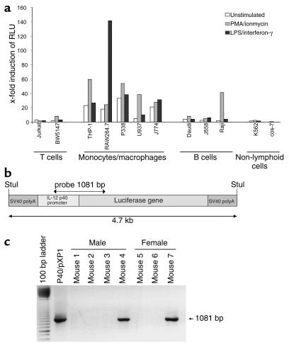
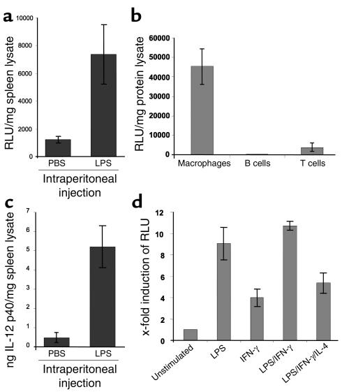
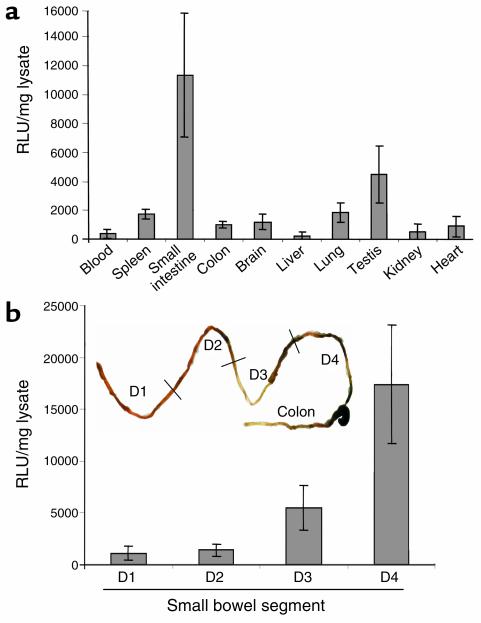
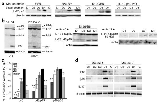
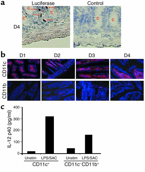
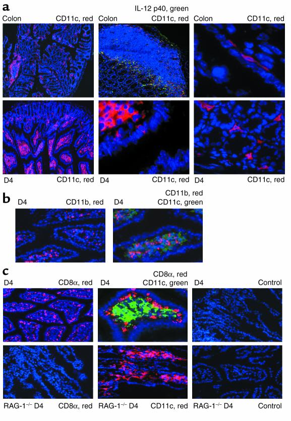
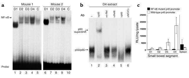
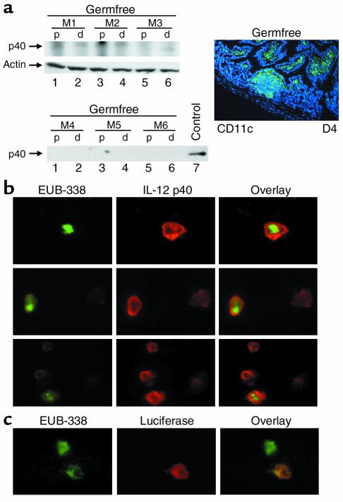
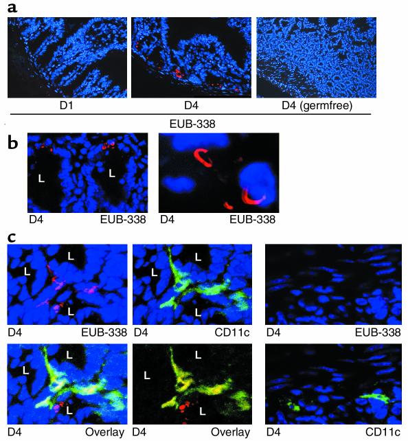
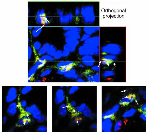
IL-12 p40-related cytokines such as IL-12 p35/p40 heterodimer and IL-23 (p19/p40) are potent regulators of adaptive immune responses. Little is known, however, about the transcriptional regulation of the p40 gene in vivo. In an attempt toward this goal, we have generated transgenic mice expressing firefly luciferase under the control of the IL-12 p40 promoter. High constitutive transgene expression was found in the small intestine only, whereas little reporter gene activity was observed in other tissues. Within the small bowel, constitutive promoter activity was restricted to the terminal ileum and associated with high expression of p40 mRNA as well as p40 and IL-23 p19/p40 proteins. The cells constitutively producing IL-12 p40 were identified as CD8alpha and CD11b double-negative CD11c+ lamina propria dendritic cells (LPDCs) that represent a major cell population in the lamina propria of the small intestine, but not in the colon. FISH directly demonstrated the uptake of bacteria by a subset of LPDCs in the terminal ileum that was associated with p40 expression. Furthermore, little or no p40 protein expression in LPDCs was found in the terminal ileum of germfree mice, indicating a key role of the intestinal flora for constitutive p40 expression. In addition, analysis of transgenic mice with a mutated NF-kappaB target site in the p40 promoter showed a critical role of NF-kappaB for constitutive transgene expression. Our data reveal important functional differences between the mucosal immune systems of the small and large bowel in healthy mice and suggest that the high bacterial load in the terminal ileum activates p40 gene transcription in LPDCs through NF-kappaB. These data suggest a predisposition of the terminal ileum to develop chronic inflammatory responses through IL-23 and thus may provide a molecular explanation for the preferential clinical manifestation of Crohn disease in this part of the gut.


Figures

Figure 1
Figure 1
Generation of IL-12 p40 promoter/luciferase transgenic mice. (a) Reporter gene analysis of the p40/pXP1 reporter gene construct in various cell lines. The IL-12 p40 promoter was cloned as a Stul restriction enzyme fragment upstream of the luciferase reporter gene into the pXP1 vector yielding the p40/pXP1 vector. P40/pXP1 was transfected in various cell lines using the DEAE transfection method. Cells were left untreated or were stimulated for 8 hours with PMA/ionomycin or LPS/IFN-γ, as indicated. Data represent average values of two independent experiments and are presented as fold induction of relative light units (RLU) as compared with the transfection of the empty pXP1 reporter gene vector. (b and c) Map of the luciferase expression cassette and generation of IL-12 p40 promoter/luciferase transgenic mice. A 4.7-kb IL-12 p40 promoter/luciferase expression cassette was used for the generation of transgenic animals. The screening of transgenic mice was performed by isolation of tail DNA and subsequent PCR analysis with a set of construct-specific primers, giving rise to a single band of 1,081 bp. Several founder mice were identified by PCR that were used for subsequent analysis.
Figure 2
Figure 2
Cell-specific and stimulation-dependent expression of the transgene. (a) Upper panel: Luciferase expression in the spleen of control and LPS-injected transgenic mice. Mice were injected intraperitoneally with 200 μg of LPS. After 4 hours, splenic luciferase expression was assessed. Data are shown as mean values of three mice per group ± SD. LPS injection led to increased luciferase activity indicating LPS-dependent p40 promoter activity in spleen cells. Lower panel: IL-12 p40 protein levels in the spleen of the same animals as above were determined by ELISA analysis of splenic cell lysates. Mean values ± SD are shown. (b) Luciferase expression in splenic macrophages, T cells, and B cells of transgenic mice upon stimulation with LPS/SAC (macrophages, B cells) or anti-CD3 plus anti-CD28 Ab’s (T cells) for 24 hours. Date represent mean values ± SD of three mice per group. (c) Luciferase activity in peritoneal macrophages of transgenic mice. Cells were isolated as described in Methods and stimulated for 24 hours as indicated. The data represent mean values ± SD of three independent experiments.
Figure 3
Figure 3
Analysis of various organs from IL-12 p40 promoter/luciferase transgenic mice. (a) Luciferase activity in different tissues of untreated, healthy transgenic mice. Luciferase expression was measured in a standard luminometer after homogenization of whole organs from transgenic mice. Results were normalized to the protein content of the homogenates and are presented as relative light units (RLU) per milligram of protein extract ± SD of five independent experiments. All founder lines of IL-12 p40 promoter/luciferase transgenic mice showed the highest constitutive activity in the small intestine, whereas little activity was seen in the spleen, liver, kidney, heart, and colon. (b) Luciferase activity in different segments of the small intestine from untreated, healthy transgenic mice upon removal of the Peyer’s patches. The small intestine was divided into four segments of equal length (from the proximal segment D1 to the distal segment D4), and luciferase expression was measured in a standard luminometer after homogenization of gut samples from transgenic mice. Results were normalized to the protein content of the homogenates and are presented as relative light units per milligram of protein extract ± SD of three independent experiments. All founder lines of IL-12 p40 promoter/luciferase transgenic mice showed the highest constitutive activity in the distal segment of the small intestine, whereas little activity was seen in proximal segments.
Figure 4
Figure 4
Nonreducing Western blot analysis of gut samples from the small intestine (D1–D4) and colon (C). Samples were analyzed for p40, p40/p19 (IL-23), p40/p35 (IL-12), and p40 homodimer (p40)2 levels. β-Actin staining served as loading control. (a) Western blot for monomeric p40 protein using extracts from different mouse strains. Extracts from IL-12 p40 S129/B6 KO mice served as negative control (far right panel). (b) Analysis of higher molecular weight p40 complexes in FVB and BALB/c mice (left panels) and S129/B6 mice (middle panel, same blot as in a). A marked increase of IL-23 p40/p19, but not IL-12 p40/p19 levels was noted in the distal small bowel as compared with the proximal segments of the small intestine. Right panel: These data were confirmed by Western blot analysis using an IL-23–specific AB (four mice per group, two shown). (c) Densitometry of above p40 Western blots. Data are reported as percentage of expression as compared with D4 (100%). Data represent average values ± SD of six to eight mice per group. Statistically significant differences (*P < 0.05, **P < 0.01, ***P < 0.001) are indicated. (d) RT-PCR analysis of RNA isolated from the small (D1–D4) and large (C) intestine of healthy FVB mice. A marked upregulation in D4 as compared to the proximal small intestine and colon was seen for the mRNAs of p19 and p40, but, importantly, also for the mRNA of IL-17, a recently identified target gene of IL-23 in memory T cells (56).
Figure 5
Figure 5
IL-12 p40 is produced largely by CD11c+ DCs located below the crypts in the lamina propria. (a) Cryosections of transgenic mice were analyzed by immunohistochemistry for luciferase expression. One representative staining for luciferase in the small intestine (D4) of a transgenic animal out of four is shown (left panel). Luciferase-expressing cells were mainly seen in the lamina propria below the crypts (arrows). No staining was seen in sections from transgenic mice treated with an isotype control Ab right panel), the proximal segments of the small intestine (D1, D2), and in healthy nontransgenic control mice (not shown). (b) Detection of CD11c+ and CD11b+ cells in the lamina propria of transgenic mice. More CD11c+ than CD11b+ cells were detected in the lamina propria, suggesting that many cells in the lamina propria of healthy mice carry surface markers of DCs. No differences in the staining patterns of CD11b+ and CD11c+ cells were noted between the proximal and the distal segments of the small bowel. (c) IL-12 p40 cytokine levels in supernatants from CD11c+ enriched DCs and CD11cCD11b+ enriched macrophages isolated from the lamina propria of healthy mice. Lamina propria cells were isolated as described in Methods and purified using the MACS system. To measure IL-12 p40 protein production, 500,000 cells/well were seeded out in 1 ml culture medium in triplicate and incubated in the presence or absence of LPS/SAC. After 24 hours, supernatants were removed and assayed for p40 concentration by ELISA. Unstim, unstimulated.
Figure 6
Figure 6
LPDCs are differentially located in the small intestine as compared with the colon and are largely CD11bCD8α. Location of LPDCs in the colon and small bowel. Cryosections were analyzed by immunofluorescence using the tyramide signal amplification Cy3 or FITC system and fluorescence microscopy. Cryosections were stained for CD11c, IL-12 p40, CD11b, and CD8a, as indicated. Nuclei were counterstained in blue. (a) Differential localization of LPDCs in the colon (upper panels) and small intestine (lower panels). The lamina propria of the small bowel contained many CD11c+ LPDCs as well as some CD11c+ subepithelial cells, whereas few LPDCs were seen in the colon. (b) Staining for CD11b+ (red), CD11c+ (green), and CD11b+ plus CD11c+ (yellow) cells in the distal small bowel of healthy mice, showing that most CD11c+ LPDCs do not coexpress CD11b. (c) Staining for CD11c (green), CD8α (red), and CD11c plus CD8α (yellow) cells in the distal small bowel (D4) of healthy FVB mice (upper panels), showing that most CD11c+ LPDCs do not coexpress CD8a. CD11c+ LPDCs did express MHC class II molecules, as determined by immunohistochemistry, however (not shown). No staining for CD8α was noted in T cell–deficient RAG knockout mice (RAG–/–; lower panels), although many CD11c+; LPDCs were detected in these animals.
Figure 7
Figure 7
Binding of NF-κB to the IL-12 p40 promoter is upregulated in the distal small intestine. (a) EMSA analysis: 40 μg of tissue lysate from the small intestine (D1–D4) of FVB mice was incubated with a radiolabeled probe corresponding to the IL-12 p40 promoter NF-κB site. Protein/DNA complexes were analyzed on a 5% native polyacrylamide gel. Two representative experiments out of four are shown. (b) For supershift analysis, D4 protein lysate was preincubated with 2 μg of Ab’s specific for the indicated transcription factors prior to the addition of radiolabeled probe. The locations of the p50/p65 complex and the p50 supershift are indicated. (c) Constitutive luciferase activity in samples D1 (proximal) to D4 (distal) of the small bowel and spleen of IL-12 p40 wild-type promoter/transgenic mice and NF-κB mutant promoter/luciferase transgenic mice. Luciferase expression was measured in a standard luminometer after homogenization of organ samples of transgenic mice. Results were normalized to the protein content of the homogenate and are presented as relative light units per milligram of protein extract ± SD of three independent experiments with independent founder mice. A striking reduction of luciferase activity was noted in the distal small intestine of mice carrying a loss-of-function mutation in the NF-κB–binding site of the IL-12 p40 promoter as compared with transgenic mice carrying the wild-type p40 promoter upstream of the luciferase gene. In contrast, no reduction of luciferase activity in spleen cell lysates was noted.
Figure 8
Figure 8
Bacteria in the distal small intestine drive constitutive intestinal IL-12 p40 expression. (a) Western blot for IL-12 p40 of gut samples (D1 plus D2: p; D3 to D4: d) derived from three mice (M1, M2, M3) bred under germfree conditions (control), no constitutive p40 protein expression was seen under germfree conditions. The number of CD11c+ LPDCs in the terminal ileum of germfree mice was comparable to that in the ileum of mice bred under specific pathogen-free conditions, as demonstrated by immunofluorescence analysis (right panel) and quantification of fluorescence-positive cells (not shown). (b) FISH analysis on CD11c-enriched lamina propria cells from the distal small intestine of healthy FVB mice using a universal, FITC-labeled eubacterial oligonucleotide probe (EUB-338) and simultaneous immunocytochemical analysis of IL-12 p40 expression. Three representative high-power fields are shown. Image arithmetic (overlay) demonstrated colocalization of bacteria and p40 protein expression in CD11c-enriched lamina propria cells. The CD11c-enriched lamina propria cells did not express CD8α or CD11b, as shown by double-staining analysis (not shown). (c) FISH analysis of CD11c enriched lamina propria cells of the distal small intestine of healthy p40 promoter transgenic FVB mice using a universal, FITC-labeled eubacterial oligonucleotide probe (EUB-338) and simultaneous immunohistochemical analysis of luciferase expression. Colocalization of bacteria and luciferase protein expression was noted in lamina propria cells. No staining was observed using a control probe (NONEUB-338) complementary to EUB-338 to exclude nonspecific binding (not shown).
Figure 9
Figure 9
Bacteria in the distal small intestine are actively taken up by LPDCs in close proximity to the crypts. (a) FISH analysis using a universal, Cy3-labeled eubacterial oligonucleotide probe (EUB-338). Whereas marked staining was seen in the distal small bowel (D4), no staining was detected in the proximal samples (D1). Furthermore, no staining was observed using a control probe (which was NONEUB-338) complementary to EUB-338 to exclude nonspecific binding (not shown). (b) Analysis of the above FISH experiment by confocal laser microscopy. Higher magnifications showed that the bacteria in the terminal ileum (D4) of healthy mice typically had a curved appearance. L, crypt lumen. (c) Immunofluorescence triple-staining analysis of CD11c (FITC: green), Hoechst 3342 (blue), and FISH (Cy3: red), using confocal laser microscopy in D4. CD11c+ plus FISH double-positive cells (yellow, image arithmetic overlay) were identified in the distal small intestine (D4), suggesting uptake of bacteria by LPDCs in vivo. (d) Immunofluorescence triple-staining analysis of CD11c (FITC: green), Hoechst 3342 (blue), and FISH (Cy3: red) using confocal laser microscopy in D4. The majority of CD11c -positive LPDC cells were negative for EUB-338.
Figure 10
Figure 10
Detailed analysis of double staining. Immunofluorescence triple-staining analysis of CD11c (FITC: green), Hoechst 3342 (blue), and FISH (Cy3: red) in D4 using confocal laser microscopy. Orthogonal projections of the sample in three dimensions are shown. Imaging was performed using a multitracking program (Carl Zeiss, Oberkochen, Germany), which eliminates bleed through of other channels. Control images with a single laser activated were collected, and these control images demonstrated no bleed through (not shown). Horizontal and vertical sections from the same images on LPDCs are presented in the upper panels followed by consecutive sections in the lower panels, showing distinct positive red and green areas (arrows) as well as double positivity for CD11c plus FISH in yellow (arrows).

Comment in

Similar articles

Cited by

References

    1. Gubler U, et al. Coexpression of two distinct genes is required to generate secreted bioactive cytotoxic lymphocyte maturation factor. Proc. Natl. Acad. Sci. U. S. A. 1991;88:4143–4147. - PMC - PubMed
    1. Kobayashi M, et al. Identification and purification of natural killer cell stimulatory factor (NKSF), a cytokine with multiple biologic effects on human lymphocytes. J. Exp. Med. 1989;170:827–845. - PMC - PubMed
    1. Stern AS, et al. Purification to homogeneity and partial characterization of cytotoxic lymphocyte maturation factor from human B-lymphoblastoid cells. Proc. Natl. Acad. Sci. U. S. A. 1990;87:6808–6812. - PMC - PubMed
    1. Wolf SF, et al. Cloning of cDNA for natural killer cell stimulatory factor, a heterodimeric cytokine with multiple biologic effects on T and natural killer cells. J. Immunol. 1991;146:3074–3081. - PubMed
    1. D’Andrea A, et al. Production of natural killer cell stimulatory factor (interleukin 12) by peripheral blood mononuclear cells. J. Exp. Med. 1992;176:1387–1398. - PMC - PubMed

Publication types

MeSH terms