. 2003 Sep;112(5):707-16.
doi: 10.1172/JCI17995.

Alimentary tract innervation deficits and dysfunction in mice lacking GDNF family receptor alpha2

Affiliations

Alimentary tract innervation deficits and dysfunction in mice lacking GDNF family receptor alpha2

Jari Rossi et al. J Clin Invest. 2003 Sep.

Abstract

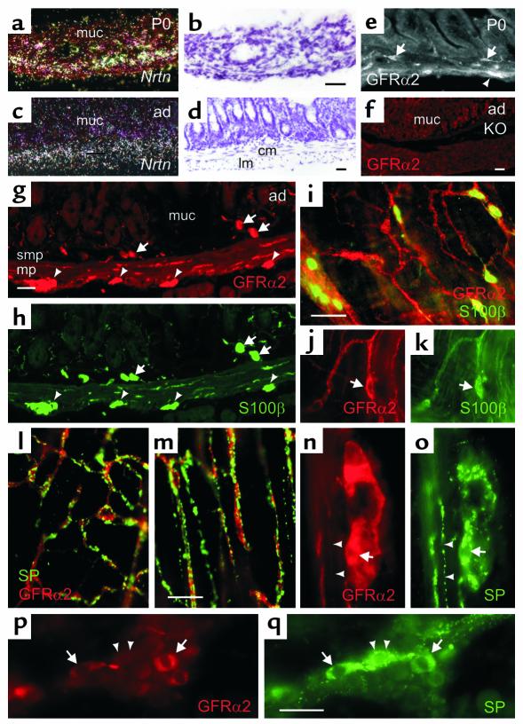
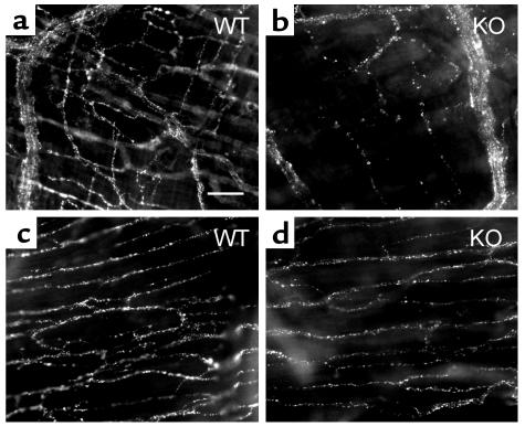
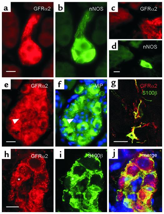
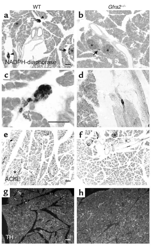
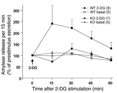
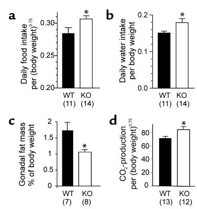
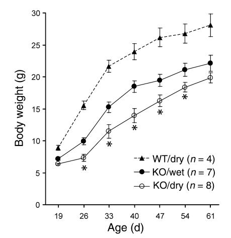
Subsets of parasympathetic and enteric neurons require neurturin signaling via glial cell line-derived neurotrophic factor family receptor alpha2 (GFRalpha2) for development and target innervation. Why GFRalpha2-deficient (Gfra2-/-) mice grow poorly has remained unclear. Here, we analyzed several factors that could contribute to the growth retardation. Neurturin mRNA was localized in the gut circular muscle. GFRalpha2 protein was expressed in most substance P-containing myenteric neurons, in most intrapancreatic neurons, and in surrounding glial cells. In the Gfra2-/- mice, density of substance P-containing myenteric ganglion cells and nerve bundles in the myenteric ganglion cell layer was significantly reduced, and transit of test material through small intestine was 25% slower compared to wild-type mice. Importantly, the knockout mice had approximately 80% fewer intrapancreatic neurons, severely impaired cholinergic innervation of the exocrine but not the endocrine pancreas, and increased fecal fat content. Vagally mediated stimulation of pancreatic secretion by 2-deoxy-glucose in vivo was virtually abolished. Retarded growth of the Gfra2-/- mice was accompanied by reduced fat mass and elevated basal metabolic rate. Moreover, the knockout mice drank more water than wild-type controls, and wet-mash feeding resulted in partial growth rescue. Taken together, the results suggest that the growth retardation in mice lacking GFRalpha2 is largely due to impaired salivary and pancreatic secretion and intestinal dysmotility.


Figures

Figure 1
Figure 1
Expression of Nrtn mRNA and GFRα2 protein in alimentary tract. (ad) Localization of Nrtn mRNA in mouse gut by in situ hybridization. (b and d) Corresponding bright-field images of dark-field images a and c, respectively. Nrtn is strongly expressed in the circular muscle layer at birth (a and b), but its levels decrease in adults (c and d). Distribution of GFRα2 protein in a cross section of the small intestine from newborn (e) and adult (g and n) wild-type mouse. No specific labeling is seen in a corresponding section from adult Gfra2–/– mice (f). (g) GFRα2 is found in myenteric (arrowheads) and to submucosal ganglia (arrows). (h) Immunostaining for S100β shows that glial cells in the muscle and mucosa are also labeled. (im) Whole-mount preparation of duodenum from adult wild-type mouse immunostained for GFRα2 (red) and S100β (i and k, green) or SP (l and m, green), focused at the same level as the myenteric ganglion cells (il) or deep muscular plexus (m). (ik) S100β-positive enteric glia (arrows) express GFRα2. (l and m) SP-containing secondary and tertiary nerve fiber bundles are GFRα2 positive. (nq) Myenteric ganglia stained for GFRα2 (n and p) and SP (o and q) in a cross section (n and o) or in colchicine-treated whole-mount preparation (p and q). Arrowheads point at SP-positive, GFRα2-negative structures. Arrows point at double-labeled neurons. Bars in ah = 100 μm; iq = 50 μm. cm, circular muscle; lm, longitudinal muscle layer; muc, mucosa; ad, adult; P0, postnatal day 0; mp, myenteric plexus; smp, submucosal plexus.
Figure 2
Figure 2
Deficient fine SP-positive fibers in the duodenum of Gfra2–/– mice. (a and b) In adult wild-type mice, in the same focal plane as the myenteric ganglion cells, SP-positive fiber bundles form a network in the spaces between the meshes of the primary plexus (a), whereas in Gfra2–/– (KO) mice, the density of these fiber bundles is severely reduced (b). (c and d) Density of SP-positive fibers in the deep muscular plexus at the innermost level of the circular muscle is comparable between wild type (c) and Gfra2–/– duodenum (d). Bar, 50 μm.
Figure 3
Figure 3
Localization of GFRα2 protein in the pancreas. (a and b) An intrapancreatic ganglion, in which all the neurons are positive for GFRα2 (red) and nNOS (green). (c and d) A single nNOS-positive neuron in the pancreas negative for GFRα2. (e and f) A large intrapancreatic ganglion, in which all VIP-positive (green) neurons express GFRα2 (red). Arrowheads mark VIP-negative, GFRα2-expressing cells that may be glial cells. DAPI-stained nuclei are blue. (g) Confocal optical section image showing colocalization of GFRα2 (red) and S100β (green) in exocrine pancreas. Black asterisk marks a terminal Schwann cell soma. GFRα2 expression is also seen in S100β-negative nerve fibers. (hj) Confocal optical section image through an intrapancreatic ganglion showing the colocalization of GFRα2 (red) and S100β (green) in satellite cells. A satellite cell nucleus is marked by white asterisks. DAPI-stained cell nuclei are blue in the merged image. Bars, 10 μm.
Figure 4
Figure 4
Profound loss of intrapancreatic neurons and cholinergic innervation in Gfra2–/– mouse pancreas. (a) In wild-type animals, intrapancreatic neurons labeled by NADPH-diaphorase histochemistry are seen in numerous ganglia throughout the exocrine pancreas (arrowheads). Single neurons (arrow) were observed also in the vicinity of islets of Langerhans (asterisks). (b) No intrapancreatic ganglia are found in Gfra2–/– mice. The remaining few labeled neurons (arrow) are mainly located near the islets (asterisks). (c) A higher magnification of a typical ganglion in wild-type pancreas that has more than ten labeled neurons. (d) A rare example of a neuron in the exocrine part of pancreas in Gfra2–/– mice. (e) In wild-type animals, AChE-positive cholinergic nerve fibers are seen as scattered fiber bundles. (f) There is a profound loss of cholinergic innervation in the Gfra2–/– pancreas. Note that the cholinergic innervation of islets (asterisks) is less affected compared with the exocrine part. (g and h) Sympathetic innervation of Gfra2–/– mouse pancreas is unchanged compared with wild-type mouse pancreas. TH, tyrosine hydroxylase. Bars, 100 μm.
Figure 5
Figure 5
Reduced 2-DG–stimulated pancreatic secretion in Gfra2–/– mice in vivo. The 2-DG, a central vagal stimulant, induces amylase secretion from wild-type mouse pancreas (filled squares). In Gfra2–/– (KO) mice, 2-DG–stimulated amylase secretion is virtually absent (filled circles). Nonstimulated, basal pancreatic secretion does not change significantly during the 60-minute period (open symbols). The number of animals in each group is indicated in parentheses. The integrated secretion response to 2-DG stimulation (area between stimulated and basal secretion curves) was significantly different between the genotypes (P = 0.01). Average amylase secretion before stimulus was set as 100%. For WT this represents 34 ± 7 MU/15 min; for KO 54 ± 13 MU/15 min.
Figure 6
Figure 6
Food and water intake, gonadal fat mass, and BMR (CO2 production) of adult wild-type and Gfra2–/– (KO) mice under standard dry-pellet feeding. Because average weights differ between the genotypes, food intake and CO2 production values are standardized to (body weight)0.75 (38). (a) Total food intake, expressed as daily intake in grams per (body weight in grams)0.75, is significantly increased. (b) Water intake per body weight is also increased. (c) Gfra2–/– mice have less proportional gonadal fat compared with wild-type mice. Fat weights were measured from 4- to 6-month-old female mice. (d) The BMR, expressed as CO2 production using the equation (μl min–1) per (body weight in grams)0.75, is significantly higher in the knockout than in the wild-type mice. The number of mice used in each experiment is shown in parenthesis below the bars. *P < 0.05.
Figure 7
Figure 7
Partial rescue of the Gfra2–/– (KO) mice growth by wet-mash feeding. Growth curves of Gfra2–/– male mice fed with dry- (open circles) or wet-mash (filled circles) food measured during an 8-week period. Wet-mash food was provided to mice starting from 2 weeks-of-age. Growth of male wild-type mice fed with dry pellets (filled triangles) is shown as a reference. Wild-type mice grew equally well with wet-mash food (not shown). Feeding with wet-mash food significantly enhanced the growth of Gfra2–/– mice compared with Gfra2–/– mice fed with dry pellets. *P < 0.05.

Similar articles

Cited by

References

    1. Manié S, Santoro M, Fusco A, Billaud M. The RET receptor: function in development and dysfunction in congenital malformation. Trends Genet. 2001;17:580–589. - PubMed
    1. Takahashi M. The GDNF/RET signaling pathway and human diseases. Cytokine Growth Factor Rev. 2001;12:361–373. - PubMed
    1. Airaksinen MS, Saarma M. The GDNF family: signalling, biological functions and therapeutic value. Nat. Rev. Neurosci. 2002;3:383–394. - PubMed
    1. Baloh RH, Enomoto H, Johnson EMJ, Milbrandt J. The GDNF family ligands and receptors — implications for neural development. Curr. Opin. Neurobiol. 2000;10:103–110. - PubMed
    1. Rossi J, Tomac A, Saarma M, Airaksinen MS. Distinct roles for GFRα1 and GFRα2 signalling in different cranial parasympathetic ganglia in vivo. Eur. J. Neurosci. 2000;12:3944–3952. - PubMed

Publication types

MeSH terms