. 2003 Sep;112(5):725-35.
doi: 10.1172/JCI17786.

Tetrahydrobiopterin-dependent preservation of nitric oxide-mediated endothelial function in diabetes by targeted transgenic GTP-cyclohydrolase I overexpression

Affiliations

Tetrahydrobiopterin-dependent preservation of nitric oxide-mediated endothelial function in diabetes by targeted transgenic GTP-cyclohydrolase I overexpression

Nicholas J Alp et al. J Clin Invest. 2003 Sep.

Abstract

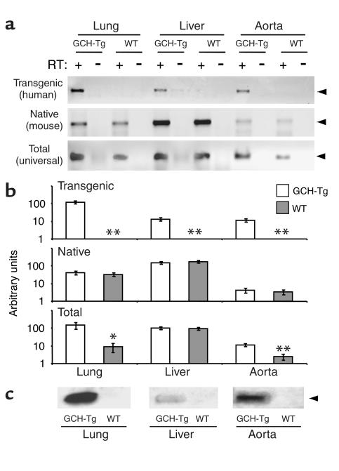
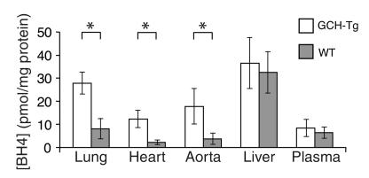
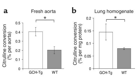
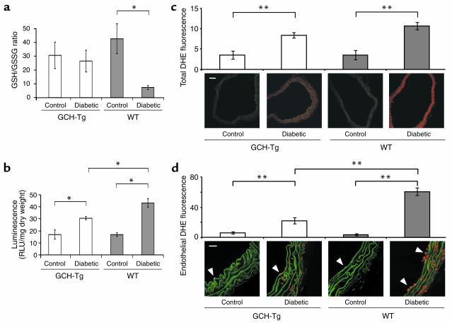
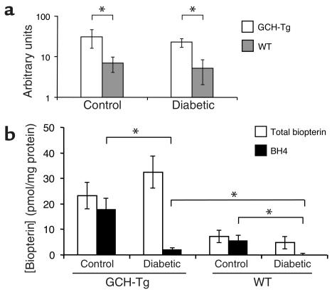
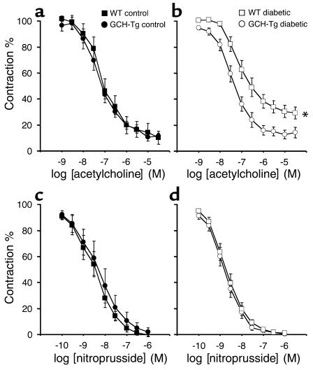
Increased production of reactive oxygen species and loss of endothelial NO bioactivity are key features of vascular disease states such as diabetes mellitus. Tetrahydrobiopterin (BH4) is a required cofactor for eNOS activity; pharmacologic studies suggest that BH4 may mediate some of the adverse effects of diabetes on eNOS function. We have now investigated the importance and mechanisms of BH4 availability in vivo using a novel transgenic mouse model with endothelial-targeted overexpression of the rate-limiting enzyme in BH4 synthesis, guanosine triphosphate-cyclohydrolase I (GTPCH). Transgenic (GCH-Tg) mice demonstrated selective augmentation of endothelial BH4 levels. In WT mice, induction of diabetes with streptozotocin (STZ) increased vascular oxidative stress, resulting in oxidative loss of BH4, forming BH2 and biopterin. Endothelial cell superoxide production in diabetes was increased, and NO-mediated endothelium-dependent vasodilatation was impaired. In diabetic GCH-Tg mice, superoxide production from the endothelium was markedly reduced compared with that of WT mice, endothelial BH4 levels were maintained despite some oxidative loss of BH4, and NO-mediated vasodilatation was preserved. These findings indicate that BH4 is an important mediator of eNOS regulation in diabetes and is a rational therapeutic target to restore NO-mediated endothelial function in diabetes and other vascular disease states.


Figures

Figure 1
Figure 1
(a) Schematic of the murine Tie2 promoter-enhancer/human GTPCH transgene. The murine Tie2 promoter and its intronic enhancer (10 kb) are depicted as gray bars, human GTPCH cDNA is depicted as a white bar, and SV40 poly A signal is depicted as a black bar. Oligonucleotide primers were used to screen genomic DNA for the presence of the transgene. Restriction endonuclease sites for SalI and XbaI are shown. GCH, GTPCH; pA, SV40 poly A; P1 and P2, oligonucleotide primers. (b) Genomic DNA analysis of potential founders. The top panel shows PCR reactions performed on DNA isolated from tail biopsies. The expected 150-bp product (filled arrowhead) was identified in founder mouse 16; linearized pTie2-GCH plasmid DNA was used as a positive control. The bottom panel shows Southern blot analysis performed to confirm the transgenic founders’ genotype and estimate transgene copy number. Founder mouse 16 showed a single hybridization fragment of 8 kb (open arrowhead), suggesting a single chromosomal integration site. The pTie2-GCH plasmid digested with XbaI was used as a positive control.
Figure 2
Figure 2
(a) Evaluation of transgenic and native GTPCH mRNA expression by RT-PCR. The top panel shows human-specific primers to detect transgenic GTPCH mRNA. A PCR product of 181 bp (arrowhead) was detected in lung and liver, and aorta from GCH-Tg but not WT littermates, and only after the RT step (RT+). The middle panel shows mouse-specific primers used to detect native GTPCH mRNA. A PCR product of 181 bp (arrowhead) was detected in lung, liver, and aorta from both GCH-Tg and WT mice. (b) Quantification of GTPCH expression by real-time fluorescent RT-PCR. Shown are means and SEMs of three samples, each quantified in triplicate. Note that arbitrary units are independent among the three graphs. In the top panel, transgenic GTPCH mRNA expression was 10-fold higher in lung than in liver and aorta among GCH-Tg animals but was not detected in WT littermates (**P < 0.01 for comparisons). In the middle panel, native GTPCH mRNA expression in liver was approximately 10-fold higher than in lung and 100-fold higher than in aorta, but similar between GCH-Tg and WT mice for each organ. In the bottom panel, total GTPCH mRNA expression in the lung was approximately 20-fold higher in GCH-Tg than in WT mice (*P < 0.05); in the liver there was no difference in GCH-Tg as compared with WT mice; in the aorta, there was a 5-fold increase in total GTPCH mRNA expression in GCH-Tg as compared with WT mice (**P < 0.01). (c) Immunoblotting with a rabbit anti-human GTPCH polyclonal antibody to detect transgenic GTPCH protein identified a specific 35-kDa band (arrowhead) in lysates from lung, liver, and aorta from GCH-Tg but not WT animals.
Figure 3
Figure 3
BH4 levels in GCH-Tg and WT mouse tissues. Organs and plasma were harvested, and BH4 levels were determined by iodine oxidation in acid and base followed by HPLC. BH4 levels were increased threefold in GCH-Tg lung, heart, and aorta as compared with WT (*P < 0.05, n = 3–4) but not in liver or plasma.
Figure 4
Figure 4
Arginine-to-citrulline conversion as a measure of eNOS activity. (a) Citrulline production was increased twofold in GCH-Tg as compared with WT aortas stimulated with Ca ionophore for 90 minutes (*P = 0.012, n = 5–6). (b) Basal citrulline production was increased in GCH-Tg as compared with WT lung protein homogenate (*P < 0.05, n = 3).
Figure 5
Figure 5
Oxidation of glutathione stores and superoxide generation in aorta from diabetic GCH-Tg and WT mice. (a) The GSH/GSSG ratio was reduced in diabetic WT mice as compared with control WT mice (*P = 0.02, n = 3), but diabetes did not alter the GSH/GSSG ratio in GCH-Tg mice. (b) Lucigenin-enhanced chemiluminescence. Superoxide production in control WT aortas was similar to that in GCH-Tg aortas. Superoxide production was increased more than twofold in diabetic WT aortas as compared with control (*P < 0.05), but this increase was significantly smaller in diabetic GCH-Tg aortas (*P < 0.05 as compared with control and with diabetic WT, n = 4–6). RLU, relative light units. (c) DHE staining for whole-vessel superoxide production. Representative sections are shown (×10), with total red DHE fluorescence expressed in arbitrary units. Aortic DHE fluorescence was increased two- to threefold in both diabetic WT and GCH-Tg mice as compared with their respective controls (**P < 0.01, n = 3). Scale bar: 100 μm. (d) DHE staining for endothelial cell superoxide production. Representative sections are shown (×60), with specific endothelial cell red DHE fluorescence (arrowheads) expressed in arbitrary units. Endothelial DHE fluorescence was increased by more than 17-fold in diabetic WT aorta, whereas endothelial superoxide production in diabetic GCH-Tg aorta was increased only 3.5-fold as compared with control (**P < 0.01, n = 3). Elastic laminas exhibit green autofluorescence. Scale bar: 20 μm.
Figure 6
Figure 6
Effect of diabetes on GTPCH expression and aortic biopterins. (a) Total GTPCH mRNA levels in aorta from diabetic or control GCH-Tg mice and WT littermates were determined by quantitative RT-PCR and plotted on a log scale. Total GTPCH mRNA levels were increased fivefold in GCH-Tg as compared with WT mice (*P < 0.05, n = 3), but diabetes did not affect GTPCH expression in either group. (b) Total biopterin and BH4 levels in aorta from diabetic or GCH-Tg transgenic mice and WT littermates. There was a threefold increase in total biopterin levels in GCH-Tg aortas as compared with WT. Diabetes had no effect on total biopterin levels as compared with control in either group. However, in WT diabetic aortas, BH4 was almost undetectable, and BH4 levels in diabetic GCH-Tg aortas were significantly reduced as compared with control (*P < 0.05, n = 4–6).
Figure 7
Figure 7
Isometric tension studies in aortic rings from diabetic and control GCH-Tg and WT mice (n = 5–8 animals per group). (a) Vessel relaxations to the endothelium-dependent agonist acetylcholine were normal in control GCH-Tg (filled circles) and WT mice (filled squares). (b) Diabetic WT mice (open squares) exhibited impaired endothelium-dependent relaxations as compared with GCH-Tg mice (open circles) (*P = 0.002) and with control WT mice (filled squares, P = 0.048). There was no difference in endothelium-dependent relaxations between diabetic and control GCH-Tg mice (open and filled circles, respectively; P = 0.468). (c and d) Vessel relaxations to the NO donor sodium nitroprusside were identical in all groups of mice.

References

    1. Harrison DG. Cellular and molecular mechanisms of endothelial dysfunction. J. Clin. Invest. 1998;100:2153–2157. - PMC - PubMed
    1. Cosentino F, Hishikawa K, Katusic ZS, Luscher TF. High glucose increases nitric oxide synthase expression and superoxide anion generation in human aortic endothelial cells. Circulation. 1997;96:25–28. - PubMed
    1. Cosentino F, Luscher TF. Tetrahydrobiopterin and endothelial nitric oxide synthase activity. Cardiovasc. Res. 1999;43:274–278. - PubMed
    1. Vasquez-Vivar J, et al. Superoxide generation by endothelial nitric oxide synthase: the influence of cofactors. Proc. Natl. Acad. Sci. U. S. A. 1998;95:9220–9225. - PMC - PubMed
    1. Stuehr DJ. Mammalian nitric oxide synthases. . Biochim. Biophys. Acta. 1999; 1411:217–230. - PubMed

Publication types

MeSH terms