. 2003 Sep 1;162(5):773-9.
doi: 10.1083/jcb.200304069.

Differential requirements for AP-2 in clathrin-mediated endocytosis

Affiliations

Differential requirements for AP-2 in clathrin-mediated endocytosis

Sean D Conner et al. J Cell Biol. .

Abstract

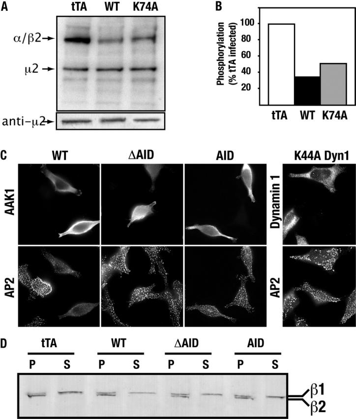
AP-2 complexes are key components in clathrin-mediated endocytosis (CME). They trigger clathrin assembly, interact directly with cargo molecules, and recruit a number of endocytic accessory factors. Adaptor-associated kinase (AAK1), an AP-2 binding partner, modulates AP-2 function by phosphorylating its mu2 subunit. Here, we examined the effects of adenoviral-mediated overexpression of WT AAK1, kinase-dead, and truncation mutants in HeLa cells, and show that AAK1 also regulates AP-2 function in vivo. WT AAK1 overexpression selectively blocks transferrin (Tfn) receptor and LRP endocytosis. Inhibition was kinase independent, but required the full-length AAK1 as truncation mutants were not inhibitory. Although changes in mu2 phosphorylation were not detected, AAK1 overexpression significantly decreased the phosphorylation of large adaptin subunits and the normally punctate AP-2 distribution was dispersed, suggesting that AAK1 overexpression inhibited Tfn endocytosis by functionally sequestering AP-2. Surprisingly, clathrin distribution and EGF uptake were unaffected by AAK1 overexpression. Thus, AP-2 may not be stoichiometrically required for coat assembly, and may have a more cargo-selective function in CME than previously thought.


Figures

Figure 1.
Figure 1.
Structural requirements for AAK1-mediated μ2 phosphorylation. (A) Diagram illustrating the AAK1 constructs used in this study. Baculovirus constructs were GST-tagged at the COOH terminus, whereas adenovirus constructs were HA-tagged at the NH2 terminus (see supplemental methods, available at http://www.jcb.org/cgi/content/full/jcb.200304069/DC1). (B) FSBA-inactivated APs (5.2 μg) were incubated with AAK1–GST fusion proteins, as indicated, in the presence of [γ32P]ATP and phosphorylated protein detected by SDS-PAGE and autoradiography. Assays were performed with 0.25 μM of WT, K74A, or D176A AAK1 and 0.65 μM (2.5×) or 2.5 μM (10×) of the ΔAID mutant. (C) The indicated AAK1–GST fusion proteins were immobilized on glutathione–agarose beads at either ∼0.25 mg/ml (1×) or ∼2.5 mg/ml (10×) and incubated with isolated APs. Bound AP-2 was detected by immunoblot analysis.
Figure 2.
Figure 2.
Full-length AAK1 inhibits Tfn uptake in vivo. tTA-HeLa cells were infected with either control adenovirus encoding the tTA transcription activator or tetracycline-regulatable adenoviruses encoding either full-length WT AAK1 or kinase-inactive mutants (K74A or D176A) (A), or AAK1 fragments (B), as indicated in the legend, and tested for Tfn endocytosis as described in the Materials and methods. Protein expression levels were tested by immunoblot analysis (insets), using pAbs against ΔAID AAK1 and/or the AID AAK1 fragment. Each time point represents the average of three independent experiments ± the standard deviation. (C) The observed inhibition is concentration dependent. Cells were infected with WT AAK1 adenovirus in the presence of increasing concentrations of tetracycline to titrate AAK1 expression, and Tfn endocytosis was assessed as described here.
Figure 3.
Figure 3.
AAK1 globally disrupts AP-2 function. (A) Adenovirally-infected tTA HeLa cells overexpressing WT AAK1, K74A AAK1, or the tTA were labeled in vivo with 32P-orthophosphate. AP-2 was then immunoprecipitated with the mAb AP.6 and analyzed by SDS-PAGE (top). Immunoblot (bottom) with antibodies specific for μ2 (provided by J. Bonifacino, National Institutes of Health, Bethesda, MD) indicates equal loading. (B) Quantitation of phosphorylated large adaptin subunits from whole cell lysates relative to the tTA control. Data shown are representative of three independent experiments. (C) tTA HeLa cells, cultured in the absence of G418, were infected with the indicated AAK1 or dynamin adenovirus constructs. All cells are virally infected, but only those cells that retain tTA express the adenovirus-encoded constructs. Infected cells were fixed with ice-cold acetone and methanol extracted before further processing for immunolocalization of AAK1 using pAbs against either the ΔAID or AID fragment and the mAb AP.6 that recognizes the α-adaptin subunit of AP-2. Samples were visualized by epifluorescence microscopy using a Zeiss Axiophot with an attached Zeiss Axiocam. (D) The distribution of AP-2 in particulate (P) and soluble (S) fractions, obtained as previously described (Damke et al., 1994), was tested in adenovirus-infected cells by immunoblot analysis, using the mAb 100/2 (Sigma-Aldrich).
Figure 4.
Figure 4.
AP-2–deficient clathrin-coated pits are functional for EGF uptake. (A) WT AAK1-infected tTA HeLa cells (cultured as described in the legend to Fig. 3 C) show no defect in the recruitment of clathrin to the plasma membrane or its ability to generate coated pits, as observed by immunolocalization using the mAb X22. (B) EGF internalization was tested in tTA HeLa cells infected with adenoviruses expressing either tTA, WT, or K74A AAK1, or K44A dynamin-1 adenoviruses, as indicated in the legend. Protein expression levels were tested by immunoblot analysis (inset) using pAbs against either the ΔAID AAK1 fragment (lanes 1–3) or against dynamin (lanes 1 and 4). (C) tTA HeLa cells, cultured on coverslips and infected with the indicated adenoviruses, were tested for their ability to internalize rhodamine-conjugated Tfn and Alexa-488–conjugated EGF, simultaneously (Molecular Probes). Cells were incubated in the presence of 2 ng/ml EGF and 4 μg/ml Tfn for 15 min at 37°C. Cells were then transferred to ice, washed to remove unbound ligand, fixed with acetone, and visualized by epifluorescence as described in the legend to Fig. 3. A significant decrease in Tfn accumulation in the endosome is observed in WT AAK1–infected cells relative to AID AAK1– or control-infected cells (arrows), whereas EGF uptake is unaffected.

References

    1. Altschuler, Y., S.M. Barbas, L.J. Terlecky, K. Tang, S. Hardy, K.E. Mostov, and S.L. Schmid. 1998. Redundant and distinct functions for dynamin-1 and dynamin-2 isoforms. J. Cell Biol. 143:1871–1881. - PMC - PubMed
    1. Brodsky, F.M., C.Y. Chen, C. Knuehl, M.C. Towler, and D.E. Wakeham. 2001. Biological basket weaving: formation and function of clathrin-coated vesicles. Annu. Rev. Cell Dev. Biol. 17:517–568. - PubMed
    1. Carter, L.L., T.E. Redelmeier, L.A. Woollenweber, and S.L. Schmid. 1993. Multiple GTP-binding proteins participate in clathrin-coated vesicle- mediated endocytosis. J. Cell Biol. 120:37–45. - PMC - PubMed
    1. Collins, B.M., A.J. McCoy, H.M. Kent, P.R. Evans, and D.J. Owen. 2002. Molecular architecture and functional model of the endocytic AP-2 complex. Cell. 109:523–535. - PubMed
    1. Conner, S.D., and S.L. Schmid. 2002. Identification of an adaptor-associated kinase, AAK1, as a regulator of clathrin-mediated endocytosis. J. Cell Biol. 156:921–929. - PMC - PubMed

Publication types