. 2003 Sep 1;162(5):789-94.
doi: 10.1083/jcb.200302124.

GPI-anchored uPAR requires Endo180 for rapid directional sensing during chemotaxis

Affiliations

GPI-anchored uPAR requires Endo180 for rapid directional sensing during chemotaxis

Justin Sturge et al. J Cell Biol. .

Abstract

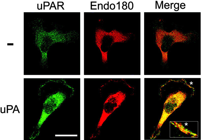
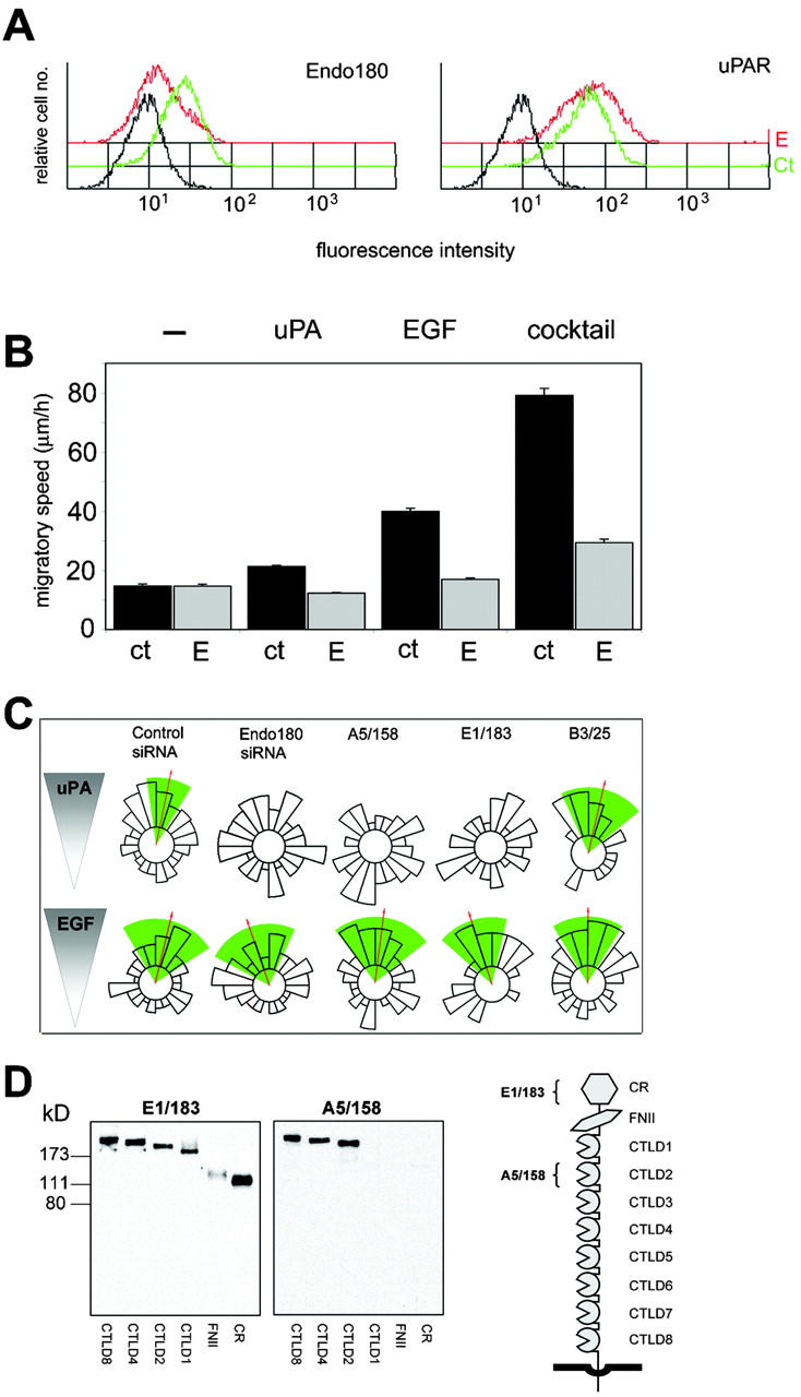
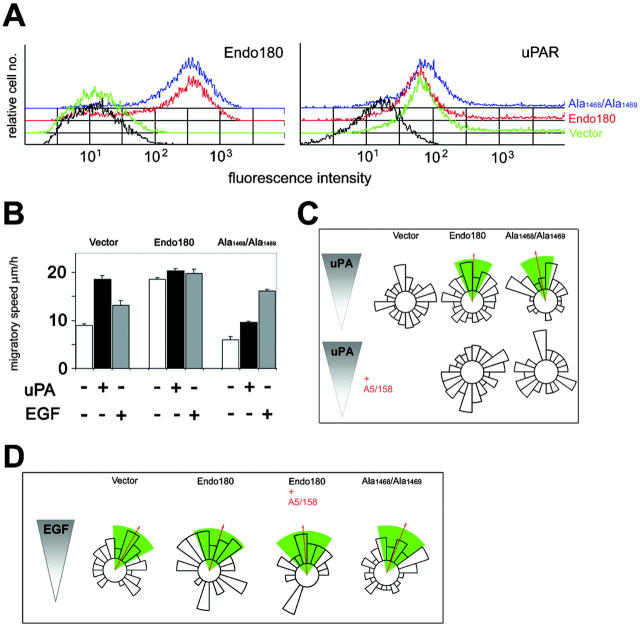
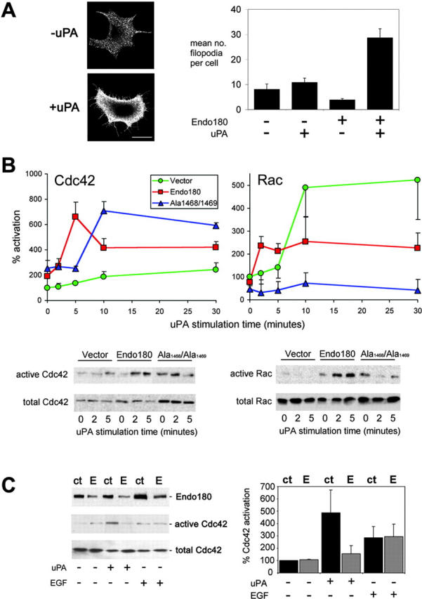
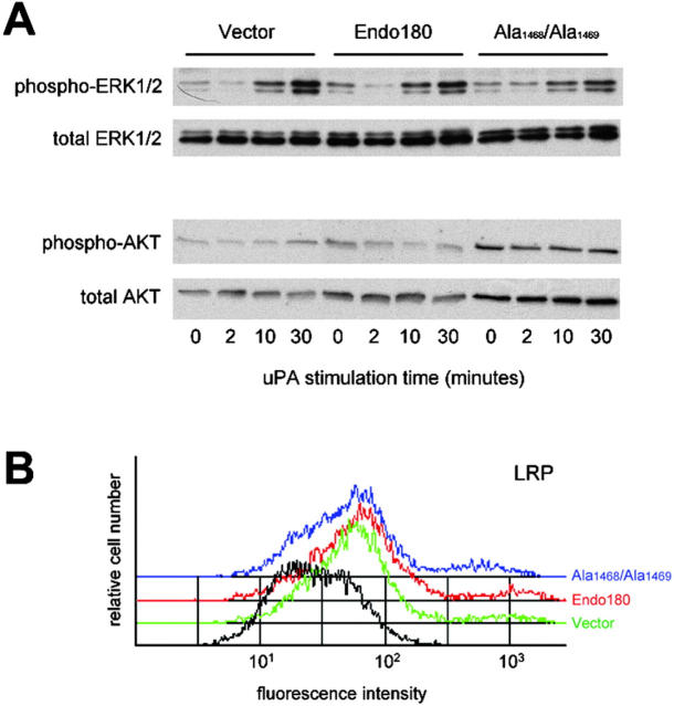
Urokinase-type plasminogen activator (uPA) and its receptor (uPAR) play an important role in cell guidance and chemotaxis during normal and pathological events. uPAR is GPI-anchored and the mechanism by which it transmits intracellular polarity cues across the plasma membrane during directional sensing has not been elucidated. The constitutively recycling endocytic receptor Endo180 forms a trimolecular complex with uPAR in the presence of uPA, hence its alternate name uPAR-associated protein. Here, we demonstrate that Endo180 is a general promoter of random cell migration and has a more specific function in cell chemotaxis up a uPA gradient. Endo180 expression was demonstrated to enhance uPA-mediated filopodia production and promote rapid activation of Cdc42 and Rac. Expression of a noninternalizing Endo180 mutant revealed that promotion of random cell migration requires receptor endocytosis, whereas the chemotactic response to uPA does not. From these studies, we conclude that Endo180 is a crucial link between uPA-uPAR and setting of the internal cellular compass.


Figures

Figure 1.
Figure 1.
Endogenous Endo180 and uPAR colocalize in response to uPA stimulation. MDA-MB-231 cells were stimulated with or without uPA for 30 min, fixed, permeabilized, and stained. Notably the colocalization of uPAR and Endo180 rich clusters in uPA-stimulated cells occurs in areas of new actin polymerization and membrane ruffling (as determined by costaining of uPAR or Endo180 with phalloidin; not depicted). Bar, 20 μm. Asterisk indicates area of interest magnified five times and shown in inset.
Figure 2.
Figure 2.
Endo180 is a general requirement for growth factor–stimulated cell motility but a specific promoter of directionality up a uPA gradient. MDA-MB-231 cells were incubated with control or Endo180 siRNA oligonucleotides or left untreated and analyzed after 48 h. (A) FACS® analysis of Endo180 and uPAR expression levels in control (green) and Endo180 (red) siRNA treated cells. Black lines show binding of second antibody alone to control siRNA treated cells. Endo180 expression in control siRNA treated cells was identical to that in untreated cells (not depicted). (B) Mean migratory speed of unstimulated, uPA- or EGF-stimulated cells, or cells cultured in growth factor cocktail (Opti-MEM medium). ct, control siRNA; E, Endo180 siRNA. Migratory speed values are given as the mean speed for all cells analyzed over the 5-h period ± SEM. (C) Chemotactic response of siRNA or mAb treated cells to a gradient of uPA or EGF. (B and C) Data shown includes analysis of >50 cells pooled from at least three separate experiments. (C) Cell directionality was determined using the Rayleigh test and a horizon distance, which included 50% of all cells assayed for each treatment group. Analysis of the same data using 85% of cells is included in Fig. S1. This highly stringent approach discounts any bias in the calculation of directionality that might be caused by alterations in cell speed. Mean direction of cell migration (red arrow)/95% confidence interval (green wedge). (D) Epitope mapping of anti-Endo180 mAbs. Endo180-Fc constructs were resolved by 10% nonreducing SDS-PAGE and subject to Western blotting. The schematic diagram of Endo180 indicates mAb E1/183 binding to the cysteine-rich domain (CR) and mAb A51/58 binding to CTLD2.
Figure 3.
Figure 3.
Expression of Endo180 is sufficient to confer a sense of direction up a concentration gradient of uPA. (A) FACS analysis of Endo180 and uPAR cell surface expression levels in MCF-7 cells transfected with vector alone (green), wild-type Endo180 (red), or Endo180(Ala1468/Ala1469) (blue). Profiles in black represent vector transfected cells incubated with secondary antibody alone. (B) Mean migratory speed of transfected MCF-7. The directional data are summarized in a circular histogram showing the number of cells lying within each 18° interval. The mean direction and its 95% confidence interval are represented as a red arrow and green sector, respectively. (C, D) Chemotactic response of untreated or mAb A5/158-treated transfected MCF-7 cells in a gradient of uPA or EGF. Data shown in B–D includes analysis of >70 cells pooled from at least three experiments. Cell directionality was determined using the Rayleigh test and a horizon distance which included 50% of all cells assayed for each treatment group. (C and D) The directional data are summarized in a circular histogram showing the number of cells lying within each 18 degree interval. The mean direction and its 95% confidence interval are represented as a red arrow and green sector, respectively.
Figure 4.
Figure 4.
Endo180 promotes filopodia formation in response to uPA and promotes rapid uPA–uPAR mediated activation of Cdc42 and Rac. (A) Endo180 transfected MCF-7 cells were stimulated with or without uPA for 5 min, fixed and immunostained with mAb A5/158 and counterstained with Alexa 555-phalloidin. Bar, 10 μm. Quantitative values shown graphically represent mean ± SEM number of filopodia per cell from a total of 50 randomly chosen cells in two independent experiments. (B) Cdc42 and Rac activation time courses in MCF-7 cells transfected with vector alone (green), Endo180 (red) or Endo180(Ala1468/Ala1469) (blue) and stimulated with uPA. Values represent the mean ± SEM percent change from levels of vector alone cells at zero time point (n = 4–6 experiments). Bottom panels show representative immunoblots. (C) Endo180 expression and Cdc42 activation in siRNA treated MDA-MB-231 cells stimulated with uPA or EGF for 5 min ct, control siRNA; E, Endo180 siRNA. Left panel shows representative blot, right panel shows a graphical representation of mean ± SD percent change from levels in control siRNA treated cells (n = 2 experiments).
Figure 5.
Figure 5.
Endo189 expression does not alter ERK1/2 and Akt phosphorylation or cell surface LRP. (A) ERK1/2 and Akt phosphorylation in MCF-7 cells transfected with vector alone, Endo180 or Endo180(Ala1468/Ala1469) and stimulated with uPA. Blots show phosphorylated ERK1/2 and Akt together with total ERK1/2 and Akt as loading controls. (B) FACS® analysis of LRP cell surface expression levels in MCF-7 cells transfected with vector alone (green), wild-type Endo180 (red) or Endo180(Ala1468/Ala1469) (blue). Profiles in black represent vector transfected cells incubated with secondary antibody alone.

References

    1. Allen, W.E., D. Zicha, A.J. Ridley, and G.E. Jones. 1998. A role for Cdc42 in macrophage chemotaxis. J. Cell Biol. 141:1147–1157. - PMC - PubMed
    1. Baggiolini, M. 2001. Chemokines in pathology and medicine. J. Intern. Med. 250:91–104. - PubMed
    1. Behrendt, N., O.N. Jensen, L.H. Engelholm, E. Mortz, M. Mann, and K. Dano. 2000. A urokinase receptor-associated protein with specific collagen binding properties. J. Biol. Chem. 275:1993–2002. - PubMed
    1. Blasi, F. 1993. Urokinase and urokinase receptor: a paracrine/autocrine system regulating cell migration and invasiveness. Bioessays. 15:105–111. - PubMed
    1. Blasi, F., and P. Carmeliet. 2002. uPAR: A versatile signalling orchestrator. Nat. Rev. Mol. Cell Biol. 3:932–943. - PubMed

Publication types

MeSH terms

Substances