. 2003 Sep;15(9):2165-80.
doi: 10.1105/tpc.013854.

A grape ASR protein involved in sugar and abscisic acid signaling

Affiliations

A grape ASR protein involved in sugar and abscisic acid signaling

Birsen Cakir et al. Plant Cell. 2003 Sep.

Abstract

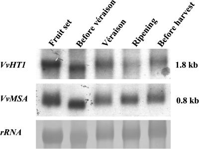
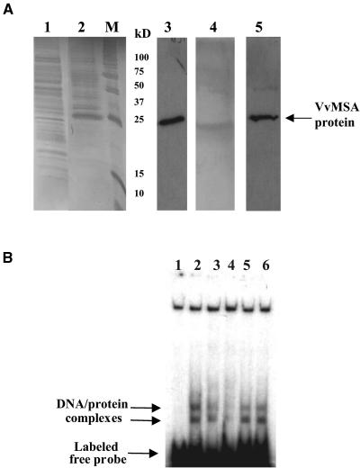
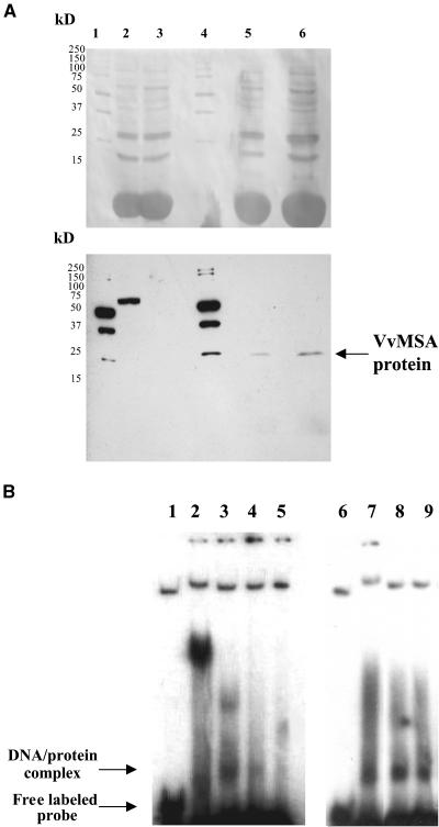
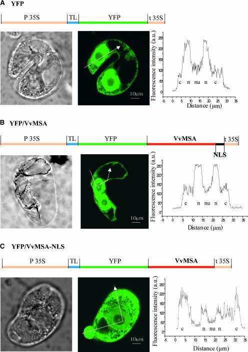
The function of ASR (ABA [abscisic acid]-, stress-, and ripening-induced) proteins remains unknown. A grape ASR, VvMSA, was isolated by means of a yeast one-hybrid approach using as a target the proximal promoter of a grape putative monosaccharide transporter (VvHT1). This promoter contains two sugar boxes, and its activity is induced by sucrose and glucose. VvMSA and VvHT1 share similar patterns of expression during the ripening of grape. Both genes are inducible by sucrose in grape berry cell culture, and sugar induction of VvMSA is enhanced strongly by ABA. These data suggest that VvMSA is involved in a common transduction pathway of sugar and ABA signaling. Gel-shift assays demonstrate a specific binding of VvMSA to the 160-bp fragment of the VvHT1 promoter and more precisely to two sugar-responsive elements present in this target. The positive regulation of VvHT1 promoter activity by VvMSA also is shown in planta by coexpression experiments. The nuclear localization of the yellow fluorescent protein-VvMSA fusion protein and the functionality of the VvMSA nuclear localization signal are demonstrated. Thus, a biological function is ascribed to an ASR protein. VvMSA acts as part of a transcription-regulating complex involved in sugar and ABA signaling.


Figures

Figure 1.
Figure 1.
Amino Acid Alignment of VvMSA and 22 Known ASR Proteins from Different Species Performed with the CLUSTAL Program.
Figure 2.
Figure 2.
Gel Blot Analysis of Grape Genomic DNA. Ten micrograms of genomic DNA was digested with different enzymes: BglII, EcoRI, HindII, or KpnI. The DNA gel blot was hybridized with VvMSA cDNA as an α-32P labeled probe. M, Smart ladder marker DNA.
Figure 3.
Figure 3.
VvMSA and VvHT1 Gene Expression during Grape Development. Gel blot hybridization of RNA from berries at different stages of ripening with VvMSA and VvHT1 probes. Twenty micrograms of total RNA was loaded in each well. Equal loading was checked by staining of 25S rRNA with methylene blue.
Figure 4.
Figure 4.
ABA Induction of VvMSA and VvHT1 Gene Expression in Grape Berry Cell Suspension. (A) RNA gel blot analysis of VvMSA messenger accumulation at 48 and 72 h after ABA treatment (10 μM) in either the presence (+S) or absence (−S) of sucrose. (B) RNA gel blot analysis of VvMSA and VvHT1 transcript amounts at 24, 48, and 72 h after ABA treatment in sucrose-supplemented medium. Twenty micrograms of total RNA was loaded in each well. VvMSA- and VvHT1-specific labeled probes were used for hybridization. Equal loading was checked by 25S rRNA staining with methylene blue.
Figure 5.
Figure 5.
Time Course of VvMSA and VvHT1 Induction by Sucrose in Grape Berry Cell Suspension. RNA gel blot analysis of RNA from grape berry cells with VvMSA and VvHT1 probes. The cells were harvested at the times indicated after transfer in fresh culture medium containing 58 mM sucrose. Twenty micrograms of total RNA was loaded in each well. Equal gel loading was confirmed by staining of 25S rRNA.
Figure 6.
Figure 6.
Production and DNA Binding Activity of 6xHis-Tagged VvMSA. (A) SDS-PAGE of 6xHis-tagged VvMSA visualized by silver staining (lanes 1, 2, and 4) and protein gel blot analysis (lanes 3 and 5) with antibody against RGS 6xHis tag. Lane 1, total protein extract of M15 bacterial cells; lane 2, production of VvMSA protein in transformed M15 cells harvested 3 h after isopropylthio-β-galactoside induction; lane M, molecular mass markers; lane 3, 6xHis-tagged VvMSA protein revealed with the anti-RGS 6xHis antibody in a total protein extract of transformed M15 cells harvested 3 h after isopropylthio-β-galactoside induction; lanes 4 and 5, SDS-PAGE (lane 4) and immunostaining (lane 5) of 6xHis-tagged VvMSA after purification on an Ni-NTA affinity column. (B) In vitro binding activity of purified VvMSA to the target VvHT1 promoter fragment. Lane 1, DNA-labeled free probe corresponding to the 160-bp fragment of the VvHT1 promoter; lane 2, DNA/protein complexes formed by the interaction of VvMSA and the labeled 160-bp fragment of the VvHT1 promoter; lanes 3 and 4, competition assays with the unlabeled 160-bp fragment of the VvHT1 promoter at 100- and 200-fold molar excess, respectively; lanes 5 and 6, competition assays with unrelated unlabeled probe (150 bp) at 100- and 200-fold molar excess, respectively.
Figure 7.
Figure 7.
Production and DNA Binding Activity of the Native VvMSA Protein. (A) SDS-PAGE of TnT proteins produced in reticulocyte lysate and visualized after protein gel blot transfer by Ponceau S staining (top gel) and after immunochemical reaction developed using streptavidin-peroxidase and enhanced chemiluminescence (bottom gel). Lane 1, molecular mass marker; lane 2, TnT-positive control based on luciferase detection; lane 3, TnT-negative control (proteins produced in the absence of the VvMSA cDNA); lane 4, molecular mass marker; lanes 5 and 6, TnT proteins produced in the presence of the VvMSA cDNA. Two microliters of TnT reaction mixture was loaded in lanes 2 and 3, 4 μL was loaded in lane 5, and 8 μL was loaded in lane 6. (B) In vitro binding activity of TnT-produced VvMSA protein to the target fragment of the VvHT1 promoter. Lanes 1 and 6, free DNA-labeled probe corresponding to the 160-bp fragment of the VvHT1 promoter; lane 2, unspecific DNA/protein complex obtained in the control binding assay with proteins produced in the VvMSA cDNA–free TnT system; lanes 3 and 7, complex corresponding to the interaction of the VvMSA protein and the labeled 160-bp fragment of the VvHT1 promoter; lanes 4 and 5, competition assays with the unlabeled 160-bp fragment of the VvHT1 promoter at 100- and 200-fold molar excess, respectively; lanes 8 and 9, competition assays with unrelated unlabeled probe (150 bp) at 100- and 200-fold molar excess, respectively.
Figure 8.
Figure 8.
Interaction of VvMSA with Some Consensus Motifs of the VvHT1 Promoter. (A) Sequences of the positive strain of double-stranded oligonucleotides used in the DNA binding assays. DNA sequences are presented from the 5′ to the 3′ end. cis element sequences are shown in boldface, and when they overlap, the second one is underlined. (B) S3S1 sequence was used as a labeled probe in all experiments, and competition assays were performed with each of three unlabeled probes at 50- and 100-fold molar excess, as indicated. Lane 1, free S3S1 labeled probe; lane 2, unspecific DNA/protein complexes obtained in a control binding assay with proteins produced in the VvMSA cDNA–free TnT system; lanes 3, 6, and 9, complex corresponding to the interaction of VvMSA and S3S1 labeled probe; lanes 4 and 5, competition assay with S3S1 unlabeled oligonucleotide in 50- and 100-fold molar excess, respectively; lanes 7 and 8, competition assay with S3 unlabeled oligonucleotide in 50- and 100-fold molar excess, respectively; lanes 10 and 11, competition assay with S1 unlabeled oligonucleotide in 50- and 100-fold molar excess, respectively. These gel-shift assay results are representative of three to five independent experiments with similar results.
Figure 9.
Figure 9.
Subcellular Localization of the VvMSA Protein. (A) Subcellular compartmentalization of YFP alone, considered as a positive control for free nuclear targeting. (B) Preferential nuclear expression of the YFP-VvMSA fusion protein (C) Strong cytoplasmic localization of the YFP-VvMSA fusion protein, deleted for the VvMSA NLS. Gene structures of the constructs used are detailed above the corresponding micrographs. From left to right are transmission micrographs, confocal fluorescence images, and profiles of fluorescence intensity obtained for different subcellular compartments as indicated by the arrows. c, cytoplasm; n, nucleus; nu, nucleolus; TL, translational enhancer.
Figure 10.
Figure 10.
In Planta Coexpression and Interaction of VvMSA and the VvHT1 Promoter. (A) Effector and reporter constructs obtained in pBI121 and pBI101.1 binary vectors, respectively, and introduced in Agrobacterium tumefaciens. (B) RNA gel blot analysis of VvMSA expression 48 h after infiltration with Agrobacteria carrying VvMSA cDNA of tobacco leaves expressing the pVvHT1-GUS chimerical gene. Two independent tobacco lines were analyzed, and agroinfiltrated areas were compared with buffer-infiltrated areas on the same leaves. (C) Induction of VvHT1 promoter–conferred GUS activity by VvMSA in two independent tobacco transformants. VvMSA's effect on the VvHT1 promoter (Agro+VvMSA) was compared with that of three different controls: untreated areas of the same leaf (Control), buffer-infiltrated areas of the same leaf (Buffer), and areas of the same leaf infiltrated with the Agrobacterium cell suspension without VvMSA cDNA (Agro). MU, 4-methylumbelliferone. (D) Absence of VvMSA's effect on 35S promoter–conferred GUS activity in two independent tobacco transformants carrying the p35S/GUS construct. In both (C) and (D), agroinfiltration results were confirmed in at least four independent experiments for each promoter. Error bars correspond to the se.

Similar articles

Cited by

References

    1. Amitai-Zeigerson, H., Scolnik, P.A., and Bar-Zvi, D. (1994). Genomic nucleotide sequences of tomato Asr2, a second member of the stress/ripening-induced Asr1 gene family. Plant Physiol. 106, 1699–1700. - PMC - PubMed
    1. Arenas-Huertero, F., Arroyo, A., Zhou, L., Sheen, J., and Léon, P. (2000). Analysis of Arabidopsis glucose insensitive mutants, gin5 and gin6, reveals a central role of the plant hormone ABA in the regulation of plant vegetative development by sugar. Genes Dev. 14, 2085–2096. - PMC - PubMed
    1. Atanassova, R., Leterrier, M., Gaillard, C., Agasse, A., Sagot, E., Coutos-Thévenot, P., and Delrot, S. (2003). Sugar-regulated expression of a putative hexose transport gene in grape. Plant Physiol. 131, 326–334. - PMC - PubMed
    1. Bernard, M., Dinet, V., and Voisin, P. (2001). Transcriptional regulation of the chicken hydroxyindole-O-methyltransferase gene by the cone-rod homeobox-containing protein. J. Neurochem. 79, 248–257. - PubMed
    1. Blouin, J., and Guimberteau, G. (2000). Maturité. In Maturation et Maturité des Raisins. (Bordeaux, France: Féret), pp. 29–31.

Publication types

MeSH terms

Associated data