. 2003 Sep;15(9):2181-91.
doi: 10.1105/tpc.012849.

The Arabidopsis NPR1 disease resistance protein is a novel cofactor that confers redox regulation of DNA binding activity to the basic domain/leucine zipper transcription factor TGA1

Affiliations

The Arabidopsis NPR1 disease resistance protein is a novel cofactor that confers redox regulation of DNA binding activity to the basic domain/leucine zipper transcription factor TGA1

Charles Després et al. Plant Cell. 2003 Sep.

Abstract

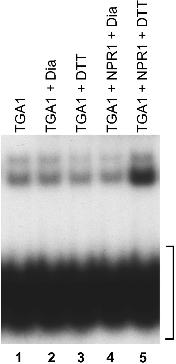
The Arabidopsis NPR1 protein is essential for regulating salicylic acid-dependent gene expression during systemic acquired resistance. NPR1 interacts differentially with members of the TGA class of basic domain/Leu zipper transcription factors and regulates their DNA binding activity. Here, we report that although TGA1 does not interact with NPR1 in yeast two-hybrid assays, treatment with salicylic acid induces the interaction between these proteins in Arabidopsis leaves. This phenomenon is correlated with a reduction of TGA1 Cys residues. Furthermore, site-directed mutagenesis of TGA1 Cys-260 and Cys-266 enables the interaction with NPR1 in yeast and Arabidopsis. Together, these results indicate that TGA1 relies on the oxidation state of Cys residues to mediate the interaction with NPR1. An intramolecular disulfide bridge in TGA1 precludes interaction with NPR1, and NPR1 can only stimulate the DNA binding activity of the reduced form of TGA1. Unlike its animal and yeast counterparts, the DNA binding activity of TGA1 is not redox regulated; however, this property is conferred by interaction with the NPR1 cofactor.


Figures

Figure 1.
Figure 1.
NPR1 Interacts with TGA1 in Arabidopsis Leaves. (A) Schemes of the constructs used for the plant two-hybrid assays. Promoters are shown in white boxes. CaMV 35S indicates the double Cauliflower mosaic virus 35S:Alfalfa mosaic virus promoter. 5X UASGAL4 indicates a promoter composed of a multimerized (five elements) Gal4 upstream activating sequence fused to a minimal TATA box and the Ω translational enhancer from the Tobacco mosaic virus. Coding sequences are shown in dark and light gray boxes. GAL4 DB indicates the GAL4 DNA binding domain. VP16 TA indicates the constitutive transactivation domain of viral particle 16. All constructs possess the polyadenylation signal from the nopaline synthase gene (not shown). The 35S:Renilla construct is an internal reference to normalize transfection efficiency. The construct containing the NPR1:VP16 TA fusion was transfected into untreated leaves along with the reporter and internal standard constructs and was given an arbitrary value of 1 relative luciferase unit ± 1 sd. (B) Histogram illustrating the level of interaction between TGA1 and NPR1 or nim1-2 in Arabidopsis leaves treated with water (white bars) or in leaves treated for 24 h with 1 mM SA (gray bars). As controls, all Gal4 DB and VP16 TA constructs also were transfected separately with the reporter and internal standard constructs. Values represent averages ± 1 sd.
Figure 2.
Figure 2.
Identification of a 30–Amino Acid Region in TGA2 That Determines the Potential for NPR1 Interaction. At left are schemes of TGA1, TGA2, and the proteins encoded by various TGA1:TGA2 chimeric genes used to assess interaction with NPR1. All proteins were expressed as fusions to the Gal4 TA. Numbers indicate amino acid positions within the wild-type TGA1 and TGA2 proteins. At right are results of the interaction with NPR1:DB (NPR1) in yeast using the X-GAL filter test, which monitors the activation of the lacZ reporter gene. “Yes” indicates that blue color was detected after 1 h, and “No” indicates that there was no blue color after 16 h.
Figure 3.
Figure 3.
Cys-260 and Cys-266 in TGA1 Determine the Potential for NPR1 Interaction. (A) Portion of a multiple alignment covering a region of TGA2 involved in interaction with NPR1. The alignment is composed of seven Arabidopsis TGA factors that have been tested in directed yeast two-hybrid assays for interaction with NPR1 (Després et al., 2000). Numbers indicate amino acid positions within wild-type TGA1. (B) At left are schemes of the region of TGA1 and TGA4 shown in (A) as well as of two site-directed mutants (TGA1 SDM and TGA4 SDM) in which Cys residues were replaced by corresponding residues present in TGA2. Numbers indicate amino acid positions within the wild-type TGA1 protein. At right are results of the interaction of the full-length wild-type or site-directed mutant proteins with NPR1 in yeast using the X-GAL filter test, which monitors the activation of the lacZ reporter gene. “Yes” indicates that blue color was detected after 1 h, and “No” indicates that there was no blue color after 16 h. (C) Histogram illustrating the level of interaction between TGA1 SDM and NPR1 or nim1-2 in Arabidopsis leaves treated with water (white bars) or in leaves treated for 24 h with 1 mM SA (gray bars). As a control, TGA1 SDM in the Gal4 DB vector was transfected separately with the reporter and internal standard constructs. Values for the VP16 TA constructs transfected separately with the reporter and internal standard constructs are presented in Figure 1B. Values represent averages ± 1 sd.
Figure 4.
Figure 4.
Determination of the in Vivo Redox Status of Cys Residues Present in TGA1. (A) Flow chart of the method used to determine the in vivo redox status of Cys residues in TGA1. P1 and P2 represent proteins 1 and 2, respectively. SH represents a reduced Cys, and S-S represents a disulfide bridge between two Cys residues. MPB is an oxidizing alkylating agent that is biotinylated; it is used to modify and biotinylate free SH groups. NEM (N-ethylmaleimide) is an oxidizing alkylating agent used to block free SH groups. DTT is a reducing agent. After Cys residues have been modified by MPB, TGA1 is purified by immunoprecipitation, and the immunoprecipitated protein is blotted and reacted with a streptavidin: Alexa Fluor 680 (AF680) conjugate that will interact with biotin moieties and fluoresce at 702 nm after excitation at 679 nm. (B) Blot analysis of TGA1 immunoprecipitate used to assess the in vivo redox status of Cys residues of TGA1 expressed in yeast cells. TGA1 was expressed as a Gal4 TA fusion, and the immunoprecipitation was performed using an anti-FLAG antibody linked to agarose beads. Lane 1, immunoprecipitate from proteins labeled for reduced Cys residues (SH); lane 2, immunoprecipitate from proteins labeled for oxidized Cys residues (S-S). (C) Blot analysis of TGA1 immunoprecipitate used to assess the in vivo redox status of Cys residues of endogenous TGA1 present in cells of Arabidopsis leaves. The immunoprecipitation was performed using an anti-TGA1/TGA4 antibody raised against a peptide common to TGA1 and TGA4. Lanes 1 and 3, immunoprecipitates from proteins labeled for reduced Cys residues (SH); lanes 2 and 4, immunoprecipitate from proteins labeled for oxidized Cys residues (S-S). Lanes 1 and 2 contain proteins from untreated Arabidopsis, whereas lanes 3 and 4 contain proteins from Arabidopsis treated for 24 h with SA. (D) Specificity of the anti-TGA1/TGA4 antibody used in the immunoprecipitations. TGA1, TGA2, TGA3, and TGA4 were translated and radiolabeled in vitro and were immunoprecipitated using the anti-TGA1/TGA4 antibody. Lanes 1 to 4, 10% of the input radiolabeled TGA factors; lanes 5 to 8, immunoprecipitates (IP).
Figure 5.
Figure 5.
An Intramolecular Disulfide Bridge in TGA1 Affects Its Electrophoretic Mobility. (A) Oxidized TGA1 was either reduced with 2-mercaptoethanol (lane 1) or left in its oxidized form (lane 2) before loading for SDS-PAGE. Reduced TGA1 migrates with an apparent molecular mass of 47.5 kD. An intermolecular disulfide bridge between two TGA1 monomers would have an apparent molecular mass of ∼95 kD. At right is a close-up of the gel at left emphasizing the different electrophoretic mobilities of reduced (lane 1) and oxidized (lane 2) TGA1. (B) Oxidized TGA1, containing a Cys-to-Asn mutation at position 260, was either reduced with 2-mercaptoethanol (lane 1) or left in its oxidized form (lane 2) before loading for SDS-PAGE. The protein migrates with an apparent molecular mass of 47.5 kD. At right is a close-up of the gel at left showing that the protein has the same electrophoretic mobility under both reduced (lane 1) and oxidized (lane 2) conditions.
Figure 6.
Figure 6.
Binding of Reduced TGA1 to the as-1 Element Is Enhanced by NPR1. Electrophoretic mobility shift assay using untreated (lane 1), diamide-treated (Dia; lanes 2 and 4), or DTT-treated (lanes 3 and 5) in vitro–translated TGA1 and the as-1 DNA element as a probe (lanes 1 to 5). NPR1 was added to the TGA1–as-1 reaction (lanes 4 and 5). The bracket indicates the position of the free probe. An equal amount of reticulocyte lysate extract was used in all lanes.
Figure 7.
Figure 7.
A Working Model Depicting the Indirect Redox Control of NPR1 on TGA1 DNA Binding Activity. In resting Arabidopsis cells, oxidized TGA1 forms an intramolecular disulfide bridge, which alters its conformation and prevents interaction with NPR1. In the absence of NPR1 interaction, the binding of TGA1 to the cognate cis element is low (dotted arrow). When SA accumulates inside Arabidopsis cells, TGA1 becomes reduced and adopts a conformation that allows interaction with NPR1. The binding of NPR1 to TGA1 stimulates its DNA binding activity to the cognate cis element. TGA1 binds DNA as a dimer; therefore, it is represented as a homodimer.

Similar articles

Cited by

References

    1. Abate, C., Patel, L., Rauscher, F.J., III, and Curran, T. (1990). Redox regulation of fos and jun DNA-binding activity in vitro. Science 249, 1157–1161. - PubMed
    1. Alvarez, M.E., Pennell, R.I., Meijer, P.J., Ishikawa, A., Dixon, R.A., and Lamb, C. (1998). Reactive oxygen intermediates mediate a systemic signal network in the establishment of plant immunity. Cell 92, 773–784. - PubMed
    1. Arabidopsis Genome Initiative (2000). Analysis of the genome sequence of the flowering plant Arabidopsis thaliana. Nature 408, 796–815. - PubMed
    1. Aravind, L., and Koonin, E.V. (1999). Fold prediction and evolutionary analysis of the POZ domain: Structural and evolutionary relationship with the potassium channel tetramerization domain. J. Mol. Biol. 285, 1353–1361. - PubMed
    1. Bayer, E.A., Safars, M., and Wilchek, M. (1987). Selective labeling of sulfhydryls and disulfides on blot transfers using avidin-biotin technology: Studies on purified proteins and erythrocyte membranes. Anal. Biochem. 161, 262–271. - PubMed

Publication types

MeSH terms