. 2003 Sep 15;31(18):e107.
doi: 10.1093/nar/gng095.

Rapid generation of gene disruption constructs by in vitro transposition and identification of a Dictyostelium protein kinase that regulates its rate of growth and development

Affiliations

Rapid generation of gene disruption constructs by in vitro transposition and identification of a Dictyostelium protein kinase that regulates its rate of growth and development

Tomoaki Abe et al. Nucleic Acids Res. .

Abstract

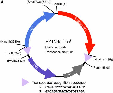
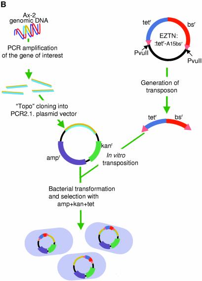
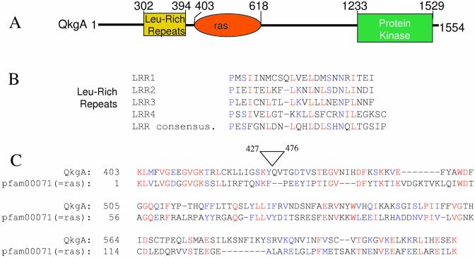
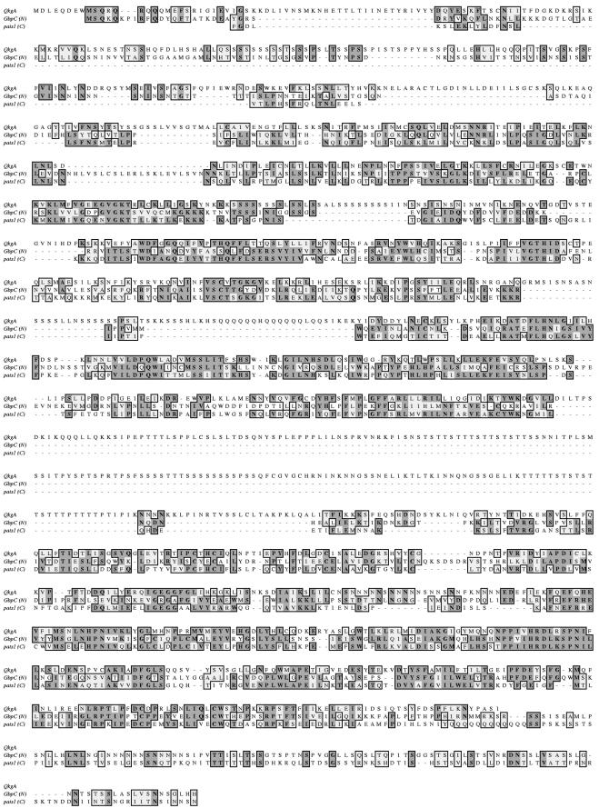
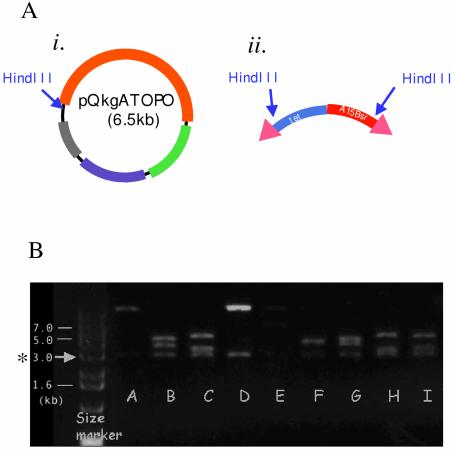
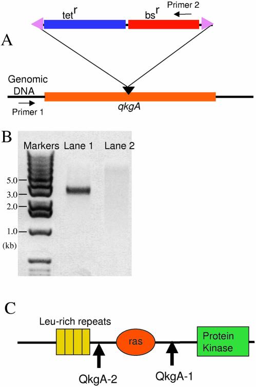
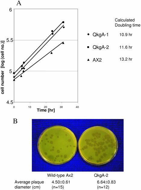
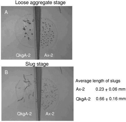
We describe a rapid method for creating Dictyo stelium gene disruption constructs, whereby the target gene is interrupted by a drug resistance cassette using in vitro transposition. A fragment of genomic DNA containing the gene to be disrupted is amplified by PCR, cloned into a plasmid vector using topoisomerase and then employed as the substrate in an in vitro Tn5 transposition reaction. The transposing species is a fragment of DNA containing a Dictyostelium blasticidin S resistance (bs(r)) cassette linked to a bacterial tetracycline resistance (tet(r)) cassette. After transposition the plasmid DNA is transformed into Escherichia coli and clones in which the bs(r)-tet(r) cassette is inserted into the Dictyostelium target DNA are identified. To demonstrate its utility we have employed the method to disrupt the gene encoding QkgA, a novel protein kinase identified from the Dictyostelium genome sequencing project. QkgA is structurally homologous to two previously identified Dictyostelium kinases, GbpC and pats1. Like them it contains a leucine-rich repeat domain, a small GTP-binding (ras) domain and a MEKK domain. Disruption of the qkgA gene causes a marked increase in growth rate and, during development, aggregation occurs relatively slowly to form abnormally large multicellular structures.


Figures

Figure 1
Figure 1
The gene disruption method. (A) Map of the plasmid EZTN:tetr-bsr. The EZTN:tetr-bsr plasmid generates a transposable blasticidin resistance (bsr) cassette that can be used to create Dictyostelium gene disruptants. In order to create EZTN:tetr-bsr, the bacterial tetr cassette, isolated from pBR322, and the Dictyostelium Actin15Bsr cassette, isolated from Actin15ΔBamBsr, were cloned into the EZ::TNpMOD2 vector (Epicentre, USA) as indicated. The 19 bp Tn5 transposon recognition sequences are represented as triangles and their sequence is shown below the map. The plasmid has PvuII sites flanking the transposable element and the transposable blasticidin resistance cassette is usually generated by PvuII digestion of the construct. (B) Diagrammatic representation of the scheme for gene disruption. A fragment of genomic DNA is isolated by PCR and inserted, by topoisomerase cloning, into pCR2.1-TOPO, a vector that confers resistance to ampicillin and kanamycin. In vitro transposition is then performed using transposon DNA prepared as in (A). The resultant DNA molecules are recovered by transformation into E.coli using a triple drug selection, with ampicillin, kanamycin and tetracycline.
Figure 1
Figure 1
The gene disruption method. (A) Map of the plasmid EZTN:tetr-bsr. The EZTN:tetr-bsr plasmid generates a transposable blasticidin resistance (bsr) cassette that can be used to create Dictyostelium gene disruptants. In order to create EZTN:tetr-bsr, the bacterial tetr cassette, isolated from pBR322, and the Dictyostelium Actin15Bsr cassette, isolated from Actin15ΔBamBsr, were cloned into the EZ::TNpMOD2 vector (Epicentre, USA) as indicated. The 19 bp Tn5 transposon recognition sequences are represented as triangles and their sequence is shown below the map. The plasmid has PvuII sites flanking the transposable element and the transposable blasticidin resistance cassette is usually generated by PvuII digestion of the construct. (B) Diagrammatic representation of the scheme for gene disruption. A fragment of genomic DNA is isolated by PCR and inserted, by topoisomerase cloning, into pCR2.1-TOPO, a vector that confers resistance to ampicillin and kanamycin. In vitro transposition is then performed using transposon DNA prepared as in (A). The resultant DNA molecules are recovered by transformation into E.coli using a triple drug selection, with ampicillin, kanamycin and tetracycline.
Figure 2
Figure 2
Domain analysis of the qkgA gene. (A) Schematic of the domain structure of QkgA. The qkgA gene contains a domain comprised of four copies of the leucine-rich repeat (LRR) sequence, a ras domain and a kinase domain. The approximate boundaries of each domain are indicated. (B) Alignment of the leucine-rich repeat (LRR) sequences of QkgA. The four leucine-rich repeats of QkgA are shown aligned with a consensus LRR sequence (9). (C) Alignment of QkgA sequences with pfam0007, a ras domain from the Pfam database. The search (query) sequence was a manually deleted version of region 427–476 of qkgA. This deletion removes a very serine-rich, ‘simple sequence’ region. The ‘subject’ sequence is pfam00071. The E value with this pfam member is 6e–07.
Figure 3
Figure 3
In a BLAST search at NCBI using the QkgA sequence (databases updated 9 April, 2003; all non-redundant GenBank CDS translations + PDB + SwissProt + PIR + PRF; 1 442 039 sequences; 463 592 631 total letters), GbpC and pats1 were the most closely related proteins. There were also many other proteins with homology to the predicted kinase domain of QkgA but the top hits were all dual specificity kinases, not (as the original annotation would have predicted) receptor tyrosine kinases. This is a Clustal W analysis showing regions of homology between QkgA, GbpC and pats1. Both the pats1 and the GbpC proteins are significantly larger than QkgA. In order to simplify the alignment by Clustal W (which is length sensitive) only the homologous subregions of pats1 [1400–2500 = pats1(C)] and GbpC [1–1300 = GbpC(N)] were used.
Figure 4
Figure 4
Transposition of the bsr cassette into qkgA. (A) A 2.6 kb fragment of qkgA (encoding amino acids M1–I980) was PCR amplified and cloned into pCR2.1-TOPO, to create the target for disruption. The orange line represents the cloned qkgA fragment and its single HindIII site is marked by an arrow (i). Two HindIII sites located near each end of the bsr cassette are also marked with arrows (ii) (described in Fig. 1A). (B) Examples of HindIII digests of minipreps prepared from the bacteria transformed with the product of an in vitro transposition of the cassette (ii) into qkgATOPO (i). Nine examples are shown in lanes A–I. Since there is a HindIII site located very near each end of the transposable bsr cassette (ii), one of the bands has a constant size of 3 kb (marked with an asterisk).
Figure 5
Figure 5
PCR confirmation of insertion of the transposable bsr cassette in the qkgA locus and determination of the transposon insertion point. (A) A schematic diagram of the qkgA locus and the inserted bsr cassette. The orange line indicates that part of the qkgA open reading frame which was used as the target for disruption. The position of insertion of the bsr cassette is marked with a black triangle. The two primers used for PCR confirmation are indicated with arrows and marked primers 1 and 2. (B) Two examples of clones. Genomic DNA was isolated from the clones grown in selective medium containing blasticidin. Lanes 1 and 2 show the results of PCR with the primers described in (A). The sample analysed in lane 1 shows successful insertion of the selection cassette into qkgA, while the absence of a band in lane 2 suggests a non-homologous insertion of the cassette elsewhere in the chromosome. (C) Following PCR confirmation of transposon insertion in the genomic target, the insertion point was estimated from the size of the amplicons. The arrows indicate the approximate insertion points of two separate clones, QkgA-1 and QkgA-2.
Figure 6
Figure 6
Growth rate comparison of qkgA and parental Ax-2 cells. (A) Cells growing in HL-5 medium. The vertical axis represents the common logarithm of cell number and the horizontal axis represents the time of culture in hours starting from the first sampling point. Triangles, Ax-2 wild-type cells; diamonds, qkgA strain1 C3; square, qkgA strain 2. The doubling time was calculated after curve fitting. (B) Cells feeding on bacterial lawns. Pre-cultured cells of the qkgA mutant clone QkgA-2 and wild-type control strain Ax-2 in HL-5 medium were harvested, washed in KK2 (16.5 mM KH2PO4, 3.8 mM K2HPO4, pH 6.2) and co-cultured with K.aerogenes on SM plates. After 5 days of incubation, the size of the plaques was measured.
Figure 7
Figure 7
Parallel development of qkgA mutant and parental Ax-2 cells. qkgA mutant strain QkgA-2 and the wild-type control Ax-2 were monitored side-by-side with time-lapse recording. The figure shows two frames of the recorded film. The left-hand side of the frame shows the QkgA-2 mutant cells and the right-hand side shows the Ax-2 cells. (A) Frame for 9 h; (B) frame for 17 h. The average lengths of the slugs were measured using the data in (B).

References

    1. Hamer L., DeZwaan,T.M., Montenegro-Chamorro,M.V., Frank,S.A. and Hamer,J.E. (2001) Recent advances in large-scale transposon mutagenesis. Curr. Opin. Chem. Biol., 5, 67–73. - PubMed
    1. Kessin R.H. (2001) Dictyostelium—Evolution, Cell Biology and the Development of Multicellularity. Cambridge University Press, Cambridge, UK.
    1. Glockner G., Eichinger,L., Szafranski,K., Pachebat,J.A., Bankier,A.T., Dear,P.H., Lehmann,D., Baumgart,C., Parra,G., Abril,J.F. et al. (2002) Sequence and analysis of chromosome 2 of Dictyostelium discoideum. Nature, 418, 79–85. - PubMed
    1. Morio T., Urushihara,H., Saito,T., Ugawa,Y., Mizuno,H., Yoshida,M., Yoshino,R., Mitra,B.N., Pi,M., Sato,T. et al. (1998) The Dictyostelium developmental cDNA project: generation and analysis of expressed sequence tags from the first-finger stage of development. DNA Res., 5, 335–340. - PubMed
    1. Kuwayama H., Obara,S., Morio,T., Katoh,M., Urushihara,H. and Tanaka,Y. (2002) PCR-mediated generation of a gene disruption construct without the use of DNA ligase and plasmid vectors. Nucleic Acids Res., 30, U14–U18. - PMC - PubMed

Publication types

MeSH terms