. 2003 Sep 3;23(22):8070-6.
doi: 10.1523/JNEUROSCI.23-22-08070.2003.

The biological clock nucleus: a multiphasic oscillator network regulated by light

Affiliations

The biological clock nucleus: a multiphasic oscillator network regulated by light

Jorge E Quintero et al. J Neurosci. .

Abstract

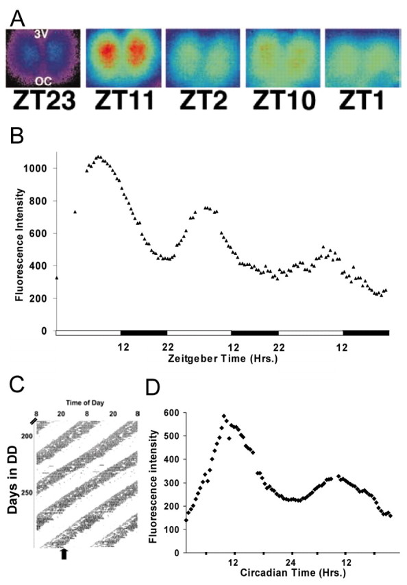
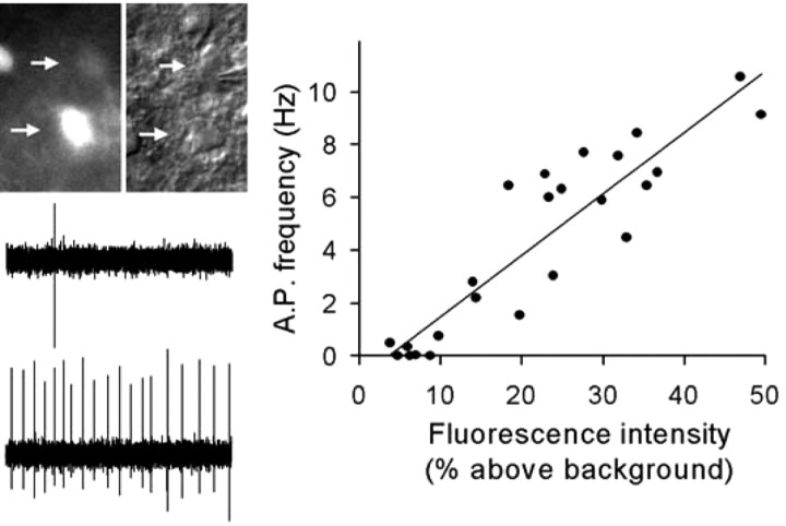
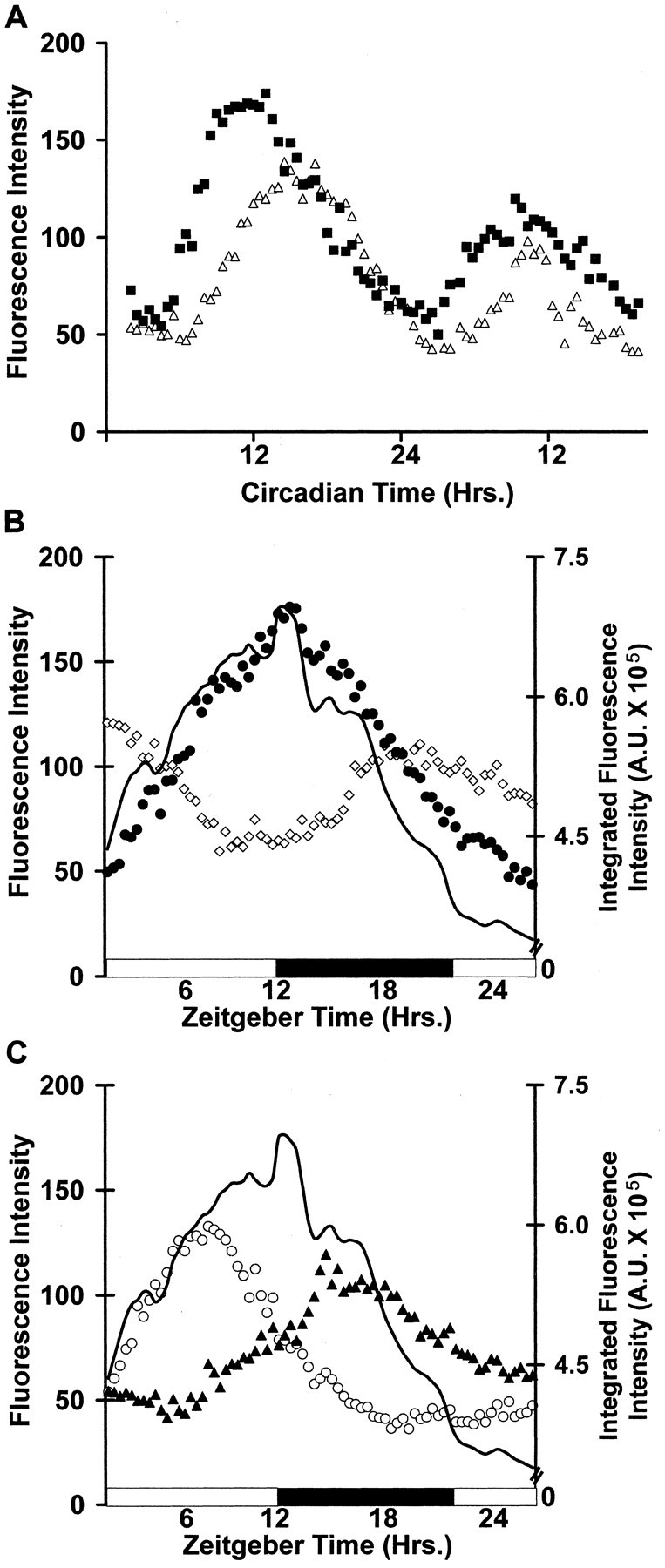
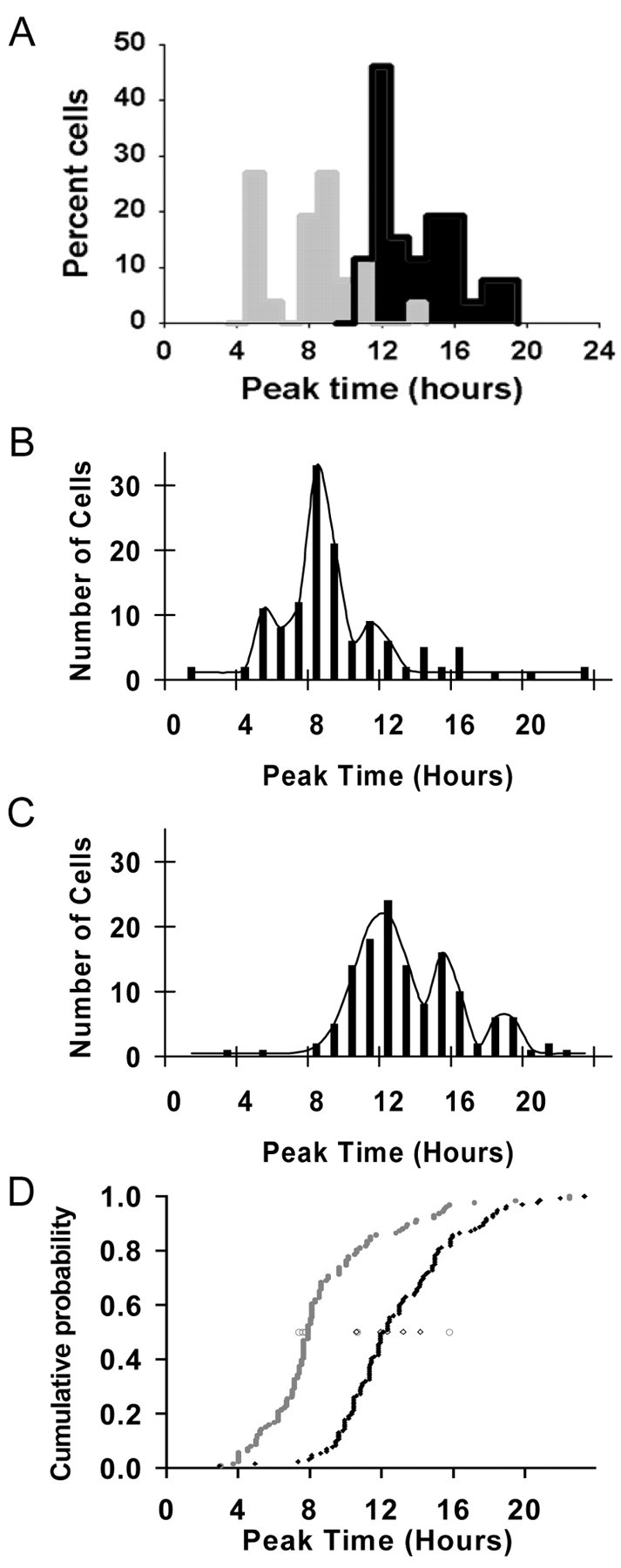
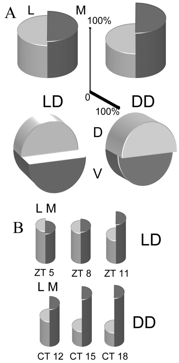
The circadian clock nucleus of the mammalian brain is composed of thousands of oscillator neurons, each driven by the cell-autonomous action of a defined set of circadian clock genes. A critical question is how these individual oscillators are organized into an internal clock that times behavior and physiology. We examined the neural organization of the suprachiasmatic nucleus (SCN) through time-lapse imaging of a short-half-life green fluorescent protein (GFP) reporter of the circadian clock gene Period 1 (Per1). Using brain slice preparations, Per1 promoter rhythms were resolved at the level of the SCN, and in individual neurons within the SCN, to determine the temporal patterns of rhythmicity resulting from exposure of mice to light/dark cycle (LD) and constant darkness (DD) conditions. Quantitative imaging and patch-clamp electrophysiology were used to define the relationship of Per1 gene expression to neurophysiological output on an individual neuron basis. We found that in both LD and DD, the overall rhythm of the clock nucleus is composed of individual cellular rhythms that peak in distinct phase groups at 3-4 hr intervals. However, the phase relationships of Per1 oscillations to locomotor activity and the phase relationships among individual neuronal oscillators within the SCN are different in LD and DD. There was a positive, linear correlation of Per1 transcription with neuronal spike frequency output, thus Per1::GFP rhythms are representative of physiological rhythmicity. Our results reveal multiple phase groupings of SCN oscillators and suggest that light regulation of oscillator interactions within the SCN underlies entrainment to the photoperiod.


Figures

Figure 1.
Figure 1.
Time-lapse imaging of SCN Per1-driven GFP rhythms. A, Individual pseudocolored fluorescence images from a time-lapse recording of Per1-driven GFP fluorescence rhythms from an SCN brain slice in vitro. B, Fluorescence rhythms in the in vitro SCN from an animal housed in a 14/10 hr LD cycle. Images were captured every 30 min. The open and filled bars indicate the previous light/dark cycle for the animal. C, Wheel-running activity, double-plotted, from an animal housed in constant darkness. Vertical marks show times when the animal was active. The filled arrow marks the time the SCN slice was prepared for imaging. D, GFP fluorescence recording of the SCN from the animal in C. Images were captured every 30 min for 2 d.
Figure 2.
Figure 2.
Per1-driven fluorescence intensity correlates with spike frequency in SCN neurons. Right, Fluorescence intensity, measured as percent above background, versus action potential (AP) frequency plotted for SCN neurons (n = 24, 3 slices). r = 0.91; p < 0.001. Left, Example of spike output from neighboring neurons; top arrow, low fluorescence intensity (6.9% above background, 0.03 Hz); bottom arrow, high fluorescence intensity (49.5% above background, 9.14 Hz).
Figure 3.
Figure 3.
Individual neuron gene expression rhythms in the SCN. A, Per1-driven GFP rhythms from two individual SCN neurons in an in vitro DD SCN obtained by confocal time-lapse imaging. B, C, Example gene expression rhythms from four individual neurons in an in vitro SCN from a LD animal. Each set of symbols represents the measured fluorescence for an individual cell during the circadian cycle. The black solid line indicates the integrated overall fluorescence rhythms of the SCN as a whole.
Figure 4.
Figure 4.
Neuronal phase relationships are dependent on light history. A, Histograms of individual neuron fluorescence peak times in an SCN slice from an LD animal (gray bars) and an SCN slice from a DD animal (black bars). Cell peak times were binned at 1 hr intervals; n = 26 neurons for the LD slice; n = 37 neurons for the DD slice. B, Histogram of individual neuron fluorescence peak times summed from SCN slices from five LD animals. Peak times are in 1 hr bins. The black line indicates the best fit curve showing phase group peak times of ZT5, ZT8, and ZT11. C, Histogram of individual neuron fluorescence peak times summed from SCN slices from five DD animals. The black line indicates the best fit curve showing phase group peak times of CT12, CT15, and CT18. D, Cumulative probability plot of peak times from animals housed in the LD cycle (filled circle, gray line) and peak times from DD (filled diamond, black line). The median time of cellular peaks for each animal is plotted as an open symbol (circle, LD; diamond, DD).
Figure 5.
Figure 5.
Rhythmic neurons by SCN region. Split cylinders shown in a three-dimensional perspective rendering represent the percentage of rhythmic neurons in the SCN mapped by SCN region. The height of each half-cylinder indicates the percentage of rhythmic SCN neurons in that region. Scale bars indicate the percentage scale for the lateral-medial axis (vertical) and the dorsal medial axis (horizontal). Total cells are n = 126 cells for LD slices and 131 cells for DD slices. A, Overall distributions of rhythmic neurons. Top row, Percentage of rhythmic SCN neurons mapped in the lateral-medial axis for LD (left) and DD (right) slices. Bottom row, Percentage of rhythmic SCN neurons mapped in the dorsal-medial axis for LD (left) and DD (right) slices. B, Distributions of rhythmic neurons by phase group. Top row, Percentage of rhythmic SCN neurons from the three LD phase groups mapped in the lateral-medial axis. Bottom row, Percentage of rhythmic SCN neurons from the three DD phase groups mapped in the lateral-medial axis.

References

    1. Albrecht U ( 2002) Functional genomics of sleep and circadian rhythm: invited review. Regulation of mammalian circadian clock genes. J Appl Physiol 92: 1348-1355. - PubMed
    1. Bae K, Jin X, Maywood ES, Hastings MH, Reppert SM, Weaver DR ( 2001)Differential functions of mPer1, mPer2, and mPer3 in the SCN circadian clock. Neuron 30: 525-536. - PubMed
    1. Davis FC, Gorski RA ( 1985) Development of hamster circadian rhythms. I. Within-litter synchrony of mother and pup activity rhythms at weaning. Biol Reprod 33: 353-362. - PubMed
    1. Hamada T, LeSauter J, Venuti JM, Silver R ( 2002) Expression of Period genes: rhythmic and non-rhythmic compartments of the suprachiasmatic nucleus pacemaker. J Neurosci 21: 7742-7750. - PMC - PubMed
    1. Herzog ED, Geusz ME, Khalsa SB, Straume M, Block GD ( 1997) Circadian rhythms in mouse suprachiasmatic nucleus explants on multimicroelectrode plates. Brain Res 757: 285-290. - PubMed

Publication types

MeSH terms