. 2003 Sep 3;23(22):8176-84.
doi: 10.1523/JNEUROSCI.23-22-08176.2003.

Slit2 guides both precrossing and postcrossing callosal axons at the midline in vivo

Affiliations

Slit2 guides both precrossing and postcrossing callosal axons at the midline in vivo

Tianzhi Shu et al. J Neurosci. .

Abstract

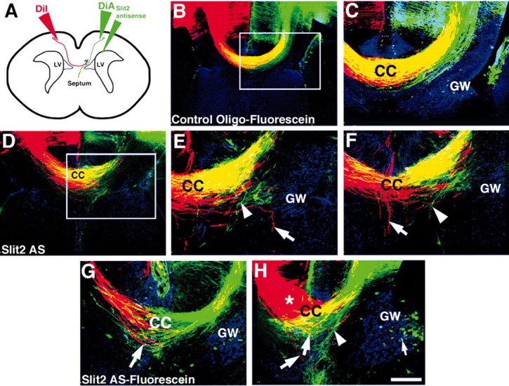
Commissural axons generally cross the midline only once. In the Drosophila nerve cord and mouse spinal cord, commissural axons are guided by Slit only after they cross the midline, where Slit prevents these axons from recrossing the midline. In the developing corpus callosum, Slit2 expressed by the glial wedge guides callosal axons before they cross the midline, as they approach the corticoseptal boundary. These data highlighted a potential difference between the role of Slit2 in guiding commissural axons in the brain compared with the spinal cord. Here, we investigate whether Slit2 also guides callosal axons after they cross the midline. Because such questions cannot be addressed in conventional gene knock-out animals, we used in utero injections of antisense oligonucleotides to specifically deplete Slit2 on only one side of the brain. We used this technique together with a novel in vitro assay of hemisected brain slices to specifically analyze postcrossing callosal axons. We find that in the brain, unlike the spinal cord, Slit2 mediates both precrossing and postcrossing axonal guidance. Depletion of Slit2 on one side of the brain causes axons to defasciculate and, in some cases, to aberrantly enter the septum. Because these axons do not recross the midline, we conclude that the principle function of Slit2 at the cortical midline may be to channel the axons along the correct path and possibly repel them away from the midline. We find no evidence that Slit2 prevents axons from recrossing the midline in the brain.


Figures

Figure 1.
Figure 1.
Robo ectodomain proteins block the chemosuppressive-repulsive effect of the glial wedge. Robo1 and Robo2 ectodomain proteins were synthesized using an in vitro transcription-translation system and visualized by Western blot analysis by the incorporation of biotinylated lysines (A). Both Robo1 and Robo2 ectodomain proteins were produced with an estimated mass of 150-160 kDa (A, lanes 2 and 3), consistent with previous findings (Brose et al., 1999). In the control lane (A, lane 1), an empty vector was added to the reaction, and, thus, no protein was produced. Collagen gel experiments were performed using explants derived from E17 brain slices. Cortical explants (Ctx) were cocultured with glial wedge (GW) explants (C) or another cortical explant (B) as controls. As described previously, the GW causes a growth suppression-repulsion of cortical axons (B). Robo ectodomain proteins were mixed and added to the growth medium of Ctx/GW cocultures at either 2 ng/μl total protein (D) or 20 ng/μl total protein (E). After 3 d in culture, explants were fixed, and DiI was injected into the cortical explant to label the axons. F, G, Significantly more, and longer, axons grew into the GW when either 2 ng/μl or 20 ng/μl of the Robo ectodomain proteins was added to the cultures (*p < 0.05; ANOVA), indicating the inhibitory effect of the GW had been blocked. Histograms represent the mean ± SEM. Scale bar (in E): B-E, 150 μm.
Figure 2.
Figure 2.
Callosal axons express Robo receptors. Robo immunohistochemistry-labeled callosal axons at E15 (A, B, arrow; B is a higher-power view of the region delineated in A), as axons approach the midline and as they cross at E16 (C, D, arrow; D is a higher-power view of the region delineated in C). Robo labeling at E17 (F) overlaps with 3A10 antibody labeling in an adjacent section (E, F, arrows). The lateral cortical projection through the internal capsule is also labeled by the Robo antibodies (C, arrow). All panels are of coronal sections. Scale bar (in F): A, C, 700 μm; B, 180 μm; D-F, 200 μm.
Figure 3.
Figure 3.
Slit2 antisense oligonucleotides are localized in glial wedge cells after in utero injection. Sequences of the Slit2 and control antisense oligonucleotides used are shown in A. The binding region of the Slit2 antisense is indicated by the alignment (A). Slit2 antisense oligonucleotides were mixed with a small amount of fast green dye to visualize the injection site. The dye remained within the injected hemisphere (B; arrow indicates the injection site). In this case, the injection was performed at E16 and is visualized after perfusion on E17. To localize the Slit2 antisense oligonucleotides, fluorescein-tagged Slit2 antisense oligonucleotides were injected at E16, and brains were perfused after 24 hr and labeled with GFAP immunohistochemistry. Slit2 antisense oligonucleotides (C, D, green dots) were present in the glial wedge, which was identified by GFAP immunohistochemistry (C, D, red processes). Slit2 antisense oligonucleotides were located in the ventricular and subventricular zones of the corticoseptal boundary within the soma of the glial wedge cells (D is a higher-power view of the region delineated in C). This distribution resembled the expression pattern of Slit2 mRNA by in situ hybridization (F is a higher-power view of the boxed region in E). B is a dorsal view, and C-F are coronal sections. Scale bar (in F): B, 6 mm; C, 400 μm; E, 200 μm; D, F, 50 μm.
Figure 5.
Figure 5.
Pathfinding errors of callosal axons in brains injected with Slit2 antisense oligonucleotides at E16. E16 brains injected with control oligonucleotides displayed normal callosal pathfinding (A, B, F, DiI-labeled red axons; B is a higher-power view of the region delineated in A). However, brains injected with Slit2 antisense oligonucleotides displayed varying degrees of pathfinding errors (C-E, G, DiI-labeled red axons). In some brains, axons deviated from the main bundle (C, arrow), and in others the majority of axons grew aberrantly into the septum (D, E, G, arrows). Some experiments were performed with fluorescein-tagged control (F) or Slit2 antisense (G) oligonucleotides and could be detected at the cortiocseptal boundary and within the glial wedge (F, G, green dots). All sections are in the coronal plane and represent different brains in each panel, except in A and B. The glial wedge is labeled with GFAP immunohistochemistry in green in A-E and in pseudo-blue in F and G. Scale bar (in G): A, 400 μm; B, D, 150 μm; C,E-G, 200 μm.
Figure 4.
Figure 4.
In utero injection of Slit2 antisense oligonucleotides disrupts precrossing callosal axon pathfinding at midline on E15. Control (A, C) or Slit2 antisense (B, D) oligonucleotides were injected into the right lateral ventricle of E15 mouse embryos. On E17, the injected embryos were perfused, and after postfixation, DiI was injected into the medial cortex to label callosal axons (axons labeled red in all panels). GFAP immunohistochemistry was used to label the glial wedge (GW) (A, B, green labeling; C, D, pseudo-blue labeling). Control oligonucleotide-injected brains developed a normal corpus callosum (CC) (A, C; two different brains). Brains injected with Slit2 antisense oligonucleotides displayed dramatic axon pathfinding errors at the midline. Many callosal axons stalled at the midline and failed to cross, sometimes forming Probst bundle-like structures (B, asterisk). Some axons projected aberrantly into the septum (B, arrow). Unlabeled oligonucleotides were used in A and B, and fluorescein-labeled oligonucleotides were used in C and D; however, the fluorescein tag could not be detected after the 2 d survival period (C, D). All sections are in the coronal plane. Scale bar (in D): A, 300 μm; B, 250 μm; C, D, 150 μm.
Figure 6.
Figure 6.
Disruption of postcrossing callosal axons in vivo by Slit2 antisense oligonucleotides. To investigate the role of Slit2 in regulating the growth of postcrossing callosal axons, we injected either control oligonucleotides (B, C) or Slit2 antisense oligonucleotides (D-H) into the right lateral ventricle of E16 embryos in utero. Embryos were perfused on E17 and later labeled with either DiA to label the precrossing axons (green axons in all panels) or DiI to label the postcrossing axons (red axons in all panels). A schematic of the experimental paradigm is shown (A). The glial wedge was labeled with GFAP and a Cy5 secondary antibody (blue labeling in all panels). In control oligonucleotide-injected brains (C is a higher-power view of the region delineated in B), both precrossing and postcrossing callosal axons crossed the midline normally and overlapped within the same tract (B, C; yellow labeling indicates overlapping axons). In brains injected with Slit2 antisense oligonucleotides, both precrossing and postcrossing callosal axons displayed pathfinding errors (D-H). Aberrant pathfinding of precrossing axons was observed (E, F, H, arrowheads; E is a higher-power view of the region delineated in D; F is another section from the same brain) as shown in Figure 5 and served as an internal control for the knockdown of Slit2. Postcrossing axons also displayed pathfinding errors. Axons left the main callosal bundle, grew into the septum or even ventrally along the midline (E-G, axons labeled with an arrow). In some cases, axons did not even cross the midline or formed Probst-like bundles (H, red axons and bundle labeled by an asterisk; G and H are examples from two additional brains). All sections are in the coronal plane. Fluorescein-labeled oligonucleotides were used in B, C, G, and H (green dots in these panels; H, small arrow); unlabeled oligonucleotides were used in D-F. Scale bar (in H): B, D, 200 μm; C, E-H, 100 μm.
Figure 7.
Figure 7.
The glial wedge supresses-repels the growth of postcrossing callosal axons in vitro. To specifically study postcrossing callosal axons, we made live coronal slices of E17 brains and bisected them along the midline to obtain slices containing only one hemisphere. These hemisected slices were grown in collagen for 3 d and then fixed and labeled with the cellular marker Sytox green (green cellular labeling in all panels). Callosal axons were visualized with DiI injected into the medial cortex of the slice (A-I, red axons). A schematic of the slice culture is shown in A. When slices were cultured alone, callosal axons crossed the midline and grew into the collagen (B, C, arrow; C is a higher-power view of the region delineated in B). Hemisected slices cocultured with cortical explants (D, E; two different examples) had callosal axons that grew into the collagen and entered the cortical explants (D, E, arrows). However, when hemisected slices were cocultured with glial wedge explants, fewer callosal axons left the slice and entered the glial wedge (F, G; two different examples). In many cases, as the callosal axons approached the glial wedge, they changed direction and turned away (F, G, arrows). To investigate whether this repulsive-suppressive effect on the growth of postcrossing callosal axons was caused by Slit2, we added 20 ng/μl Robo1 and Robo2 ectodomain proteins to the cultures. Two examples of these cultures are shown in H and I, where callosal axons were now able to grow into the glial wedge explant (H, I, arrows). Quantitation of these effects is shown in J-L; the mean and SEM are shown. The number of axons entering the explant is shown in J. The number and the length of axons that exited the hemisected slice (regardless of whether they entered the opposing slice) are shown in K and L, respectively. *p < 0.01 (in J, K); *p < 0.001 (in L). GW, Glial wedge explant; Ctx, cortex explant. Scale bar (in I): B, 400 μm; C-I, 150 μm.

Similar articles

Cited by

References

    1. Bagri A, Marin O, Plump AS, Mak J, Pleasure SJ, Rubenstein JLR, Tessier-Lavigne M (2002) Slit proteins prevent midline crossing and determine the dorsoventral position of major axonal pathways in the mammalian forebrain. Neuron 33:233-248. - PubMed
    1. Brose K, Bland KS, Wang KH, Arnott D, Henzel W, Goodman CS, Tessier-Lavigne M, Kidd T (1999) Slit proteins bind Robo receptors and have an evolutionary conserved role in repulsive axon guidance. Cell 96:795-806. - PubMed
    1. Cummings DM, Malun D, Brunjes PC (1997) Development of the anterior commissure in the opossum: midline extracellular space and glia coincide with early axon decussation. J Neurobiol 32:403-414. - PubMed
    1. Erskine L, Williams SE, Brose K, Kidd T, Rachel RA, Goodman CS, Tessier-Lavigne M, Mason CA (2000) Retinal ganglion cell axon guidance in the mouse optic chiasm: expression and function of Robos and slits. J Neurosci 20:4975-4982. - PMC - PubMed
    1. Goodhill GJ (2003) A theoretical model of axon guidance by the Robo code. Neural Comput 15:549-564. - PubMed

Publication types

MeSH terms

Substances

LinkOut - more resources