. 2003 Sep 16;100(19):10848-53.
doi: 10.1073/pnas.1831408100. Epub 2003 Sep 3.

Specificity in the activation and control of transcription factor E2F-dependent apoptosis

Affiliations

Specificity in the activation and control of transcription factor E2F-dependent apoptosis

Timothy C Hallstrom et al. Proc Natl Acad Sci U S A. .

Abstract

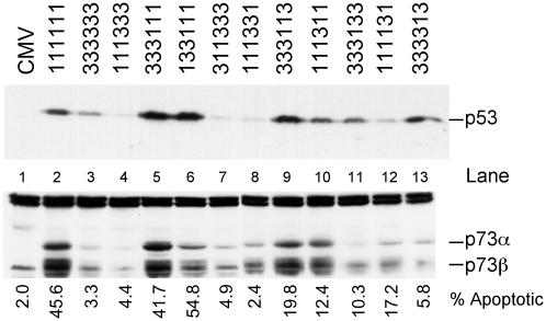
Previous work has demonstrated a role for the E2F1 gene product in signaling apoptosis, both as a result of the deregulation of the Rb/E2F pathway as well as in response to DNA damage. We now show that the ability of cells to suppress the apoptotic potential of E2F1, as might occur during the course of normal cellular proliferation, requires the action of the Ras-phosphoinositide 3-kinase-Akt signaling pathway. In addition, we also identify a domain within the E2F1 protein, previously termed the marked-box domain, that is essential for the apoptotic activity of E2F1 and that distinguishes the E2F1 protein from E2F3. We also show that the E2F1-marked-box domain is essential for the induction of both p53 and p73 accumulation. Importantly, a role for the marked-box domain in the specificity of E2F1-mediated apoptosis coincides with recent work demonstrating a role for this domain in achieving specificity in the activation of transcription. We conclude that the unique capacity of E2F1 to trigger apoptosis reflects a specificity of transcriptional activation potential, and that this role for E2F1 is regulated through the action of the Akt protein kinase.


Figures

Fig. 1.
Fig. 1.
A serum-activated PI3K-signaling pathway blocks E2F1-induced apoptosis. (A) E2F1-induced apoptosis is blocked by serum. REF52 cells were deprived of serum (quiescent) or grown in serum (stimulated) as described in Materials and Methods. REF52 cells were infected on the same day with E2F-expressing adenoviruses at an moi of 75 focus-forming units per cell. CMV, cytomegalovirus control. Samples were harvested 40 h after infection and assayed for active caspase-3 levels by fluorescence-activated cell sorter (FACS; Becton Dickinson) analysis. (B) REF52 cells were grown in 10% serum-containing medium for 24 h. After infection with the indicated E2F adenoviruses, medium containing 10% serum and either 100 μM mitogen-activated protein kinase kinase (MEK) inhibitor (PD 98059) or 50 μM PI3K inhibitor (LY-294002) was added back to the cells. Cells were harvested 40 h after infection and processed for active caspase-3 levels by FACS analysis. (C) Equal expression of E2F-producing HA-E2Fs. Protein extracts were prepared from REF52 cells infected with E2F adenoviruses 24 h after infection. Extracts were analyzed by Western blotting using antiserum against HA (E2Fs) or actin to verify equal protein loading.
Fig. 2.
Fig. 2.
Constitutively active Akt attenuates E2F1-induced apoptosis and p53 accumulation. (A) Constitutively active Akt blocks E2F1-induced apoptosis in REF52 cells. Serum-deprived REF52 cells were infected with E2F1 adenovirus-expressing (150 moi) and increasing mois (150, 300, and 500) of vAkt-producing adenovirus. Cells were harvested 40 h after infection and assayed for active caspase-3. (B) Constitutively active Akt blocks E2F1-dependent p53 protein accumulation. Serum-starved REF52 cells were infected at an moi of 150 with control (CMV), E2F3-, E2F1-, or E2F1-expressing adenoviruses, along with increasing amounts of adenovirus that expresses hyperactive Akt (moi 150 and 300). A control adenovirus (vector lacking an insert) was used in place of the Akt virus for the samples shown in lanes 3 (moi 300) and 4 (moi 150). Whole cell lysates were prepared 24 h after infection, and equivalent protein amounts from each infection were separated by SDS/PAGE and probed with anti-p53 antiserum.
Fig. 3.
Fig. 3.
Construction of chimeric E2Fs. (A) Restriction sites were introduced into the cDNAs of HA-E2F1 and HA-E2F3 to allow the cloning of individual domains between the genes. The introduced restriction sites are outlined in Materials and Methods. (B) Diagram of the chimeric E2Fs described in this study. The nomenclature describes the identity of individual domains in the chimeras: first digit, N terminus/cyclin A binding; second digit, DNA-binding domain; third digit, DP1 dimerization domain; fourth digit, marked box; fifth digit, marked-box-adjacent region; sixth digit, transactivation/Rb-binding domain. Resultant E2F cDNAs were used to generate recombinant adenovirus expressing the HA-E2Fs. (C) HA-E2F protein expression in REF52 cells. Serum-starved REF52 cells were infected with the noted HA-E2F-expressing adenovirus and processed for Western blotting 24 h after infection. Equal amounts of protein were separated by SDS/PAGE and probed with anti-HA antiserum. Actin is shown as a loading control. (D) Induction of BrdUrd incorporation. Serum-starved REF52 cells were infected with the chimeric E2F-expessing adenoviruses and returned to 0.25% serum-containing medium for 8 h. Cells were then labeled with 50 μM BrdUrd for 15 h, and BrdUrd incorporation was assayed by indirect immunofluorescence.
Fig. 4.
Fig. 4.
The E2F1 marked-box domain is required for the induction of apoptosis. (Left) REF52 cells were incubated in 0.25% serum medium for 48 h and then infected with the indicated chimeric E2F-producing adenovirus at an moi of 75 focus-forming units per cell. After infection, cells were returned to starvation medium for 40 h before harvesting for detection of active caspase-3 levels by fluorescence-activated cell sorter (FACS; Becton Dickinson) analysis. (Right) The flow cytometry data shown in Left are presented here in graph format.
Fig. 5.
Fig. 5.
The E2F1 marked-box and adjacent domain are required for the induction of p53 and p73 protein accumulation. (Upper) Serum-starved REF52 cells were infected with the indicated E2F adenovirus (75 focus-forming units per cell) and returned to starvation medium for 24 h. Whole cell extracts were prepared and separated by SDS/PAGE. The levels of p53 protein were assessed by Western blotting. (Lower) We were unable to detect rat p73 with the available antiserum, so we used Saos-2 cells to assess expression of human p73 in response to the E2F chimeras. Saos-2 cells were plated at a density of 3 × 10⁵ cells per 60-mm-diameter dish and grown in 10% serum for 24 h, then switched to 0.25% serum-containing medium for 48 h. Cells were infected with chimeric E2F-producing virus and were then returned to starvation medium. Nuclear extracts were prepared 24 h after infection, and 100 μg of protein was separated by SDS/PAGE and probed with anti-p73 antisera (Oncogene).

References

    1. Dyson, N. (1998) Genes Dev. 12, 2245–2262. - PubMed
    1. Nevins, J. R. (1998) Cell Growth Differ. 9, 585–593. - PubMed
    1. Leone, G., DeGregori, J., Yan, Z., Jakoi, L., Ishida, S., Williams, R. S. & Nevins, J. R. (1998) Genes Dev. 12, 2120–2130. - PMC - PubMed
    1. Humbert, P. O., Verona, R., Trimarchi, J. M., Rogers, C., Dandapani, S. & Lees, J. A. (2000) Genes Dev. 14, 690–703. - PMC - PubMed
    1. Humbert, P. O., Rogers, C., Ganiatsas, S., Landsberg, R. L., Trimarchi, J. M., Dandapani, S., Brugnara, C., Erdman, S., Schrenzel, M., Bronson, R. T., et al. (2000) Mol. Cell 6, 281–291. - PubMed

Publication types