. 2003 Sep 16;100(19):10776-81.
doi: 10.1073/pnas.1831718100. Epub 2003 Sep 3.

The PDZ-binding motif of the beta2-adrenoceptor is essential for physiologic signaling and trafficking in cardiac myocytes

Affiliations

The PDZ-binding motif of the beta2-adrenoceptor is essential for physiologic signaling and trafficking in cardiac myocytes

Yang Xiang et al. Proc Natl Acad Sci U S A. .

Abstract

beta1- and beta2-adrenergic receptors (AR) regulate cardiac myocyte function through distinct signaling pathways. In addition to regulating cardiac rate and contractility, beta1AR and beta2AR may play different roles in the pathogenesis of heart failure. Studies on neonatal cardiac myocytes from beta1AR and beta2AR knockout mice suggest that subtype-specific signaling is determined by subtype-specific membrane targeting and trafficking. Stimulation of beta2ARs has a biphasic effect on contraction rate, with an initial increase followed by a sustained Gi-dependent decrease. Recent studies show that a PDZ domain-binding motif at the carboxyl terminus of human beta2AR interacts with ezrin-binding protein 50/sodium-hydrogen exchanger regulatory factor, a PDZ-domain-containing protein. The human beta2AR carboxyl terminus also binds to N-ethylmaleimide-sensitive factor, which does not contain a PDZ domain. We found that mutation of the three carboxyl-terminal amino acids in the mouse beta2AR (beta2AR-AAA) disrupts recycling of the receptor after agonist-induced internalization in cardiac myocytes. Nevertheless, stimulation of the beta2AR-AAA produced a greater contraction rate increase than that of the wild-type beta2AR. This enhanced stimulation of contraction rate can be attributed in part to the failure of the beta2AR-AAA to couple to Gi. We also observed that coupling of endogenous, wild-type beta2AR to Gi in beta1AR knockout myocytes is inhibited by treatment with a membrane-permeable peptide representing the beta2AR carboxyl terminus. These studies demonstrate that association of the carboxyl terminus of the beta2AR with ezrin-binding protein 50/sodium-hydrogen exchanger regulatory factor, N-ethylmaleimide-sensitive factor, or some related proteins dictates physiologic signaling specificity and trafficking in cardiac myocytes.


Figures

Fig. 1.
Fig. 1.
Functional expression of Flag-β2AR or Flag-β2AR-AAA in β12AR-KO myocytes. (A) The expression of Flag-β2AR and Flag-β2AR-AAA protein in the β1β2AR-KO myocytes was examined by Western blot analysis (Left) and saturation binding (Right) to determine Bmax.(B) The expression of Flag-β2AR and Flag-β2AR-AAA was characterized by competition binding assays with β2AR agonist isoproterenol (IC50 = 284 and 294 nM, respectively) and the selective antagonist ICI118551 (IC50 = 3 and 3.5 nM, respectively). The data represent the mean ± SE of five experiments (triplicates) from different myocyte preparations. [3H]DHA, [3H]dihydroalprenolol; Iso, isoproterenol; ICI, ICI118551.
Fig. 2.
Fig. 2.
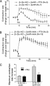
The β2AR PDZ mutation alters receptor regulation of contraction rate in cardiac myocytes. (A) The effect of 10 μM isoproterenol on the contraction rate of the β12AR-KO myocytes expressing Flag-β2AR or of the β1AR-KO myocytes with endogenous β2AR. (B) The effect of 10 μM isoproterenol on the contraction rate of β12AR-KO myocytes expressing Flag-β2AR-AAA or of the β1AR-KO myocytes with endogenous β2AR. (C) Disruption of caveolae with filipin enhances the increase in contraction rate after β2AR-AAA stimulation. The data represent the mean ± SE of N experiments from at least three different myocyte preparations. *, P < 0.05; time-course curves were found to be significantly different by two-way analysis of variance.
Fig. 3.
Fig. 3.
Disruption of the β2AR PDZ motif inhibits receptor coupling to Gi. PTX (0.75 μg/ml) treatment selectively affected the contraction rate of the β12AR-KO myocytes expressing Flag-β2AR (A) but not Flag-β2AR-AAA (B). (C) PKI partially inhibits the myocyte contraction-rate increase mediated by Flag-β2AR-AAA, but not by Flag-β2AR. The data represent the mean ± SE of four experiments from at least three different myocyte preparations. *, P < 0.05; time-course curves were found to be significantly different by two-way analysis of variance.
Fig. 4.
Fig. 4.
Agonist-induced internalization of Flag-β2AR, Flag-β2AR-AAA, and HA-β1AR-PDZmut in neonatal cardiac myocytes. (A) Flag-β2AR and Flag-β2AR-AAA are localized on the cell surface in neonatal myocytes at steady state. Punctate intracellular staining is observed after agonist stimulation of both receptors. (B) Flag-β2AR and HA-β1AR-PDZmut efficiently recycle back to the myocyte cell surface after removal of isoproterenol, while the Flag-β2AR-AAA remains inside the cell. (C) The cell-surface receptor level was measured by ELISAs after agonist-induced internalization and recycling. Cell-surface HA-β1AR-PDZmut (11%), Flag-β2AR (22%), and Flag-β2AR-AAA (23%) decreased significantly after isoproterenol stimulation. Although the surface density of HA-β1AR-PDZmut and Flag-β2AR was restored by 60 min after removal of isoproterenol, the surface density of Flag-β2AR-AAA remained low. Iso, isoproterenol; Alp, alprenolol.
Fig. 5.
Fig. 5.
Cell-permeable β2AR carboxyl-terminal peptides affect the contraction rate in β1AR-KO myocytes. β1AR-KO neonatal myocytes were cultured and treated with peptide (1 μM) for 25 min before contraction-rate experiments. The basal contraction rate of myocytes was not altered significantly by peptide treatment. Pretreatment with Tat-β2-DSPL (A) and Tat-β2-DSAL (D), but not Tat-β2-DAAA (B) or Tat-β2-ASPL (C), significantly changed the isoproterenol (Iso)-stimulated contraction-rate increase in β1AR-KO myocytes. *, P < 0.05; time-course curves were found to be significantly different by two-way analysis of variance.

References

    1. Koch, W. J., Milano, C. A. & Lefkowitz, R. J. (1996) Circ. Res. 78, 511–516. - PubMed
    1. Lefkowitz, R. J., Rockman, H. A. & Koch, W. J. (2000) Circulation 101, 1634–1637. - PubMed
    1. Gauthier, C., Langin, D. & Balligand, J. L. (2000) Trends Pharmacol. Sci. 21, 426–431. - PubMed
    1. Devic, E., Xiang, Y., Gould, D. & Kobilka, B. (2001) Mol. Pharmacol. 60, 577–583. - PubMed
    1. Chruscinski, A. J., Rohrer, D. K., Schauble, E., Desai, K. H., Bernstein, D. & Kobilka, B. K. (1999) J. Biol. Chem. 274, 16694–16700. - PubMed

Publication types

MeSH terms

Substances

LinkOut - more resources