. 2003 Sep 16;100(19):10617-22.
doi: 10.1073/pnas.1833120100. Epub 2003 Sep 3.

A tale of two TUTases

Affiliations

A tale of two TUTases

Ruslan Aphasizhev et al. Proc Natl Acad Sci U S A. .

Abstract

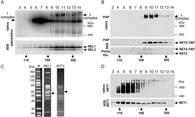
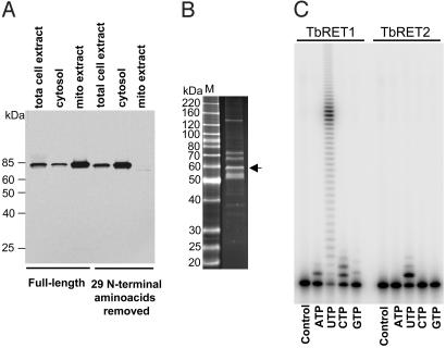
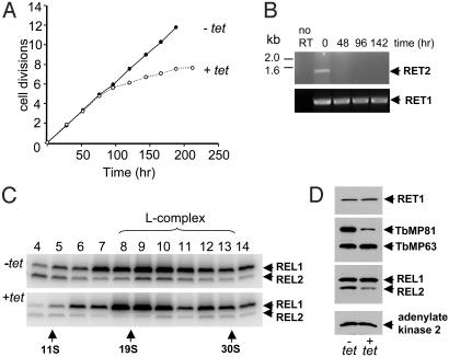
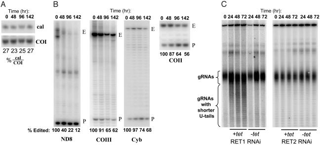
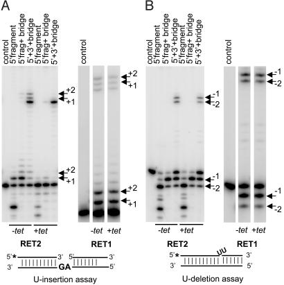
The insertion and deletion of U residues at specific sites in mRNAs in trypanosome mitochondria is thought to involve 3' terminal uridylyl transferase (TUTase) activity. TUTase activity is also required to create the nonencoded 3' oligo[U] tails of the transacting guide RNAs (gRNAs). We have described two TUTases, RET1 (RNA editing TUTase 1) and RET2 (RNA editing TUTase 2) as components of different editing complexes. Tandem affinity purification-tagged Trypanosoma brucei RET2 (TbRET2) was expressed and localized to the cytosol in Leishmania tarentolae cells by removing the mitochondrial signal sequence. Double-affinity isolation yielded tagged TbRET2, together with a few additional proteins. This material exhibits a U-specific transferase activity in which a single U is added to the 3' end of a single-stranded RNA, thereby confirming that RET2 is a 3' TUTase. We also found that RNA interference of RET2 expression in T. brucei inhibits in vitro U-insertion editing and has no effect on the length of the 3' oligo[U] tails of the gRNAs, whereas down-regulation of RET1 has a minor effect on in vitro U-insertion editing, but produces a decrease in the average length of the oligo[U] tails. This finding suggests that RET2 is responsible for U-insertions at editing sites and RET1 is involved in gRNA 3' end maturation, which is essential for creating functional gRNAs. From these results we have functionally relabeled the previously described TUT-II complex containing RET1 as the guide RNA processing complex.


Figures

Fig. 1.
Fig. 1.
RET1 and RET2 are components of different complexes. (A) Mitochondrial extract from the RET2-TAP L. tarentolae cell line was fractionated on a glycerol gradient. Each fraction was incubated with [α-32P]ATP and separated on a 4–12% native gel (Upper) or an 8–16% SDS gel (Lower), which were blotted onto filters for PhosphorImager analysis. The L-complex is identified by the labeled REL1 and REL2 ligase bands (Lower) and the single-labeled band (arrow) (Upper). (B) RET2 is present only in the L-complex. The filters were either treated with the PAP reagent to detect the RET2-protein A fusion or with the monoclonal Penta-His antibody (Qiagen) to detect RET2. (C) Comparison of protein compositions of L-complexes purified through TAP-tagged RET2 TUTase or REL1 RNA ligase. Arrows indicated positions of tagged proteins. (D) Distribution of RET1 in gradient. Filter A was treated with polyclonal antibodies against RET1.
Fig. 2.
Fig. 2.
3′ RNA uridylyl transferase activity of TbRET2. (A) Deletion of mitochondrial signal peptide results in the cytosolic localization of the TbRET2. Ten micrograms of protein from total cell extract, soluble fraction, and the S100 mitochondrial extract were fractionated on an 8–16% SDS gel, transferred onto nitrocellulose membrane, and treated with the PAP reagent to detect protein A fusion proteins. (B) Isolation of recombinant TbRET2 from L. tarentolae. The fraction eluted from the calmodulin column (see Materials and Methods) was analyzed on a 10–20% SDS gel followed by staining with Sypro ruby. Arrow indicates position of the tagged TbRET2. (C) Uridylyl transferase activity of recombinant TbRET1 and TbRET2 enzymes. The 5′ end-labeled 5′ fragment used in the precleaved assay was incubated with the respective enzyme in the presence of 0.3 mM ribonucleotide triphosphates, and the products were separated on a 14% polyacrylamide/urea sequencing gel. Control, no NTP added.
Fig. 3.
Fig. 3.
Effects of RNAi down-regulation of TbRET2 expression in procyclic T. brucei on cell growth and stability of the L-complex. (A) RNAi was induced by addition of 1 μg/ml tetracycline. Cell growth is affected after ≈100 h of RNAi. (B) RT-PCR analysis of RET1 and RET2 mRNAs during RET2 RNAi. (C) Glycerol gradients of mitochondrial extract from uninduced and 3-day RNAi-induced cells. Fractions were labeled with [α-32P]ATP and separated in an SDS gel. The location of the L-complex is indicated. (D) Mitochondrial extracts from uninduced or 3-day RNAi-induced cells were fractionated in an SDS gel, and the blot reacted with antisera against the indicated proteins.
Fig. 4.
Fig. 4.
Effect of RET1 and RET2 RNAi on in vivo mRNA editing and on length of gRNA oligo[U] tails. (A) The never-edited COI and cytosolic calmodulin mRNAs were analyzed in the same reactions as internal loading controls. (B) The relative abundance of fully edited and preedited ND8, COIII, Cyb, and COII transcripts was analyzed by primer extension. E, fully edited; P, preedited. Relative percentages of edited transcripts are shown below. (C) gRNAs were 5′ labeled with [α-32P]GTP in the presence of guanylyltransferase and separated on 12% polyacrylamide/urea gel. (Left) RET1 RNAi. (Right) RET2 RNAi. The band at the top of the gel is a cytosolic RNA that labels with [α-32P]GTP and is used as a loading control.
Fig. 5.
Fig. 5.
Effect of RET1 RNAi and RET2 RNAi on in vitro precleaved editing activity. (A) RNAi was induced for 3 days, and mitochondrial extract was fractionated in a glycerol gradient. The L-complex fractions were used for the U-insertion assay. The RNA substrates are diagrammed. Positions of extended 5′ fragments and the corresponding ligation products are indicated by arrows. (B) U-deletion assay. See A for description. The 5′ fragments with one or two Us removed and corresponding ligation products are indicated by arrows.

Comment in

References

    1. Blum, B., Bakalara, N. & Simpson, L. (1990) Cell 60, 189–198. - PubMed
    1. Seiwert, S. D. & Stuart, K. (1994) Science 266, 114–117. - PubMed
    1. Seiwert, S. D., Heidmann, S. & Stuart, K. (1996) Cell 84, 831–841. - PubMed
    1. Cruz-Reyes, J. & Sollner-Webb, B. (1996) Proc. Natl. Acad. Sci. USA 93, 8901–8906. - PMC - PubMed
    1. Maslov, D. A. & Simpson, L. (1992) Cell 70, 459–467. - PubMed

Publication types

MeSH terms

LinkOut - more resources