. 2003 Sep 4;22(38):6107-10.
doi: 10.1038/sj.onc.1206732.

Human DF3/MUC1 carcinoma-associated protein functions as an oncogene

Affiliations

Human DF3/MUC1 carcinoma-associated protein functions as an oncogene

Yongqing Li et al. Oncogene. .

Abstract

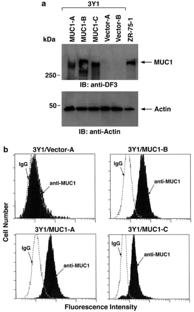
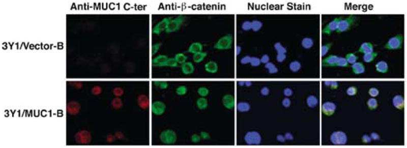
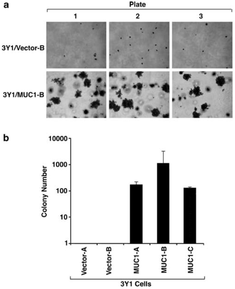
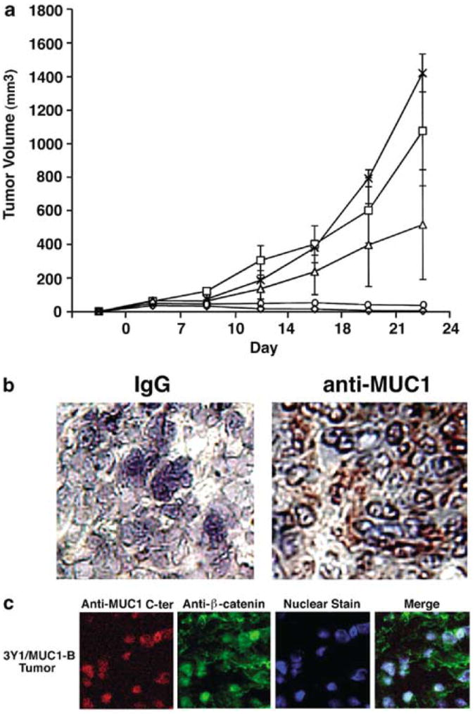
The human DF3/MUC1 mucin-like glycoprotein is aberrantly overexpressed by most carcinomas of the breast and other epithelia. The contribution of MUC1 overexpression to the malignant phenotype is, however, not known. In the present studies, we have stably expressed MUC1 in rat 3Y1 fibroblasts. MUC1-positive cells were selected from independent transfections. The results demonstrate that, as found in human carcinomas, MUC1 is expressed on the cell surface and as a complex with beta-catenin in the nucleus of the transfectants. Colony formation in soft agar demonstrates that cells expressing MUC1, but not the empty vector, exhibit anchorage-independent growth. The results also show that MUC1 expression confers tumor formation in nude mice. These findings provide the first evidence that MUC1 induces cellular transformation.


Conflict of interest statement

DK has a financial interest in ILEX.

Figures

Figure 1
Figure 1
Cell surface expression of MUC1 in 3Y1 cell transfectants. (a) Lysates from 3Y1/MUC1-A, 3Y1/MUC1-B, 3Y1/MUC1-C, 3Y1/Vector-A and 3Y1/Vector-B cells were subjected to immunoblotting (IB) with anti-MUC1 (MAb DF3) (upper panel) and anti-actin (lower panel) antibodies. ZR-75-1 cell lysate was used as positive control. (b) The indicated cells were incubated with anti-MUC1 (solid bars) or a control mouse IgG (open panels) and analysed by flow cytometry
Figure 2
Figure 2
Nuclear colocalization of the MUC1 C-ter and β-catenin. 3Y1/Vector-B and 3Y1/MUC1-B cells were fixed, permeabilized and incubated with anti-MUC1 C-ter and anti-β-catenin as described (Li et al., 2003). Nuclei were stained with TO-PRO-3 (Molecular Probes, Eugene, OR, USA). The cells were analysed by confocal microscopy as described (Li et al., 2003)
Figure 3
Figure 3
MUC1 confers anchorage-independent 3Y1 cell growth in soft agar. (a) 3Y1/Vector-A, 3Y1/Vector-B, 3Y1/MUC1-A, 3Y1/MUC1-B and 3Y1/MUC1-C cells (1 × 10⁴/plate) were suspended in soft agar and incubated for 3 weeks. Representative photomicrographs are shown for 3Y1/Vector-B and 3Y1/MUC1-B cells each seeded into triplicate plates. Similar results were obtained with the other independently selected cells expressing the empty vector or MUC1. (b) Colonies larger than 25 cells were counted for each of the indicated cells. The results are expressed as the number of colonies (mean±s.d.) obtained in three independent experiments
Figure 4
Figure 4
MUC1 induces tumorigenicity of 3Y1 cells in nude mice. (a) 3Y1/Vector-A (◇), 3Y1/Vector-B (○), 3Y1/MUC1-A (□), -B (X) and -C (Δ) cells (1 × 10⁷) were injected subcutaneously into the posterior flank of nude mice. Tumor volumes were calculated from bidimensional measurements at each time point. The results are expressed as the tumor volume (mean±s.d.) obtained from two independent experiments that each included six to eight mice per group. (b) Frozen tumor tissue was sectioned and stained with a control IgG or the DF3 anti-MUC1 antibody. (c) Fixed sections from a 3Y1/MUC1-B tumor were permeabilized and incubated with anti-MUC1 C-ter and anti-β-catenin. Nuclei were stained with TO-PRO-3. Confocal microscopy was performed as described (Li et al., 2003). Little if any staining was obtained when the Texas Red- and fluorescein-conjugated secondary antibodies were used alone as controls (data not shown)

References

    1. Bromberg JF, Wrzeszczynska MH, Devgan G, Zhao Y, Pestell RG, Albanese C, Darnell JE., Jr Cell. 1999;98:295–303. - PubMed
    1. Gendler S, Taylor-Papadimitriou J, Duhig T, Rothbard J, Burchell JA. J Biol Chem. 1988;263:12820–12823. - PubMed
    1. Guilford P, Hopkins J, Harraway J, McLeod M, McLeod N, Harawira P, Taite H, Scoular R, Miller A, Reeve AE. Nature. 1998;392:402–405. - PubMed
    1. Jemal A, Murray T, Samuels A, Ghafoor A, Ward E, Thun M. CA Cancer J Clin. 2003;53:5–26. - PubMed
    1. Kintner C. Cell. 1992;69:225–236. - PubMed

Publication types

MeSH terms