. 2003 Sep;41(9):4328-35.
doi: 10.1128/JCM.41.9.4328-4335.2003.

Isolation of an Anaplasma sp. organism from white-tailed deer by tick cell culture

Affiliations

Isolation of an Anaplasma sp. organism from white-tailed deer by tick cell culture

Ulrike G Munderloh et al. J Clin Microbiol. 2003 Sep.

Abstract

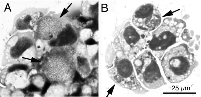
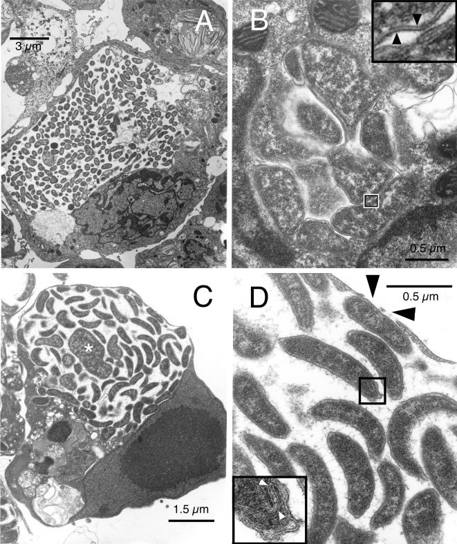
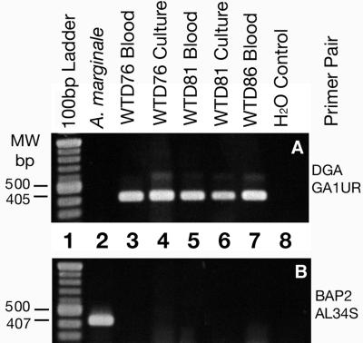
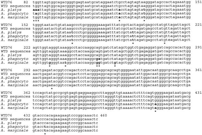
We used tick cell culture to isolate a bacterium previously referred to as the "white-tailed deer (WTD) agent" from two captive fawns inoculated with blood from wild WTD (Odocoileus virginianus). Buffy coat cells were added to ISE6 tick cell cultures and incubated at 34 degrees C, and 8 days later, Anaplasma-like inclusions were demonstrated in Giemsa-stained culture samples. The microbes became established and could be continuously passaged in tick cells. The identity of a culture isolate designated WTD76 was verified as the WTD agent by using specific PCR primers and by DNA sequencing. Comparison with sequences available in GenBank indicated that the isolate was most closely related first to Anaplasma platys and second to Anaplasma phagocytophilum, supporting its placement in the genus Anaplasma. Transmission electron microscopy of this Anaplasma sp. organism in tick cell cultures revealed large inclusions filled with pleomorphic and rod-shaped bacteria. Tick cells infected with the Anaplasma sp. organism were used to successfully infect a naive deer, thereby proving the infectivity of the isolate for deer.


Figures

FIG. 1.
FIG. 1.
Light microscopic appearance of the WTD-Anaplasma isolate in Giemsa-stained cytocentrifuge preparations of tick cell culture. (A) WTD-Anaplasma isolate from WDT76 in its eighth passage in tick cells. Arrows indicate large intracytoplasmic inclusions filled with numerous bacteria. (B) WTD-Anaplasma reisolated from WTD86. Arrows point to intracytoplasmic morulae. The scale bar in panel B gives the magnification for both panels.
FIG. 2.
FIG. 2.
Ultrastructure of the WTD-Anaplasma in tick cell culture. (A) Inclusion that appears to occupy most of its host cell. Note the lack of association of the bacteria with the endosomal membrane. (B) Image of a small morula with pleomorphic bacteria that tightly abut each other and the endosomal membrane. Inset, high magnification of the boxed-in area, showing the double-layered profile of the cell wall (arrowheads). (C) Third type of inclusion containing primarily slightly curved rods but also larger and more irregular organisms (asterisk). Note the intimate association of Anaplasma with the endosomal membrane. (D) Higher magnification of panel C. Black arrowheads indicate gaps in the endosomal membrane; white arrowheads within the inset point to folded membrane structures at one end of a curved rod, enlarged from the boxed-in area.
FIG. 3.
FIG. 3.
Agarose gel of PCR products amplified by use of primers DGA and GA1UR, specific for a 405-bp portion of the 16S rDNA of WTD-Anaplasma (A), and primers BAP2 and AL34S, specific for a 407-bp portion of the msp1β gene of A. marginale (B).
FIG. 4.
FIG. 4.
Alignment of the partial 16S rDNA sequence from the WDT-Anaplasma (WTD76) isolate with 16S rDNA sequences available in GenBank. Bases are numbered with reference to sequence AY055469 representing complete codons for the 16S rDNA of A. phagocytophilum. Five sequences from “Ehrlichia” sp. strains found in WTD (accession numbers U27104, U52514, U27103, U27102, and U27101) were used to arrive at the consensus sequence designated “WTD sequences.” Five sequences from A. platys (AF303467, AY077619, AY040853, U54806, and AF318023) were aligned to arrive at the sequence labeled A. platys, five A. phagocytophilum sequences (AY055469, AF481855, AF189153, U02521, and AF482761) were aligned and are represented as “A. phagocyto,” and a further five A. marginale sequences (AF309868, AF309867, AF309866, AF414877, and AF414873) were likewise aligned and are shown in the line labeled A. marginale. With the exception of eight unidentifiable bases in the five sequences from Ehrlichia sp. strains in WTD, there was no difference in the sequences from the respective sets within the gene segments aligned, and the WTD76 sequence was identical to the consensus WTD sequence. Differences between the WTD76 sequences and the other sequences are indicated by boldface and asterisks.

Similar articles

Cited by

References

    1. Anderson, B. E., C. E. Greene, D. C. Jones, and J. E. Dawson. 1992. Ehrlichia ewingii sp. nov., the etiologic agent of canine granulocytic ehrlichiosis. Int. J. Syst. Bacteriol. 42:299-302. - PubMed
    1. Anziani, O. S., S. A. Ewing, and R. W. Barker. 1990. Experimental transmission of a granulocytic form of the tribe Ehrlichieae by Dermacentor variabilis and Amblyomma americanum to dogs. Am. J. Vet. Res. 51:929-931. - PubMed
    1. Azad, A. F., and C. B. Beard. 1998. Rickettsial pathogens and their arthropod vectors. Emerg. Infect. Dis. 4:179-186. - PMC - PubMed
    1. Bakken, J. S., J. S. Dumler, S. M. Chen, M. R. Eckman, L. L. Van Etta, and D. H. Walker. 1994. Human granulocytic ehrlichiosis in the upper Midwest United States. A new species emerging? JAMA 272:212-218. - PubMed
    1. Bell-Sakyi, L., E. A. Paxton, U. G. Munderloh, and K. J. Sumption. 2000. Growth of Cowdria ruminantium, the causative agent of heartwater, in a tick cell line. J. Clin. Microbiol. 38:1238-1240. - PMC - PubMed

Publication types

LinkOut - more resources