. 2003 Sep 16;100(19):11076-81.
doi: 10.1073/pnas.1934001100. Epub 2003 Sep 5.

Coordination of central odor representations through transient, non-oscillatory synchronization of glomerular output neurons

Affiliations

Coordination of central odor representations through transient, non-oscillatory synchronization of glomerular output neurons

Thomas A Christensen et al. Proc Natl Acad Sci U S A. .

Abstract

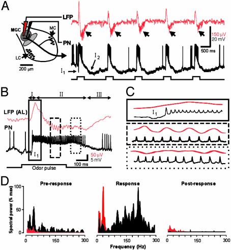
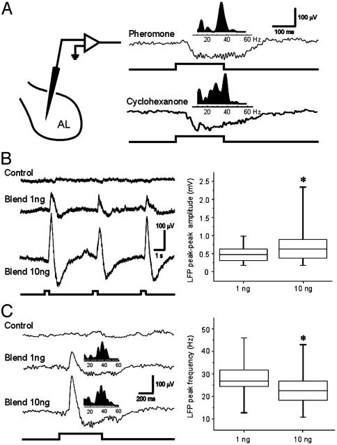
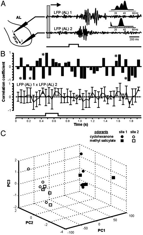
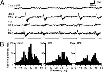
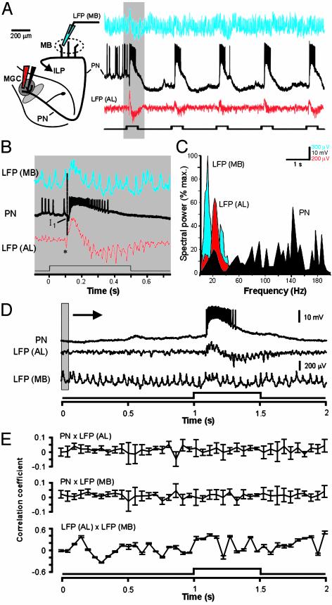
At the first stage of processing in the olfactory pathway, the patterns of glomerular activity evoked by different scents are both temporally and spatially dynamic. In the antennal lobe (AL) of some insects, coherent firing of AL projection neurons (PNs) can be phase-locked to network oscillations, and it has been proposed that oscillatory synchronization of PN activity may encode the chemical identity of the olfactory stimulus. It remains unclear, however, how the brain uses this time-constrained mechanism to encode chemical identity when the stimulus itself is unpredictably dynamic. In the olfactory pathway of the moth Manduca sexta,we find that different odorants evoke gamma-band oscillations in the AL and the mushroom body (a higher-order network that receives input from the AL), but oscillations within or between these two processing stages are not temporally coherent. Moreover, the timing of action potential firing in PNs is not phase-locked to oscillations in either the AL or mushroom body, and the correlation between PN synchrony and field oscillations remains low before, during, and after olfactory stimulation. These results demonstrate that olfactory circuits in the moth are specialized to preserve time-varying signals in the insect's olfactory space, and that stimulus dynamics rather than intrinsic oscillations modulate the uniquely coordinated pattern of PN synchronization evoked by each olfactory stimulus. We propose that non-oscillatory synchronization provides an adaptive mechanism by which PN ensembles can encode stimulus identity while concurrently monitoring the unpredictable dynamics in the olfactory signal that typically occur under natural stimulus conditions.


Figures

Fig. 1.
Fig. 1.
Simultaneous intracellular and field potential recordings illustrate that spiking activity in glomerular PNs (black) is not dependent on LFP oscillations (red). (A) Schematic drawing of the moth AL (Left) showing the approximate placement of the extracellular (LFP) and intracellular (PN) electrodes in the MGC. (Right) LFP and PN responses to five consecutive pulses of the pheromone blend (10 ng of BAL plus 10 ng of C15) are shown (stimulus duration = 500 ms; interval = 2 s). Note how both the LFP and PN responses are closely time-locked to each stimulus pulse. In the PN, this is facilitated by the large but brief IPSP (I1). Oscillations in the LFP appear only late in the response (arrowheads). (B) In another experiment, an expanded view of the first response epoch shows the details of the temporal relationship between the multiphasic LFP (phases I–III) and the PN spike train. PN spiking activity is initiated during phase I (solid outline), but LFP oscillations do not appear until phase II (dashed outline). Moreover, PN spikes are not phase-locked to the LFP at any time during the olfactory response. (C) Expanded view of boxed areas outlined in B.(D) Frequency spectra (smoothed with a 5-ms Gaussian function and normalized to maximum) computed for the PN (black) and LFP (red) before (Pre-response), during (Response), and after (Post-response) the olfactory response. Note that, during the odor-evoked response, the peak frequencies derived from the two recorded signals do not match.
Fig. 2.
Fig. 2.
Effects of odor chemistry and stimulus intensity on evoked LFPs. (A) LFP recorded near the center of the AL (Left). Raw LFP recordings (Right) show that different olfactory stimuli evoke similar temporal patterns in the LFP. Fourier analysis of the two responses (Insets) also reveals similar spectral peaks of 34 and 38 Hz for the pheromone blend (10 ng of BAL plus 10 ng of C15) and cyclohexanone (10 μg), respectively. (B) Effect of stimulus intensity on the LFP response to pheromone. Raw LFP traces (Left) reveal a dose-dependent effect on response amplitude along with a small reduction in peak spectral frequency (36 Hz for 1 ng of blend; 34 Hz for 10 ng of blend). Box plots based on pooled data from 35 trials (Right) illustrate the significant effect of stimulus intensity on peak-to-peak LFP amplitude (Wilcoxon rank sum test, P < 0.01). Boxes mark the upper and lower quartiles, the line inside each box marks the sample median, and vertical bars show the sample range. (C) Recordings from a different preparation illustrate the spectral changes in the LFP responses triggered by different stimulus intensities. Frequency analysis (Insets above raw LFP traces) reveals a spectral peak at 38 Hz for the 1-ng blend, but a reduction to 34 Hz for the 10-ng blend. (Right) Box plots based on pooled data (n = 35 trials) confirm a small but significant decrease in the mean LFP peak frequency with increasing stimulus intensity (P < 0.01).
Fig. 3.
Fig. 3.
LFPs evoked by the same olfactory stimulus but recorded from spatially distinct sites in the same neuropil are not temporally coherent. (A) Spatial variation in stimulus-evoked LFP responses recorded simultaneously from two different glomerular sites separated by 270 μm. Cyclohexanone evokes different LFP patterns across the AL, and these patterns show different spectral properties (Insets). Gray box depicts the 50-ms sliding window used to calculate the running correlations in B. LFPs were band-passed from 5 to 55 Hz. (B) Sliding-window plots of the time course of correlation between the two LFPs reveal no evidence for global coherence before, during, or after a 200-ms odor pulse. Sliding windows of 20, 50, and 100 ms were used to calculate running correlations; all showed the same outcome; thus, only the results using the 50-ms window are shown. The upper trace shows the covariation for the first of a series of five odor pulses separated by 2 s; the lower trace shows the data averaged over the five pulses (mean ± SD). Note that the strongest correlations (asterisks) did not occur during peak LFP activity. (C) Principal components analysis (PCA) showed that, irrespective of the stimulus, LFP responses recorded at the same site showed considerably less temporal variation than those recorded at different sites. The LFP responses evoked by two nonpheromonal stimuli (cyclohexanone and methyl salicylate, four trials each) were recorded simultaneously at two separate sites outside of the MGC, as shown in A. PCA revealed a clear separation between the clusters of LFP responses recorded at the two sites (99% of the variation was associated with the first three eigenvectors in the 3D plot). The strong segregation of clusters between the two sites suggests that LFP responses may not propagate globally, but are instead localized to different regions of the AL, perhaps even to different glomeruli (see text).
Fig. 4.
Fig. 4.
The temporal structure of the LFP depends on whether the glomerulus receives direct input from the olfactory stimulus. (A) LFP was recorded from the toroid, a large glomerulus in the male's MGC that receives specific input from one component of the female sex pheromone, BAL. The complete pheromone (Blend) and BAL evoked multiphasic LFPs, whereas C15 (a mimic of the second pheromone component that selectively stimulates the adjacent glomerulus) evoked only oscillations. (B) Power-spectral analysis performed over the five trials shows that the distribution of frequency peaks is different for BAL and C15, and different still for the blend of the two olfactory stimuli.
Fig. 5.
Fig. 5.
Lack of coincidence structure between PN spike trains (black) recorded simultaneously with LFPs from the MB and AL. (A) Each stimulus pulse modulates network activity at both sites: LFP (MB) is shown in blue; LFP (AL) is shown in red. The stimulus protocol was five consecutive pulses of cyclohexanone, each 500 ms in duration and separated by 2 s (lower trace). (B) Expanded view of traces from shaded area in A. The downward deflection (asterisk) in LFP (AL) likely represents the synchronous arrival of excitatory afferent input to the glomerulus. (C) Spectral analysis reveals no overlap in the peak frequencies between LFPs, and no correspondence between either LFP and peak PN activity. (D) Lack of correlation between PN spiking and LFPs recorded simultaneously in the AL and MB. Raw PN and LFP traces illustrate the response to a single 500-ms pulse of sex pheromone. The shaded area depicts the sliding window (50 ms) used to calculate moving correlations in E. (E) Sliding-window correlations calculated between PN membrane potential and LFPs, measured before, during, and after olfactory stimulation; results using the 50-ms window are shown. Sliding correlations between PN spike activity and LFPs in the AL (Top) or MB (Middle) reveal no peaks at any time before, during, or after the stimulus (P = 0.31 and 0.42 for top and middle traces, respectively). There also was no significant correlation (P = 0.09) between LFPs in the MB and AL (Bottom).

Similar articles

Cited by

References

    1. Gelperin, A. (1999) J. Exp. Biol. 202, 1855–1864. - PubMed
    1. Mori, K., Nagao, H. & Yoshihara, Y. (1999) Science 286, 711–715. - PubMed
    1. Laurent, G. (1999) Science 286, 723–728. - PubMed
    1. Christensen, T. A. & White, J. (2000) in The Neurobiology of Taste and Smell, eds. Finger, T. E., Silver, W. L. & Restrepo, D. (Wiley, New York), pp. 201–232.
    1. Lam, Y.-W., Cohen, L. B., Wachowiak, M. & Zochowski, M. R. (2000) J. Neuroscience 20, 749–762. - PMC - PubMed

Publication types

LinkOut - more resources